SciELO - Scientific Electronic Library Online

 
vol.38 issue4Endoscopic Ultrasound-Directed Transgastric Endoscopic Retrograde Cholangiopancreatography in a Patient with Gastric Bypass. Report of the First Local CaseManganese Associated with Non-Wilsonian Hepatolenticular Degeneration as a Rare Cause of Encephalopathy: Case Report author indexsubject indexarticles search
Home Pagealphabetic serial listing  

Services on Demand

Journal

Article

Indicators

Related links

  • On index processCited by Google
  • Have no similar articlesSimilars in SciELO
  • On index processSimilars in Google

Share


Revista colombiana de Gastroenterología

Print version ISSN 0120-9957On-line version ISSN 2500-7440

Rev. colomb. Gastroenterol. vol.38 no.4 Bogotá Oct./Dec. 2023  Epub Feb 26, 2024

https://doi.org/10.22516/25007440.993 

Report of cases

Primary Anorectal Melanoma: Case Report and Review of a Rare Neoplasm

Hernando Marulanda-Fernández1 
http://orcid.org/0000-0003-3941-0704

Felipe Vera-Polanía2 
http://orcid.org/0000-0002-4509-6873

Juan Antonio Trejos-Naranjo3 
http://orcid.org/0000-0002-5644-1820

Julián Ernesto Parga-Bermúdez4 
http://orcid.org/0000-0001-8988-2887

William Otero-Regino5  * 
http://orcid.org/0000-0002-6825-9014

1Gastroenterologist, Universidad Nacional de Colombia, Hospital Universitario Nacional de Colombia. Bogotá, Colombia.

2Gastroenterology fellow, Universidad Nacional de Colombia. Internal medicine specialist, Universidad del Valle. Bogotá, Colombia.

3Internist. Gastroenterology fellow, Hospital Universitario Nacional de Colombia. Bogotá, Colombia.

4Gastroenterologist. Coordinator of the gastroenterology service, Hospital El Tunal. Bogotá, Colombia.

5Gastroenterologist. Professor of Medicine, Gastroenterology Unit, Universidad Nacional de Colombia, Hospital Universitario Nacional de Colombia. Bogotá, Colombia.


Abstract

Primary anorectal melanoma is a rare malignant melanocytic neoplasm; its principal manifestation is rectal bleeding. It has an ominous prognosis with a five-year survival rate of 10%. The case of a 56-year-old woman with rectal bleeding and the sensation of a rectal mass is presented. A polypoid lesion, resected transanally, was documented in the distal rectum during the colonoscopy. The histological study confirmed a primary anorectal melanoma.

Keywords: Malignant melanoma; anal neoplasms; rectal neoplasms; gastrointestinal bleeding; colonoscopy

Resumen

El melanoma anorrectal primario es una neoplasia melanocítica maligna poco frecuente, su principal manifestación es el sangrado rectal. Tiene un pronóstico ominoso con una tasa de sobrevida del 10% a 5 años. Se presenta el caso de una mujer de 56 años con rectorragia y sensación de masa rectal. Durante la colonoscopia se documentó una lesión polipoide en el recto distal, que se resecó por vía transanal. El estudio histológico confirmó la presencia de un melanoma anorrectal primario.

Palabras clave: Melanoma maligno; neoplasias del ano; neoplasias del recto; hemorragia gastrointestinal; colonoscopia

Introduction

Primary anorectal melanoma (PAM) is a rare pathology that represents less than 1% of all colorectal tumors1. The primary gastrointestinal mucosal location is the third most common after the skin and retina and mainly affects women between the fifth and sixth decade of life2. At the anorectal level, the lesion can appear anywhere melanocytes are found (perianal region, transitional zone, or rectal mucosa)3. The most frequently described clinical manifestations are rectal bleeding, the sensation of mass, and alterations in defecation. It is not uncommon for these lesions to be confused with complicated hemorrhoidal disease, which delays the diagnosis and negatively impacts the prognosis4.

Clinical case

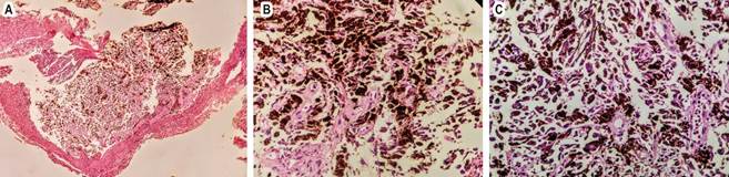
A 56-year-old woman with a history of rectal bleeding and hematochezia was referred with a diagnosis of thrombosed internal hemorrhoids. During the gastroenterology evaluation, a rectal examination and anoscopy were performed, identifying an indurated violaceous mass that protruded through the anal canal. The colonoscopy revealed a 3-cm violaceous polypoid lesion with ulcerated foci and stigmata of recent bleeding (Figure 1). In the thoracoabdominal computed tomography (CT), no lesions suspicious of metastasis or lymph node involvement were found. Subsequently, a transanal surgical resection of the lesion was performed, and the pathology was reported as a semi-pedunculated polypoid formation covered by grayish mucosa, limited to the submucosa without vascular or perineural invasion. Histology describes a malignant cell tumor with hyperchromatic pleomorphic nuclei with nucleolus, abundant brown pigment in their cytoplasm, and extensive superficial ulceration (Figure 2). Finally, the patient was assessed by dermatology and ophthalmology, who found no skin or ocular lesions suggestive of melanoma. The patient continues to be followed up by gastroenterology and oncology; there is no evidence of local or imaging recurrence.

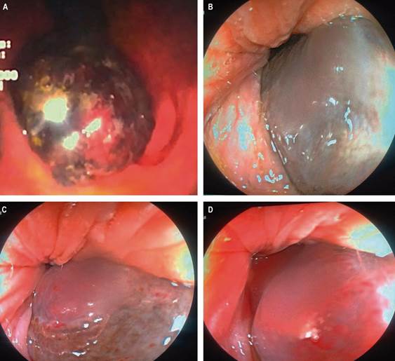
Source: Case photos, authors’ archive.

Figure 1 Colonoscopy imaging. A. In retroflexion, a hyperpigmented polyploid-looking lesion involving the distal rectum is observed. B, C, and D. A polyploid-looking lesion is observed that protrudes through the anal canal.  

Source: case photos, authors’ archive.

Figure 2 A, B and C. Histology demonstrates a malignant tumor of cells with pleomorphic and hyperchromatic nuclei with nucleoli and abundant brown pigment in their cytoplasm.  

Discussion

PAM was first described in 1857 by Moore; it is a rare pathology, with an incidence of 1.7 cases per million inhabitants5. Mucosal melanoma (MM) represents the third most common site of primary melanomas after the skin and eyes, and the incidence varies depending on its location6 (Table 1). Compared to cutaneous melanoma (CM), primary gastrointestinal melanoma is sporadic; its most frequent location is the anorectum (more than 50%), followed by the stomach, small intestine, and colon. Rectal location is more frequent than anal7. Countries with a high incidence of CM have similar incidence rates of MM compared to other geographic regions8. It is more common in women than men (2:1 ratio), and the average age at diagnosis is 54.5 years9, coinciding with the case presented. Overall survival is poor and ranges between 10% and 20% at five years, probably because of late diagnosis10. Most cases arise from the mucocutaneous junction; however, they can also arise from the skin of the anal verge, the transitional epithelium of the anal canal, or the rectal mucosa11.

Table 1 Epidemiology of primary mucosal melanoma 

Primary mucosal melanoma Cases/million/year
Conjunctival 0.5/million/year
Sinonasal 0.5/million/year
Anorectal 0.4/million/year
Oral 0.2/million/year

Taken and modified from Micu E. J Eur Acad Dermatol Venereol. 2011;25(12):1444-9.

Solar radiation is the stimulus that causes the changes necessary for malignant transformation in the case of CM; in the case of anal melanoma, the causes are unknown. Melanin synthesis is the primary function of melanocytes; their role in mucosal areas is mainly characterized by antioxidant and adjuvant activity in the local immune response12.

Malignant transformation in anorectal areas may be related to environmental and genetic factors, oxidative stress, immunosuppression, or viral infections13, and human immunodeficiency virus (HIV) infection is considered an associated risk factor14. Another hypothesis is hormonal, possibly due to the participation of estrogens in the pathogenesis of melanoma, increasing the number of melanocytes and their melanin content15. These lesions may be an incidental finding during an endoscopic study or the result of histological analysis of a rectal polyp or hemorrhoidectomy16.

Nodal involvement is found in approximately 60% of patients at diagnosis and distant metastases in 30% of cases17. The diagnosis should be suspected when observing a pigmented lesion in the anal canal, although 10% to 30% of these neoplasms are amelanotic18. An exhaustive search should be performed to identify malignant skin lesions with the potential to develop GI tract metastases to differentiate and clarify the diagnosis of the primary lesion19.

The clinical manifestations can be very varied; the most frequent symptoms of this neoplasia are rectal bleeding, proctalgia, pruritus, tenesmus, sensation of anal mass, or alteration of intestinal habit; the lesion can protrude through the anal sphincter either during defecation or with the Valsalva maneuver, becoming a reason for consultation20. In a recent series, only 0.8% of patients manifested bleeding symptoms21. Regarding the endoscopic findings, the manifestation may be a melanotic polypoid lesion, as occurred in this case, or a mass that simulates colorectal cancer22. Histologically, the melanin pigment is characteristic, although immunohistochemistry with positivity for HMB-45 and S-100 protein is much more specific23.

Treatment

Surgery is the most effective treatment for PAM if proper staging is performed. Contrast-enhanced magnetic resonance imaging (MRI) of the abdomen and pelvis allows us to clarify the degree of invasion24. PAM has an aggressive behavior; most patients have metastases at the time of diagnosis, mainly to the lungs25. Local resection (LR) has greater immediate benefits for the patient, such as early recovery, shorter hospital stays, and minimal impact on intestinal function. Abdominoperineal resection (APR) offers better disease-free survival and greater locoregional control26. Neither of these two techniques is a definitive cure; APR has given fewer recurrences than LR (29% versus 58%)27. The role of lymphadenectomy in surgical treatment is controversial26. Bilateral inguinal lymphadenectomy in patients with PAM without palpable lymph nodes increases the risk of complications without improvement in survival28. Control of local primary disease can be achieved with adjuvant radiotherapy after surgical resection29. Non-surgical treatment of PAM includes the administration of chemotherapy and radiotherapy. Moozar et al. evaluated the usefulness of radiotherapy for palliative purposes in a series of patients with PAM, noting an improvement in survival (16 months versus five months) compared to patients who underwent surgery alone30.

Regarding systemic therapies, there is currently no consensus on the most appropriate treatment for this type of melanoma. Some protocols, previously aimed at patients with CM, have been studied in the context of MM, including PAM31. A multicenter case-control study evaluating the effect of a four-drug combination known as the Dartmouth regimen (dacarbazine, cisplatin, carmustine, and tamoxifen) versus dacarbazine found a better response rate (19% vs. 10%)32. Singhal et al. reported on the efficacy of taxanes and showed a median overall survival of 11 months33.

Follow-up

There is no clarity regarding how follow-up should be performed after treatment of melanoma with primary rectal involvement. Its approach is based on the extrapolation of patients with CM34. A retrospective study that included 81 patients described the most frequent locations of metastases, regardless of the primary site of the disease, and reported extension to the liver (57%), lungs (41%), lymph nodes (38%), soft tissues (22%), bone (12%), and the central nervous system (7%). With these data, the best follow-up option likely includes brain, chest, and abdominopelvic CT. Regarding endoscopic follow-up, the surveillance intervals are not defined; some referral centers recommend following closely (every three months) during the first year and then a colonoscopy every six months35.

Conclusion

PAM is a rare malignant neoplasm that poses a diagnostic challenge. This tumor is characterized by aggressive behavior due to biological differences in the melanocytes in this anatomical area compared to other locations. Factors such as inadequate discernment of endoscopic findings, which are confused with benign pathologies such as hemorrhoidal disease, provide a poor short-term prognosis. Due to its low incidence, few studies report what the best therapeutic and surveillance strategy is; most data are extrapolations from CM. The survival of patients depends on the stage at the time of diagnosis. In this case, the patient underwent local resection and was referred to the oncology service. Therefore, it is deemed essential that gastroenterologists become familiar with this type of lesion, which will allow a timely diagnosis and adequate treatment.

Referencias

1. Hicks CW, Pappou EP, Magruder JT, Gazer B, Fang S, Wick EC, et al. Clinicopathologic presentation and natural history of anorectal melanoma: a case series of 18 patients. JAMA Surg. 2014;149(6):608-611. https://doi.org/10.1001/jamasurg.2013.4643Links ]

2. Callahan A, Anderson WF, Patel S, Barnholtz-Sloan JS, Bordeaux JS, Tucker MA, et al. Epidemiology of anorectal melanoma in the United States: 1992 to 2011. Dermatol Surg. 2016;42(1):94-9. https://doi.org/10.1097/DSS.0000000000000579Links ]

3. Seetharamu N, Ott PA, Pavlick AC. Mucosal melanomas: a review of the literature. Oncologist. 2010;15(7):772-781. https://doi.org/10.1634/theoncologist.2010-0067Links ]

4. Edelman A, Brown T, Gandhi R, Gandhi R. Anorectal Melanoma Misdiagnosed as Hemorrhoids: A Case Report and Review of the Literature. J Clin Aesthet Dermatol. 2021;14(4):32-35. [ Links ]

5. Parra RS, Almeida AL, Badiale BG, Moraes MM, Rocha JJ, Féres O. Melanoma of the anal canal. Clinics (Sao Paulo). 2010;65(10):1063-65. https://doi.org/10.1590/S1807-59322010001000026Links ]

6. Micu E, Juzeniene A, Moan J. Comparison of the time and latitude trends of melanoma incidence in anorectal region and perianal skin with those of cutaneous malignant melanoma in Norway. J Eur Acad Dermatol Venereol. 2011;25(12):1444-9. https://doi.org/10.1111/j.1468-3083.2011.04023.xLinks ]

7. Ragnarsson-Olding BK, Nilsson PJ, Olding LB, Nilsson BR. Primary ano-rectal malignant melanomas within a population-based national patient series in Sweden during 40 years. Acta Oncol. 2009;48(1):125-31. https://doi.org/10.1080/02841860802120861Links ]

8. Miller BJ, Rutherford LF, McLeod GR, Cohen JR. Where the sun never shines: Anorectal melanoma. Aust N Z J Surg. 1997;67(12):846-8. https://doi.org/10.1111/j.1445-2197.1997.tb07609.xLinks ]

9. Postow MA, Hamid O, Carvajal RD. Mucosal melanoma: Pathogenesis, clinical behavior, and management. Curr Oncol Rep. 2012;14(5):441-8. https://doi.org/10.1007/s11912-012-0244-xLinks ]

10. Cagir B, Whiteford MH, Topham A, Rakinic J, Fry RD. Changing epidemiology of anorectal melanoma. Dis Colon Rectum. 1999;42(9):1203-08. https://doi.org/10.1007/BF02238576Links ]

11. Row D, Weiser MR. Anorectal melanoma. Clin Colon Rectal Surg. 2009;22(2):120-6. https://doi.org/10.1055/s-0029-1223844Links ]

12. Zecca L, Zucca FA, Wilms H, Sulzer D. Neuromelanin of the substantia nigra: A neuronal black hole with protective and toxic characteristics. Trends Neurosci. 2003;26(11):578-80. https://doi.org/10.1016/j.tins.2003.08.009Links ]

13. Curtin JA, Fridlyand J, Kageshita T, Patel HN, Busam KJ, Kutzner H, et al. Distinct sets of genetic alterations in melanoma. N Engl J Med. 2005;353(20):2135-47. https://doi.org/10.1056/NEJMoa050092Links ]

14. Jensen C, Kin C. Black is the new black: Prolapsing primary anorectal melanoma. Dig Dis Sci. 2017;62(11):2991-3. https://doi.org/10.1007/s10620-017-4527-9Links ]

15. Smith MA, Fine JA, Barnhill RL, Berwick M. Hormonal and reproductive influences and risk of melanoma in women. Int J Epidemiol. 1998;27(5):751-7. https://doi.org/10.1093/ije/27.5.751Links ]

16. Bullard KM, Tuttle TM, Rothenberger DA, Madoff RD, Baxter NN, Finne CO, et al. Surgical therapy for anorectal melanoma. J Am Coll Surg. 2003;196(2):206-11. https://doi.org/10.1016/S1072-7515(02)01538-7Links ]

17. Smith HG, Glen J, Turnbull N, Peach H, Board R, Payne M, et al. Less is more: A systematic review and meta-analysis of the outcomes of radical versus conservative primary resection in anorectal melanoma. Eur J Cancer. 2020;135:113-120. https://doi.org/10.1016/j.ejca.2020.04.041Links ]

18. Pessaux P, Pocard M, Elias D, Duvillard P, Avril MF, Zimmerman P, et al. Surgical management of primary anorectal melanoma. Br J Surg. 2004;91(9):1183-7. https://doi.org/10.1002/bjs.4592Links ]

19. Nam S, Kim CW, Baek SJ, Hur H, Min BS, Baik SH, et al. The clinical features and optimal treatment of anorectal malignant melanoma. Ann Surg Treat Res. 2014;87(3):113-17. https://doi.org/10.4174/astr.2014.87.3.113Links ]

20. Zhou HT, Zhou ZX, Zhang HZ, Bi JJ, Zhao P. Wide local excision could be considered as the initial treatment of primary anorectal malignant melanoma. Chin Med J (Engl). 2010;123(5):585-8. [ Links ]

21. David AW, Perakath B. Management of anorectal melanomas: a 10-year review. Trop Gastroenterol. 2007;28(2):76-8. [ Links ]

22. Slingluff CL Jr, Vollmer RT, Seigler HF. Anorectal melanoma: clinical characteristics and results of surgical management in twenty-four patients. Surgery. 1990;107(1):1-9. [ Links ]

23. Das G, Gupta S, Shukla PJ, Jagannath P. Anorectal melanoma: a large clinicopathologic study from India. Int Surg. 2003;88(1):21-4. [ Links ]

24. Chae WY, Lee JL, Cho D-H, Yu SK, Roh, J, Kim JC. Preliminary suggestion about staging of anorectal malignant melanoma may be used to predict prognosis. Cancer Res Treat. 2016;48(1):240-9. https://doi.org/10.4143/crt.2014.305Links ]

25. Podnos YD, Tsai NC, Smith D, Ellenhorn JD. Factors affecting survival in patients with anal melanoma. Am Surg. 2006;72(10):917-20. https://doi.org/10.1177/000313480607201017Links ]

26. Yeh JJ, Shia J, Hwu WJ, Busam KJ, Paty PB, Guillem JG, et al. The role of abdominoperineal resection as surgical therapy for anorectal melanoma. Ann Surg. 2006;244(6):1012-17. https://doi.org/10.1097/01.sla.0000225114.56565.f9Links ]

27. Falch C, Mueller S, Kirschniak A, Braun M, Koenigsrainer A, Klumpp B. Anorectal malignant melanoma: Curative abdominoperineal resection: patient selection with 18F-FDG-PET/CT. World J Surg Oncol. 2016;14(1):185. https://doi.org/10.1186/s12957-016-0938-xLinks ]

28. Ceccopieri B, Marcomin AR, Vitagliano F. Primary anorectal malignant melanoma: report of two cases. Tumori. 2000;86(4):356-8. https://doi.org/10.1177/030089160008600430Links ]

29. Bujko K, Nowacki MP, Liszka-Deleck P. Radiation therapy for anorectal melanoma - a report of three cases. Acta Oncol. 1998;37(5):497-9. https://doi.org/10.1080/028418698430485Links ]

30. Moozar KL, Wong CS, Couture J. Anorectal malignant melanoma: treatment with surgery or radiation therapy, or both. Can J Surg. 2003;46(5):345-9. [ Links ]

31. Tacastacas JD, Bray J, Cohen YK, Arbesman J, Kim J, Koon HB, et al. Update on primary mucosal melanoma. J Am Acad Dermatol. 2014;71(2):366-753. https://doi.org/10.1016/j.jaad.2014.03.031Links ]

32. Chapman PB, Einhorn LH, Meyers ML, Saxman S, Destro AN, Panageas KS, et al. Phase III multicenter randomized trial of the Dartmouth regimen versus dacarbazine in patients with metastatic melanoma. J Clin Oncol. 1999;17(9):2745-51. https://doi.org/10.1200/JCO.1999.17.9.2745Links ]

33. Singhal NK, Ostwal VS, Shrikhande SV, Saklani A, Arya S, Ramadvar M. Systemic therapy in anorectal melanoma: Does choice of systemic therapy matter? ASCO. J Clin Oncol. 2016;34(suppl 4S; abstr 731). https://doi.org/10.1200/jco.2016.34.4_suppl.731Links ]

34. Thibault C, Sagar P, Nivatvongs S, Ilstrup DM, Wolff BG. Anorectal melanoma--an incurable disease? Dis Colon Rectum. 1997;40(6):661-8. https://doi.org/10.1007/BF02140894Links ]

35. Shoushtari AN, Bluth MJ, Goldman DA, Bitas C, Lefkowitz RA, Postow MA, et al. Clinical features and response to systemic therapy in a historical cohort of advanced or unresectable mucosal melanoma. Melanoma Res. 2017;27(1):57-64. https://doi.org/10.1097/CMR.0000000000000306Links ]

Citation: Marulanda-Fernández H, Felipe Vera-Polanía F, Trejos-Naranjo JA, Parga-Bermúdez JE, Otero-Regino W. Primary Anorectal Melanoma: Case Report and Review of a Rare Neoplasm. Revista. colomb. Gastroenterol. 2023;38(4):504-509. https://doi.org/10.22516/25007440.993

Received: November 24, 2022; Accepted: May 26, 2023

*Correspondence: William Otero-Regino. waoteror@gmail.com

Conflict of interest

The authors declare no conflict of interest.

Creative Commons License This is an open-access article distributed under the terms of the Creative Commons Attribution License