SciELO - Scientific Electronic Library Online

 
vol.38 número4Severe Hypocalcemia as an Atypical Manifestation of Seronegative Celiac Disease in a Patient with Systemic Lupus Erythematosus: Case ReportGiant Submucosal Lipoma of the Right Colon Spontaneously Expelled Rectally: Case Report índice de autoresíndice de assuntospesquisa de artigos
Home Pagelista alfabética de periódicos  

Serviços Personalizados

Journal

Artigo

Indicadores

Links relacionados

  • Em processo de indexaçãoCitado por Google
  • Não possue artigos similaresSimilares em SciELO
  • Em processo de indexaçãoSimilares em Google

Compartilhar


Revista colombiana de Gastroenterología

versão impressa ISSN 0120-9957versão On-line ISSN 2500-7440

Rev. colomb. Gastroenterol. vol.38 no.4 Bogotá out./dez. 2023  Epub 26-Fev-2024

https://doi.org/10.22516/25007440.941 

Report of cases

Infiltrating Lobular Carcinoma of the Breast Metastatic to the Duodenum, about a Case

Yirdley Gisella Sandoval-Vargas1 
http://orcid.org/0000-0002-0283-3650

Iván Enrique González-Mendoza2  * 
http://orcid.org/0000-0002-8894-7834

Raúl Antonio Cañadas-Garrido3 
http://orcid.org/0000-0003-2938-7731

Diana Valentina Ariza-Muñoz4 
http://orcid.org/0000-0003-4102-3893

1Physician and surgeon, internal medicine resident, Hospital Universitario San Ignacio. Pontificia Universidad Javeriana. Bogotá, Colombia.

2Physician, internal medicine specialist, gastroenterology fellow, Hospital Universitario San Ignacio. Pontificia Universidad Javeriana. Bogotá, Colombia.

3Physician, internal medicine and gastroenterology specialist, Gastroenterology Unit, Hospital Universitario San Ignacio. Associate professor, Pontificia Universidad Javeriana. Bogotá, Colombia.

4Physician and surgeon, pathology resident, Hospital Universitario San Ignacio. Pontificia Universidad Javeriana. Bogotá, Colombia.


Abstract

Introduction:

Gastric outlet obstruction or pyloric syndrome can occur secondary to neoplastic involvement, and metastasis as an etiology is unusual. Breast neoplasms generally cause bone, liver, and lung metastases, rarely involving the gastrointestinal tract.

Case presentation:

A 69-year-old female patient with infiltrating lobular carcinoma of the right breast consulted for abdominal pain and postprandial emetic episodes with oral intolerance and dyspnea. Bilateral neoplastic breast involvement and dilation of the gastric chamber with thickening of the pylorus were recorded. She required antiemetic management and placement of a nasogastric tube. She was taken to an upper digestive tract endoscopy, which found an ulcerated lesion with an infiltrative appearance at the postpyloric level that circumferentially compromised the duodenal lumen. Then, a biopsy was taken, which was compatible with a breast carcinoma of a lobular type. This entity, called pyloric syndrome due to neoplasia, can be managed with gastrojejunostomy or an enteral prosthesis that improves the quality of life of patients with an ominous short-term prognosis. The patient in our case expressed advance directives not to receive invasive procedures, for which an uncovered metal prosthesis was placed for palliative purposes, achieving the re-establishment of the feeding route and resolution of dyspnea due to restriction.

Conclusions:

The metastatic involvement of neoplasms of the breast to the gastrointestinal tract is rare; however, it should be suspected in elderly patients with previously documented neoplasms.

Keywords: Breast neoplasms; metastasis of neoplasms; pylorus; case reports; duodenal obstruction

Resumen

Introducción:

La obstrucción al tracto de salida gástrico o síndrome pilórico puede presentarse de manera secundaria a compromiso neoplásico, y es inusual el compromiso metastásico como etiología. Las neoplasias de la mama generalmente originan metástasis óseas, hepáticas y pulmonares, y es infrecuente el compromiso del tracto digestivo.

Presentación del caso:

Se trata de una paciente de 69 años con carcinoma lobulillar infiltrante de mama derecha que consultó por dolor abdominal y episodios eméticos posprandiales con intolerancia a la vía oral y disnea. Se registró un compromiso neoplásico mamario bilateral y dilatación de la cámara gástrica con engrosamiento del píloro. Se dio manejo antiemético y colocación de sonda nasogástrica. Fue llevada a una endoscopia de vías digestivas altas en la que se encontró una lesión ulcerada de aspecto infiltrativo a nivel pospilórico que comprometía de forma circunferencial la luz duodenal, y luego se tomó una biopsia a este nivel, que fue compatible con un carcinoma mamario de tipo lobulillar. Esta entidad, denominada síndrome pilórico por neoplasias, puede manejarse con gastroyeyunostomía o con prótesis enteral que mejore la calidad de vida en pacientes con pronóstico ominoso a corto plazo. La paciente de nuestro caso manifestó voluntades anticipadas para no recibir procedimientos invasivos, por lo cual se ofreció la colocación de una prótesis metálica descubierta con fines paliativos y se logró el restablecimiento de la vía de alimentación y resolución de la disnea por restricción.

Conclusiones:

El compromiso metastásico de neoplasias de la mama al tracto digestivo es poco frecuente; sin embargo, debe sospecharse en pacientes de edad avanzada y con neoplasias previamente documentadas.

Palabras clave: Neoplasias de la mama; metástasis de las neoplasias; píloro; informes de caso; obstrucción duodenal

Introduction

Pyloric syndrome or obstruction of the gastric outlet tract is an entity that can occur secondary to neoplastic involvement and adversely affect patients’ quality of life1,2. Metastatic involvement as an etiology is unusual and even less frequently associated with a primary breast tumor3,4, which makes it reasonable that in cases like the one described, various differential diagnoses should be suspected and explored.

Clinical case

A 69-year-old woman was diagnosed with infiltrating lobular carcinoma of the right breast one month before admission. She was admitted for a week of diffuse abdominal pain associated with nausea, postprandial emesis, and dyspnea at rest. On admission, she was found to be in a fair general condition, tachycardic and tachypneic, with ambient saturation of 57%, and she had no other relevant history. Management was given with supplemental oxygen, and a study for dyspnea and emetic syndrome was initiated. Contrast-enhanced CT studies of the chest and abdomen were performed, and the results were negative for pulmonary thromboembolism, with findings of basal subsegmental atelectasis and pericardial effusion without hemodynamic compromise. Additionally, bilateral breast neoplastic involvement and dilation of the gastric chamber with thickening of the pylorus were evident (Table 1).

Table 1 Paraclinical studies 

Study Result
Transthoracic echocardiogram Left ventricle of average size, concentric remodeling, preserved systolic function, EF of 60% without segmental disorders; moderate pericardial effusion with left pleural effusion and signs of collapsed or infiltrated lung segments; no significant valvular heart disease
Contrast-enhanced CT of the chest Bilateral breast masses predominantly on the left concerning a known neoplastic history, with suspicious-looking lymph nodes in both axillary regions and mixed polyostotic involvement attributed to a secondary neoplastic disease; subsegmental atelectasis in the middle lobe and lingula
Contrast-enhanced CT of the abdomen Moderate hepatic steatosis; dilation of the gastric chamber with apparent thickening of the pylorus walls; uncomplicated diverticular disease of the colon; polyostotic involvement concerning secondary neoplastic disease
Breast biopsy Classic invasive lobular carcinoma

EF: ejection fraction. Prepared by the authors.

With these findings, the hypoxemia was attributed to atelectatic involvement. Regarding the gastrointestinal condition, it was considered compatible with a pyloric syndrome, and management was initiated with a nasogastric tube and antiemetic, resulting in partial improvement. The patient was taken for esophagogastroduodenoscopy, which showed changes in esophagitis due to food retention and content in the gastric chamber, which, in turn, revealed an infiltrative-looking lesion with an ulcerated surface and easy bleeding at the postpyloric level that circumferentially compromised the duodenal lumen (Figure 1).

Source: Authors’ archive.

Figure 1 Prepyloric region with edematous, friable mucosa easily bleeding upon passage of the equipment with circumferential involvement.  

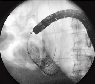
Biopsies were taken from the lesion in the duodenum, which reported involvement by a group of atypical and discohesive cells with immunohistochemistry compatible with a lobular-type carcinoma of breast origin (negative for cytokeratin 20, CDX-2, and E-cadherin) (Figure 2). The findings were explained; however, the patient expressed advance directives not to receive invasive procedures, for which a 22 mm × 60 mm uncovered metal prosthesis was inserted to reestablish the feeding route and improve dyspnea and abdominal pain (Figure 3).

Source: Authors’ archive.

Figure 2 A. (Hematoxylin-eosin). Duodenal epithelium with preserved architecture and fragments of submucosa compromised by a tumor infiltrate (black arrow). B. (Hematoxylin-eosin). The tumor cells are discohesive, small to medium in size, with nuclear pleomorphism and hyperchromasia. C. (GATA-3). Positivity of tumor cells. D. (E-cadherin). Tumor cells are negative for E-cadherin (red arrow); there is adequate internal control in the intestinal epithelium (green arrow).  

Source: Authors’ archive.

Figure 3 The duodenal prosthesis is positioned under fluoroscopy and takes the classic hourglass-shaped image.  

Discussion

Obstruction of the gastric outlet tract can be secondary to intrinsic or extrinsic causes that explain the mechanical blockage of gastric emptying; it can be located at the distal level of the stomach, pylorus, or duodenum and usually presents with nausea, vomiting, and abdominal pain1.

The etiology is usually divided into malignant (62%) and benign (38%), including gastric neoplasms, duodenal adenocarcinoma, carcinomas of the ampulla of Vater, neoplasms of the gallbladder, lymphomas, and metastases, among the malignant causes1,2. Benign etiologies include peptic ulcers, acute and chronic pancreatitis, inflammatory entities (such as Crohn’s disease), infiltrative entities (such as amyloidosis), and foreign bodies2-5.

Regarding infiltrating lobular carcinoma of the breast, it is characterized by discohesive cells and mutations in genes that code for E-cadherin since its absence supports the diagnosis6-8. However, there seems to be no association between this characteristic and the appearance of metastasis7,9. Multiple metastatic involvements of the bone, liver, and lung have been described but are rare in the peritoneum (14.6%) and the stomach (2.8%)10.

In the case of the patient, duodenal metastatic involvement due to already-known breast carcinoma was documented. While metastases are not frequent within tumor lesions of the duodenum, there are case series that describe prevalences of 32%, of which up to 12% are metastases from squamous cell carcinoma, clear cell carcinoma (8%), melanoma (8%), and pleomorphic liposarcoma (4%)3,11. Yim et al., in a series of cases of patients with pyloric syndrome, reported a metastatic etiology close to 27.6%12. Unusual manifestations also include metastatic involvement due to adenocarcinoma of the cecum, squamous cell carcinoma of the cervix and anus12-14, and pleomorphic lung carcinoma15,16. Regarding duodenal involvement due to breast carcinomas, episodes of gastric outlet obstruction secondary to or as the first manifestation of neoplasia are described, such as that reported by Khairy et al.17.

Among the therapeutic alternatives for outlet obstructions of benign etiology, pharmacological therapy and some endoscopic procedures, such as balloon dilations, are listed, leaving surgical management as the last option. Regarding the management of malignant obstructions, the insertion of antroduodenal prostheses, gastrojejunostomies (GJ), and endoscopic ultrasound-guided gastroenterostomy (EUS-GE) are described2,17-20.

Antroduodenal prostheses are metallic, whether or not covered by plastic or silicone membranes. The covered ones have a higher risk of migration but a lower stricture rate. The leading cause of stricture is intra-stent tumor growth (8-25.4%), and argon plasma therapy or a new intraluminal stent is required. Prosthesis migration occurs in 0% to 19.4%, mainly in covered ones; they are rearranged or removed endoscopically or surgically. Bleeding associated with the insertion occurs in 1%, and conservative or endoscopic management is usually given. They have a limited useful life and an in situ duration of up to 85 days. They are generally employed in patients with a poor short-term prognosis to improve quality of life and bring the oral route back, which allows longer relief of symptoms and lower frequency of reintervention and costs12,20.

GJ can be performed using the conventional approach or by laparoscopy in those patients with a life expectancy greater than or equal to two months19,20.

EUS-GE is a safe and effective alternative, which uses metallic prostheses that oppose the lumen covered with a device that prevents migration. The insertion is carried out through the stomach, communicating with the distal duodenum or the proximal jejunum; a technical success of 87% to 96% and clinical success of 81% to 92% have been reported. Complications include pneumoperitoneum, gastric fistula, bleeding, peritonitis, or abdominal pain, which are usually infrequent. When comparing this technique with antroduodenal prostheses, both have similar technical success, hospital stay, and safety19,20.

GJ has better technical success with symptom recurrence at a frequency similar to the endoscopic approach20. EUS-GE has a higher clinical success rate with greater obstruction and is a minimally invasive alternative with technical difficulty and less availability, which favors the other options19,20.

In the case of our patient, who expressed her advance directive to refuse invasive maneuvers and, taking into account her clinical condition, the insertion of an uncovered duodenal prosthesis was considered as management.

Conclusion

The pyloric syndrome is a widely known entity with a significant percentage of malignant etiology. It can be suspected in patients like the one mentioned, taking into account their advanced age and previously known neoplastic involvement. Although metastatic involvement of breast neoplasms to the digestive tract is uncommon, and even less so for lobular carcinoma, immunohistochemical markers can help establish an etiological diagnosis.

Management will depend on the patient’s context and clinical condition. Performing a GJ could be considered in patients with adequate functionality and medium-term prognosis. In those with an ominous prognosis, an enteral prosthesis could be inserted to improve the patient’s quality of life.

Referencias

1. Koop AH, Palmer WC, Stancampiano FF. Gastric outlet obstruction: A red flag, potentially manageable. Cleve Clin J Med. 2019;86(5):345-53. https://doi.org/10.3949/ccjm.86a.18035Links ]

2. Storm AC, Ryou M. Advances in the endoscopic management of gastric outflow disorders. Curr Opin Gastroenterol. 2017;33(6):455-60. https://doi.org/10.1097/MOG.0000000000000403Links ]

3. Rosas-Marcos E, Frisancho-Velarde O, Yàbar-Berrocal A. Neoplasia maligna duodenal: pérfil clínico-patológico. Rev. gastroenterol. Perú. 2003;23(2):99-106. [ Links ]

4. Lambert LA. 3-27 Breast cancer: Presentation and intervention in women with gastrointestinal metastasis and carcinomatosis. Breast Dis. 2006;17(3):250-1. https://doi.org/10.1016/S1043-321X(06)80509-6 Links ]

5. Santa-Botero DA, Luna-Meza Á, Castañeda-Cardona C, Rosselli D. Metástasis duodenogástrica por cáncer de mama: reporte de caso. Rev. colomb. Gastroenterol. 2017;32(4):379-81. https://doi.org/10.22516/25007440.182 Links ]

6. Dossus L, Benusiglio PR. Lobular breast cancer: incidence and genetic and non-genetic risk factors. Breast Cancer Res. 2015;17:37. https://doi.org/10.1186/s13058-015-0546-7 Links ]

7. Ferlicot S, Vincent-Salomon A, Médioni J, Genin P, Rosty C, Sigal-Zafrani B, et al. Wide metastatic spreading in infiltrating lobular carcinoma of the breast. Eur J Cancer. 2004;40(3):336-41. https://doi.org/10.1016/j.ejca.2003.08.007Links ]

8. Desmedt C, Zoppoli G, Gundem G, Pruneri G, Larsimont D, Fornili M, et al. Genomic Characterization of Primary Invasive Lobular Breast Cancer. J Clin Oncol. 2016;34(16):1872-81. https://doi.org/10.1200/JCO.2015.64.0334Links ]

9. Engstrøm MJ, Opdahl S, Vatten LJ, Haugen OA, Bofin AM. Invasive lobular breast cancer: the prognostic impact of histopathological grade, E-cadherin and molecular subtypes. Histopathology. 2015;66(3):409-19. https://doi.org/10.1111/his.12572 Links ]

10. Ovalle-Hernández AF, Vargas-Rubio RD. Experience in the management of neoplastic gastric outlet obstruction in patients at the Hospital Universitario San Ignacio in Bogotá, Colombia. Rev Gastroenterol Mex (Engl Ed). 2022;87(1):35-43. https://doi.org/10.1016/j.rgmxen.2021.10.002Links ]

11. Vargas-Rubio RD, Ovalle-Hernández AF, Ursida-Serrano V, Torres-Arciniegas SC, Hani de Ardila AC. Experiencia con los stents metálicos autoexpandibles para el manejo de cáncer gástrico distal. Rev. Gastroenterol. Peru. 2018;38(4):331-9. [ Links ]

12. Yim HB, Jacobson BC, Saltzman JR, Johannes RS, Bounds BC, Lee JH, et al. Clinical outcome of the use of enteral stents for palliation of patients with malignant upper GI obstruction. Gastrointest Endosc. 2001;53(3):329-32. https://doi.org/10.1016/S0016-5107(01)70407-5Links ]

13. Sebastián JJ, Zaragozano R, Vicente J, Gallego O, Trufero JM. Duodenal obstruction secondary to a metastasis from an adenocarcinoma of the cecum: a case report. Am J Gastroenterol. 1997;92(6):1051-2. [ Links ]

14. Marra E, Quassone P, Tammaro P, Cardalesi C, D’Avino R, Cipolletta F, et al. Malignant Gastric Outlet Obstruction Caused by Duodenal Cervix Metastasis in a Young Woman: Rendezvous Technique. Medicina (Kaunas). 2021;57(8):765. https://doi.org/10.3390/medicina57080765Links ]

15. Yuridullah R, Kaur P, Estifan E, Sanchez J, Nanavati S, Singhal M. Anal squamous cell carcinoma with metastasis to duodenum causing duodenal stricture and gastric outlet obstruction. AME Case Rep. 2019;3:33. https://doi.org/10.21037/acr.2019.07.12Links ]

16. Matsuda M, Kai Y, Harada S, Suzuki K, Hontsu S, Muro S. Duodenal Metastasis of Pulmonary Pleomorphic Carcinoma: A Case Report. Case Rep Oncol. 2021;14(3):1511-15. https://doi.org/10.1159/000519664 Links ]

17. Khairy S, Azzam A, Mohammed S, Suleman K, Khawaji A, Amin T. Duodenal Obstruction as First Presentation of Metastatic Breast Cancer. Case Rep Surg. 2015;2015:605719. https://doi.org/10.1155/2015/605719Links ]

18. Kouanda A, Binmoeller K, Hamerski C, Nett A, Bernabe J, Watson R. Endoscopic ultrasound-guided gastroenterostomy versus open surgical gastrojejunostomy: clinical outcomes and cost effectiveness analysis. Surg Endosc. 2021;35(12):7058-67. https://doi.org/10.1007/s00464-020-08221-zLinks ]

19. Ye BW, Lee KC, Hou MC. Endoscopic management of malignant gastric outlet obstruction. J Chin Med Assoc. 2021;84(4):346-53. https://doi.org/10.1097/JCMA.0000000000000502Links ]

20. Troncone E, Fugazza A, Cappello A, Del Vecchio Blanco G, Monteleone G, Repici A, et al. Malignant gastric outlet obstruction: Which is the best therapeutic option? World J Gastroenterol. 2020;26(16):1847-60. https://doi.org/10.3748/wjg.v26.i16.1847Links ]

Citation: Sandoval-Vargas YG, González-Mendoza IE, Cañadas-Garrido RA, Ariza-Muñoz DV. Infiltrating Lobular Carcinoma of the Breast Metastatic to the Duodenum, about a Case. Revista. colomb. Gastroenterol. 2023;38(4):534-538. https://doi.org/10.22516/25007440.941

Received: July 20, 2022; Accepted: June 27, 2023

*Correspondence: Iván Enrique González-Mendoza. ivangonzalez@javeriana.edu.co

Creative Commons License This is an open-access article distributed under the terms of the Creative Commons Attribution License