SciELO - Scientific Electronic Library Online

 
vol.39 issue1Seronegative Autoimmune Hepatitis: Case ReportBiliary Obstructive Syndrome as an Atypical Manifestation of Weil’s Disease: A Case Series author indexsubject indexarticles search
Home Pagealphabetic serial listing  

Services on Demand

Journal

Article

Indicators

Related links

  • On index processCited by Google
  • Have no similar articlesSimilars in SciELO
  • On index processSimilars in Google

Share


Revista colombiana de Gastroenterología

Print version ISSN 0120-9957On-line version ISSN 2500-7440

Rev. colomb. Gastroenterol. vol.39 no.1 Bogotá Jan./Mar. 2024  Epub May 16, 2024

https://doi.org/10.22516/25007440.1074 

Report of cases

Todani Type I Common Bile Duct Cyst in an Adult Patient: Clinical Case and Literature Review

Marco Antonio Medina-Ortega1  * 
http://orcid.org/0000-0002-5047-5229

Guillermo Vallejo-Vallecilla2 
http://orcid.org/0000-0002-4456-5746

Ana Valeria Gómez-Mosquera3 
http://orcid.org/0000-0003-3140-115X

1General Practitioner, Clínica La Estancia in Popayán. Gastroenterology Fellow, Universidad de Cartagena. Cartagena, Colombia.

2Physician, Hepatobiliary Surgeon, Hospital Universitario San José de Popayán, Clínica La Estancia. Department of Surgical Sciences, Universidad del Cauca. Popayán, Colombia.

3Medical Student, School of Health Sciences, Universidad del Cauca. Popayán, Colombia.


Abstract

Bile duct cysts are characterized by intrahepatic or extrahepatic bile duct dilation. It is a rare congenital pathology, diagnosed mainly in children. The clinical manifestation in adults is usually nonspecific but essential due to their increased risk of developing carcinoma. We present the case of a 37-year-old female patient who consulted for pain in the epigastrium radiating to the right hypochondrium, associated with emesis and choluria. The hepatobiliary ultrasound was normal, but due to the risk of choledocholithiasis, a magnetic resonance cholangiopancreatography was performed, revealing a cystic dilation of the proximal common bile duct of approximately 2 cm, classified as Todani type I. Bile duct cysts have been associated with several complications, and cholangiocarcinoma is the most important. The probability of malignancy is higher in adults and Todani type I cysts.

Keywords: Common bile duct cyst; common bile duct; abdominal pain; Roux-en-Y anastomosis; case reports

Resumen

Los quistes de vía biliar se caracterizan por la dilatación de los conductos biliares intrahepáticos o extrahepáticos. Es una patología congénita poco frecuente, diagnosticada principalmente en niños. La presentación clínica en adultos suele ser inespecífica, pero importante debido a su riesgo aumentado de desarrollar carcinoma. Se presenta el caso de una paciente de 37 años que consulta por dolor en epigastrio irradiado al hipocondrio derecho, asociado a emesis y coluria. La ecografía hepatobiliar resultó normal, pero debido al riesgo de coledocolitiasis se realizó una colangiopancreatografía por resonancia magnética en la que se evidenció una dilatación quística del colédoco proximal de aproximadamente 2 cm, clasificada como Todani tipo I. Los quistes de vía biliar se han asociado a varias complicaciones, y el colangiocarcinoma es la más importante. La probabilidad de malignización es mayor en adultos y en los quistes Todani tipo I.

Palabras clave: Quiste del colédoco; conducto del colédoco; dolor abdominal; anastomosis en Y de Roux; informes de casos

Introduction

Choledochal cysts, also known as bile duct cysts, are congenital abnormalities characterized by the dilation of the intrahepatic or extrahepatic biliary ducts. They account for 1% of hepatic anomalies with estimated incidences ranging from 1 in 100,000 to 1 in 150,000 in Western populations and as frequent as 1 in 1,000 in certain Asian populations1,2, with a female predominance3. Although typically identified in childhood, accounting for approximately 25% of cases4, the incidence in adults is on the rise5,6.

The underlying cause of persistent choledochal cysts is unclear, but the most widely accepted hypothesis is that proposed by Babbit in 19697. This theory posits an anomalous junction of the pancreatic duct and the bile duct, which merge 1.5-4 cm distal to the sphincter of Oddi, forming a “common channel” that permits the reflux of pancreatic secretions into the biliary duct, a condition known as “pancreatobiliary reflux”. The resulting activation of pancreatic enzymes leads to inflammation and thickening of the bile duct walls, prompting dilation. This is supported by reports of elevated amylase in the bile in patients with this conditions compared to those without it8,9. Babbit’s theory accounts for 50%-80% of cases since not every patient with this long common channel develops a cyst; likewise, not every patient with a choledochal cyst has such a channel10. Alternative theories propose the absence of ganglion cells, dysfunction of the sphincter of Oddi, and a weakened bile duct wall11,12.

In 1959, Alonso-Lej13 categorized choledochal cysts; subsequently, Todani and colleagues14 revised this classification, which is now widely adopted. Five types of choledochal cysts are delineated based on location and morphology (Table 1)15. Clinical presentation varies according to age groups.

Table 1 Todani’s Classification of Choledochal Cysts and Their Frequency. 

Type Description Frequency
IB

Isolated dilation of the extrahepatic bile duct

IA: Cystic

IB: Saccular

IC: Fusiform

80%-90%
II Diverticulum 2%
III Choledochocele affecting the intraduodenal portion of the bile duct 4%-5%

IVA

IVB

Dilation of intra- and extrahepatic ducts

IVA: Intra- and extrahepatic cysts

IVB: Multiple extrahepatic cysts

10%
IV Saccular dilation limited to the intrahepatic biliary tree (Caroli’s disease) Rare

Author’s own research.

Prenatal ultrasound diagnoses have been made at an average gestational age of 20 weeks16,17. The classical triad of pain, right hypochondrial mass, and jaundice is primarily observed in children18. However, studies suggest that this triad’s prevalence is relatively low18-20. In adults, symptom presentation is typically vague and nonspecific, with abdominal pain being the most frequently reported symptom1,20. When more specific symptoms do occur, they tend to be associated with complications such as cholangitis and pancreatitis1,8,21. Laboratory parameters are not diagnostic, though findings may include hyperamylasemia, raised levels of direct and total bilirubin, aminotransferases, and alkaline phosphatase22,23.

Diagnosis of a bile duct cyst relies on identifying biliary dilation once other causes have been excluded, such as neoplasia or stones24. Ultrasound often serves as the initial imaging modality, proving highly effective in detecting cystic structures with a sensitivity rate between 71%-97%, particularly in pediatric cases2,11,23,25. Computed tomography is instrumental in illustrating the cyst’s continuity with the biliary tree, as well as assessing its relations and potential malignancy26. Endoscopic retrograde cholangiopancreatography (ERCP) has largely been superseded by magnetic resonance cholangiopancreatography (MRCP), which remains useful in diagnosing choledochoceles8,27,28. MRCP is lauded for its precise delineation of anatomical pathology, including abnormal pancreatobiliary junctions, and for detecting ductal stones and tumors, rendering it the gold standard without the associated risks of ERCP29,30.

Case presentation

We present the case of a 37-year-old female with a surgical history inclusive of hysterectomy and three prior hospitalizations for epigastric pain, which ultrasound assessments had interpreted as acid peptic disease. The patient sought consultation for epigastric pain radiating to the dorsolumbar region described as a burning sensation, rated 8/10 in intensity, accompanied by multiple episodes of food and bilious vomiting, oral intake intolerance, and choluria. Physical examination did not reveal signs indicative of an acute abdomen. Laboratory tests indicated positive C-reactive protein (CRP), with elevated total bilirubin at 3.16 mg/dL, direct bilirubin at 2.90 mg/dL, alkaline phosphatase at 175 U/L, alanine aminotransferase (ALT) at 300.7 U/L, aspartate aminotransferase (AST) at 248.2 U/L, and urinalysis displaying bilirubin at 1 mg/dL with no additional anomalies.

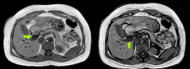
Subsequent abdominal ultrasound reported a gallbladder containing small calculi, with normal-caliber intra- and extrahepatic bile ducts and a common bile duct measuring 4.5 mm. Owing to the risk of choledocholithiasis, an MRCP was performed, which depicted a normal-sized gallbladder devoid of calculi, cystic dilation of the proximal common bile duct approximately 2 cm below the hepatic duct bifurcation extending to the ampulla of Vater, absence of stones in the common bile duct, and non-dilated intrahepatic bile duct (Figure 1).

Author’s own research.

Figure 1 Magnetic resonance cholangiopancreatography (coronal section) with a green arrow pointing to the choledochal cyst.  

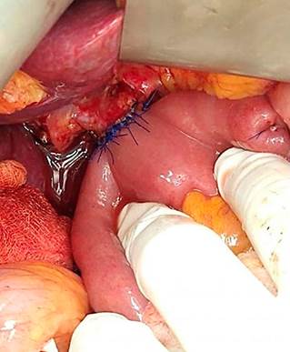
The patient was diagnosed with a choledochal cyst and underwent cyst resection followed by bilio-digestive bypass via Roux-en-Y hepaticojejunostomy (Figure 2).

Author’s own research.

Figure 2 Roux-en-Y hepaticojejunostomy.  

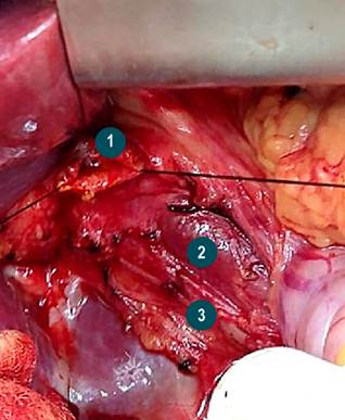
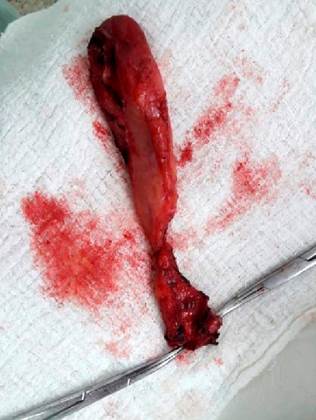
The surgical exploration revealed a distended gallbladder, a 3 mm cystic duct, and a choledochal cyst measuring 5 x 4 cm with a proximal neck situated 5 mm from the bifurcation of the hepatic ducts and a distal retro-duodenal neck with thickened walls adherent to the portal vein (Figure 3). Two pathological samples were taken: one from the choledochal cyst and one from the gallbladder (Figure 4).

Author’s own research.

Figure 3 Anatomical structures. 1. Confluence of the hepatic ducts. 2. Portal vein. 3. Right hepatic artery originating from the superior mesenteric artery.  

Author’s own research.

Figure 4 Pathological specimen of the choledochal cyst.  

Results

The surgical procedure was carried out without any adverse events, and the patient’s postoperative recovery was free of complications. At a follow-up visit fifteen days post-surgery, the patient presented a pathology report which confirmed acute cholecystitis and a benign choledochal cyst.

Discussion

The diagnosis of choledochal cysts in the adult population is becoming increasingly frequent1,19,24,30. In the case under discussion, the definitive diagnosis was established via MRCP after the initial ultrasound yielded inconclusive results. The clinical symptoms and laboratory findings were indicative of cholestasis, prompting the decision to proceed with more definitive imaging. While ultrasound remains the most accessible and cost-effective imaging technique, its variable sensitivity ranging from 71% to 97% can miss choledochal cysts, unlike MRCP, which boasts a detection rate of 96% to 100%29. Prompt diagnosis and intervention are paramount given the elevated risk of complications associated with choledochal cysts in adults, which include obstructive jaundice, symptomatic choledocholithiasis, pancreatitis, secondary biliary cirrhosis, and notably, cholangiocarcinoma1,31,32. The malignancy risk is reported to be approximately 10%-30%6,29,30, and it escalates with age-from 0.7% in the first decade to over 10% beyond the second decade, with more than half of the patients older than 60 years with choledochal cysts showing malignancy1,22. The cyst type is another risk factor, with type I presenting the highest incidence at 68%, followed by type IV at 21%6,14,33.

Consequently, the management of choledochal cysts consists of complete surgical excision6,34. Resection of the cyst along with Roux-en-Y hepaticojejunostomy is the procedure of choice for type I and IV cysts29,30, and partial hepatectomy may be necessary for type IVA. Type II cysts warrant excision and possibly biliary-enteric repair2,29,35. Treatment for type III cysts may involve sphincteroplasty, sphincterotomy36, or the insertion of a drainage tube into the duodenum11. For Todani type V, options include segmental liver resection, liver transplantation, or close patient monitoring to preempt complications29,37,38.

The emergence of minimally invasive surgery has established laparoscopic surgery as a safe and valid alternative. When compared to open surgery, the laparoscopic approach reduces the need for transfusions and shortens the recovery period, despite a typically longer operative duration39,40.

Conclusions

Diagnosing biliary cysts is a significant challenge due to the nonspecific nature of the symptoms and signs associated with the condition. Ultrasound is an effective and cost-effective first-line imaging modality for diagnosing biliary cysts; however, as evidenced in the presented case, a negative ultrasound does not eliminate the possibility of disease. Hence, MRCP is crucial for definitive diagnosis, serving as the gold standard in the detection of choledochal cysts and in ruling out lithiasis and neoplasia. Cyst resection remains the preferred treatment approach due to the significant risk of malignant transformation associated with these cysts.

Acknowledgments

This research did not receive any specific grant funding.

Referencias

1. Baison GN, Bonds MM, Helton WS, Kozarek RA. Choledochal cysts: Similarities and differences between Asian and Western countries. World J Gastroenterol. 2019;25(26):3334-3343. https://doi.org/10.3748/wjg.v25.i26.3334Links ]

2. Farías-Molina SM, Castillo-Machado RL, Sanhueza-Palma NC, Calzadilla-Riveras J.A. Quiste de colédoco Todani tipo II: caso clínico y revisión. Medwave. 2016;16(9):e6583. https://doi.org/10.5867/medwave.2016.09.6583Links ]

3. Yamaguchi M. Congenital choledochal cyst. Analysis of 1,433 patients in the Japanese literature. Am J Surg, 1980;140(5):653-657. https://doi.org/10.1016/0002-9610(80)90051-3Links ]

4. Bhavsar MS, Vora HB, Giriyappa VH. Choledochal cysts: a review of literature. Saudi J Gastroenterol. 2012;18(4):230-236. https://doi.org/10.4103/1319-3767.98425 Links ]

5. Honnavara-Srinivasan P, Anbalagan A, Shanmugasundaram R, Obla Lakshmanamoorthy N. Management of Choledochal Cysts at a Tertiary Care Centre: A Nine-Year Experience from India. Surg Res Pract. 2020;2020:8017460. https://doi.org/10.1155/2020/8017460Links ]

6. Moslim MA, Takahashi H, Seifarth FG, Walsh RM, Morris-Stiff G. Choledochal Cyst Disease in a Western Center: A 30-Year Experience. J Gastrointest Surg. 2016;20(8):1453-1463. https://doi.org/10.1007/s11605-016-3181-4 Links ]

7. Babbitt DP. Congenital choledochal cysts: new etiological concept based on anomalous relationships of the common bile duct and pancreatic bulb. Ann Radiol (Paris). 1969:12(3):231-240. [ Links ]

8. Sánchez JA, Gómez S, Morales C, Hoyos SI. Quistes del colédoco. Rev Colomb Cir. 2015;30(4):296-305. https://doi.org/10.30944/20117582.379Links ]

9. Miron A, Popa LG, Toma EA, Calu V, Parvuletu RF, Enciu O. The Curious Case of the Choledochal Cyst-Revisiting the Todani Classification: Case Report and Review of the Literature. Diagnostics (Basel). 2023;13(6):1059. https://10.3390/diagnostics13061059Links ]

10. Ye Y, Lui VCH, Tam PKH. Pathogenesis of Choledochal Cyst: Insights from Genomics and Transcriptomics. Genes (Basel). 2022;13(6):1030. https://doi.org/10.3390/genes13061030Links ]

11. Kumar M, Rajagopalan S. Choledochal cyst. Med J Armed Forces India. 2012;68(3):296-298. https://doi.org/10.1016/j.mjafi.2012.04.011Links ]

12. Marcos A. Diagnóstico prenatal de quiste de colédoco: presentación de un caso. Rev Argent Ultrason. 2008;7(1):53-56. [ Links ]

13. Alonso-Lej F, Rever WB, Pessagno DJ. Congenital choledochal cyst, with a report of 2, and an analysis of 94, cases. Int Abstr Surg. 1959;108(1):1-30. [ Links ]

14. Todani T, Watanabe Y, Narusue M, Tabuchi K, Okajima K. Congenital bile duct cysts: Classification, operative procedures, and review of thirty-seven cases including cancer arising from choledochal cyst. Am J Surg. 1977;134(2):263-269. https://doi.org/10.1016/0002-9610(77)90359-2Links ]

15. Sureka B, Rastogi A, Bihari C, Bharathy KGS, Sood V, Alam S. Imaging in ductal plate malformations. Indian J Radiol Imaging. 2017;27(1):6-12. https://doi.org/10.4103/0971-3026.202966Links ]

16. Sosa R, Hernández E, López A, Bolaños J, González F. Diagnóstico y Tratamiento del Quiste del Colédoco Neonatal. Reporte de Casos. Rev Guatem Cir. 2016;22(1):25-28. [ Links ]

17. Tannuri AC, Hara LA, Paganoti GF, Andrade WC, Tannuri U. Choledochal cysts in children: How to Diagnose and Operate on. Clinics (Sao Paulo). 2020;75:e1539. https://doi.org/10.6061/clinics/2020/e1539Links ]

18. Machado NO, Chopra PJ, Al-Zadjali A, Younas S. Choledochal Cyst in Adults: Etiopathogenesis, Presentation, Management, and Outcome-Case Series and Review. Gastroenterol Res Pract. 2015;2015:602591. https://doi.org/10.1155/2015/602591Links ]

19. Pandit N, Deo KB, Yadav TN, Gautam S, Dhakal Y, Awale L, et al. Choledochal Cyst: A Retrospective Study of 30 Cases From Nepal. Cureus. 2020;12(11):e11414. https://doi.org/10.7759/cureus.11414Links ]

20. Talini C, DE-Carvalho BC, Antunes LA, Schulz C, Sabbaga CC, Andrade SG, et al. Choledochal cyst in the pediatric population: experience of 13 laparoscopic procedures in two years at a single institution. Rev Col Bras Cir. 2018;10;45(3):e1854. https://doi.org/10.1590/0100-6991e-20181854Links ]

21. Arroyo-Martínez L, Montero-Carvajal R, Ayi-Wong J. Quiste del colédoco como causa de dolor abdominal: revisión de un caso y bibliografía. Acta Med Costarric. 2004;46(3):149-151. https://doi.org/10.51481/amc.v46i3.150Links ]

22. Søreide K, Søreide JA. Bile duct cyst as precursor to biliary tract cancer. Ann Surg Oncol. 2007;14(3):1200-11. https://doi.org/10.1245/s10434-006-9294-3 Links ]

23. Cención JD, Chanis R; Méndez J. Características clínicas y demográficas de los pacientes con quiste de colédoco atendidos en el Hospital del Niño Dr. José R. Esquivel en Panamá durante los años 2005 a 2018. Pediátr Panamá. 2019;48(2):4-10. https://doi.org/10.37980/im.journal.rspp.20191574Links ]

24. Monteiro VV, Pinheiro F, Costa K, Seixas Z, Da Silva R. Choledochal cyst- unusual presentation in the adult phase: Case report. Int J Surg Case Rep. 2020;70:33-36. https://doi.org/10.1016/j.ijscr.2020.03.014Links ]

25. Singham J, Schaeffer D, Yoshida E, Scudamore C. Choledochal cysts: analysis of disease pattern and optimal treatment in adult and paediatric patients. HPB (Oxford). 2007;9(5):383-387. https://doi.org/10.1080/13651820701646198Links ]

26. Tello de Meneses A, Riggen L, Orozco E. Quiste de colédoco tipo I: Diagnóstico por imagen y anastomosis hepático-duodenal como medida terapéutica. Bol Med Hosp Infant Mex. 2013;70(6):482-487. [ Links ]

27. Lourenço AL, Proença A, Gil P.A Rare Case of Todani Type-III Choledochal Cyst. Acta Radiologica Portuguesa. 2020;32(2):23-25. https://doi.org/10.25748/arp.19632Links ]

28. Sacher VY, Davis JS, Sleeman D, Casillas J. Role of magnetic resonance cholangiopancreatography in diagnosing choledochal cysts: Case series and review. World J Radiol. 2013;5(8):304-312. https://doi.org/10.4329/wjr.v5.i8.304Links ]

29. Katabathina VS, Kapalczynski W, Dasyam AK, Anaya-Baez V, Menias CO. Adult choledochal cysts: current update on classification, pathogenesis, and cross-sectional imaging findings. Abdom Imaging. 2015;40(6):1971-1981. https://doi.org/10.1007/s00261-014-0344-1Links ]

30. Lewis VA, Adam SZ, Nikolaidis P, Wood C, Wu JG, Yaghmai V, et al. Imaging of choledochal cysts. Abdom Imaging. 2015;40(6):1567-80. https://doi.org/10.1007/s00261-015-0381-4Links ]

31. Gyawali S, Adhikari G, Shrestha S, Pradhan S, Bhandari RS. Concomitant hilar cholangiocarcinoma with choledochal cyst and cholelithiasis in an asymptomatic patient: A case report. Int J Surg Case Rep. 2021;84:106094. https://doi.org/10.1016/j.ijscr.2021.106094Links ]

32. López-Ruiz R, Aguilera-Alonso D, Muñoz-Aguilar G, Fonseca-Martín R. Quistes de colédoco, una causa inusual de ictericia en pediatría. Presentación de serie de casos. Rev Chil Pediatr. 2016;87(2):137-142. https://doi.org/10.1016/j.rchipe.2015.08.009Links ]

33. Ten-Hove A, Meijer VE, Hulscher BF, de Kleine HJ. Meta-analysis of risk of developing malignancy in congenital choledochal malformation. Br J Surg. 2018;105(5):482-490. https://doi.org/10.1002/bjs.10798 Links ]

34. Gadelhak N, Shehta A, Hamed H. Diagnosis and management of choledochal cyst: 20 years of single center experience. World J Gastroenterol. 2014;20(22):7061-7066. https://doi.org/10.3748/wjg.v20.i22.7061Links ]

35. Ouaïssi M, Kianmanesh R, Belghiti J, Ragot E, Mentha G, Adham M, et al. Todani Type II Congenital Bile Duct Cyst: European Multicenter Study of the French Surgical Association and Literature Review. Ann Surg. 2015;262(1):130-8. https://doi.org/10.1097/SLA.0000000000000761Links ]

36. Calderón M, Abarzúa J, Quiroga C, Gana J, de Barbieri F. Coledococele: caso diagnosticado por resonancia magnética. Rev Chil Radiol. 2020;26(2):76-78. http://dx.doi.org/10.4067/S0717-93082020000200076Links ]

37. Liu Y, Sun J, Guo S, Liu Z, Zhu M, Zhang ZL. The distal classification and management of choledochal cyst in adults: Based on the relation between cyst and pancreatic duct. Medicine (Baltimore). 2017;96(12):e6350. https://doi.org/10.1097/MD.0000000000006350Links ]

38. Vargas-Vásquez N, Jiménez-Luna G, Quiñones J, Sánchez-Temoche C. Enfermedad de Caroli: a propósito de un caso. Revista. colomb. Gastroenterol.. 2020;35(3):390-393. https://doi.org/10.22516/25007440.387Links ]

39. Zhen C, Xia Z, Long L, Lishuang M, Pu Y, Wenjuan Z, et al. Laparoscopic excision versus open excision for the treatment of choledochal cysts: a systematic review and meta-analysis. Int Surg. 2015;100(1):115-122. https://doi.org/10.9738/INTSURG-D-14-00165.1Links ]

40. Liu Y, Yao X, Li S, Liu W, Liu L, Liu J. Comparison of therapeutic effects of laparoscopic and open operation for congenital choledochal cysts in adults. Gastroenterol Res Pract. 2014;2014:670260. https://doi.org/10.1155/2014/670260Links ]

Citation: Medina-Ortega MA, Vallejo-Vallecilla G, Gómez-Mosquera AV. Todani Type I Common Bile Duct Cyst in an Adult Patient: Clinical Case and Literature Review . Revista. colomb. Gastroenterol. 2024;39(1):105-111. https://doi.org/10.22516/25007440.1074

Received: April 28, 2023; Accepted: June 05, 2023

*Correspondence: Marco Antonio Medina-Ortega. mmedinao@unicartagena.edu.co

Creative Commons License This is an open-access article distributed under the terms of the Creative Commons Attribution License