SciELO - Scientific Electronic Library Online

 
vol.30 issue2Human T-lymphotropic virus type 1 (HTLV-1) infection and spastic paraparesis. Advances and diagnosis 35 years after its DiscoveryResearch and development of causal chemoprophylactics in malaria author indexsubject indexarticles search
Home Pagealphabetic serial listing  

Services on Demand

Journal

Article

Indicators

Related links

  • On index processCited by Google
  • Have no similar articlesSimilars in SciELO
  • On index processSimilars in Google

Share


Iatreia

Print version ISSN 0121-0793

Iatreia vol.30 no.2 Medellín Apr./June 2017

https://doi.org/10.17533/udea.iatreia.v30n2a05 

Artículos de revisión

Mechanisms of skin aging

Mecanismos de envejecimiento de la piel

Mecanismos de envelhecimento da pele

Ana Cecilia Mesa-Arango1 

Sindy Viviana Flórez-Muñoz2 

Gloria Sanclemente1 

1Docente, Grupo de Investigación Dermatológica, Facultad de Medicina. Universidad de Antioquia, Medellín, Colombia.

2Estudiante, Grupo de Investigación Dermatológica. Facultad de Medicina. Universidad de Antioquia, Medellín, Colombia.


SUMMARY

Skin aging is an inevitable biological phenomenon of human life that results from either the age-dependent decline of cell function (intrinsic aging) or from cumulative exposure to external harmful influences (extrinsic aging). Intrinsic and extrinsic factors act synergistically to induce skin changes that manifest clinically as burns, erythema, hyperpigmentation, telangiectasia, skin dryness or sagging, coarse wrinkles, skin texture changes or eventually as skin cancer. The molecular mechanisms of both types of skin aging are similar. This review focuses on intrinsic and extrinsic mechanisms of skin aging, and on current and new perspectives for prevention and treatment options extracted from natural products.

KEY WORDS: DNA Damage; Natural Products; Photoaging; Photoprotection; Reactive Oxygen Species (ROS); Ultraviolet Radiation (UV)

RESUMEN

El envejecimiento cutáneo es un fenómeno biológico inevitable y puede ser el resultado de la disminución de la función celular como consecuencia del proceso normal de envejecimiento (envejecimiento intrínseco) o de la acumulación de los efectos dañinos debido a la exposición a factores nocivos externos (envejecimiento extrínseco). Los factores intrínsecos y extrínsecos actúan sinérgicamente para inducir cambios en la piel que se manifiestan clínicamente como quemaduras, eritema, hiperpigmentación, telangiectasias, sequedad de la piel o flacidez, arrugas profundas, cambios de textura o cáncer de piel. Los mecanismos moleculares de ambos tipos de envejecimiento de la piel son similares. Esta revisión se centra en una descripción general de los principales mecanismos intrínsecos y extrínsecos del envejecimiento de la piel así como en las medidas actuales de fotoprotección y en las nuevas perspectivas en cuanto a la prevención y al tratamiento basados en productos naturales.

PALABRAS CLAVE: Daño del ADN; Especies de Oxígeno Reactivas (EOR); Fotoenvejecimiento; Fotoprotección; Productos Naturales; Radiación Ultravioleta (UV)

RESUMO

O envelhecimento cutâneo e um fenômeno biológico inevitável e pode ser o resultado da diminuição da função celular como consequência do processo normal do envelhecimento (envelhecimento intrínseco) ou do cúmulo dos efeitos daninhos devido à exposição com fatores nocivos externos (envelhecimento extrínseco). Os fatores intrínsecos e extrínsecos atuam sinergicamente para induzir mudanças na pele, que clinicamente manifestam-se como queimaduras, eritema, hiperpigmentação, telangiectasias, pele seca, rugas profundas, perda da tonalidade natural ou câncer de pele. Os mecanismos moleculares dos dois tipos de envelhecimento são similares. Esta revisão, centra-se na descrição geral dos principais mecanismos intrínsecos e extrínsecos do envelhecimento da pele, assim como nas medidas atuais de foto proteção e nas perspectivas referentes à prevenção e tratamento baseados no uso de produtos naturais.

PALAVRAS CHAVE: Dano do ADN; Espécies Reativas de Oxigênio (ERO); Foto Envelhecimento; Foto Proteção; Produtos Naturais; Radiação Ultravioleta (UV)

INTRINSIC FACTORS ASSOCIATED WITH AGING

Cellular senescence (also known as natural aging) is caused by intrinsic factors and is determined by both, physiological factors and genetic predisposition1. Different signs of aging occur as cellular proliferation and hormone levels decrease, or as a result of diverse factors: telomere shortening and accumulation of dysplastic keratinocytes; degradation of the extracellular matrix; mutations in nuclear and mitochondrial genes, and multiple lipid or amino acid metabolic aberrations2)(3)(4. Such disturbances cause functional and physical changes due to abnormal water distribution or a lack of hygroscopic substances, resulting in a dry appearance of the skin. In addition, there is an increase in skin surface pH and a continuous production of reactive oxygen species (ROS) in the mitochondria that results from an oxidative cell metabolism and a decrease in antioxidant activity5)( 6). Also in vivo studies have shown that the expression of connective tissue growth factor (CTGF) and a reduced signaling of the transforming growth factor (TGF)-beta/ Smad are probably responsible for type I procollagen expression loss in intrinsically aged skin7. Finally, alterations in the cutaneous immunological barrier are also part of the aging process that frequently relate to or cause certain skin disorders8.

EXTRINSIC FACTORS ASSOCIATED WITH AGING

Environmental factors such as smoking, automobile contaminants, alcohol consumption, dietary habits, industrial waste and lifestyle options can contribute to aging6)(9)(10. Ultraviolet radiation (UVR) from the sun or artificial sources has a deleterious effect on skin functions and keratinocyte survival, a process known as photoaging10)(11. Additionally, according to recent data, visible light and infrared radiation also can lead to skin damage.

SUNLIGHT RADIATION SPECTRUM

Sunlight is composed of three different types of UV radiation: UVC (100-290 nm) which is largely blocked by the ozone layer and therefore has little impact on the skin; UVB (290-320 nm) which penetrates mainly into the epidermis (stratum corneum) and is responsible for the erythema associated with a sunburn, and UVA (320-400 nm), which penetrates into the dermis, and has the main role in chronic skin damage. These types of radiation represent about 10 % of the total solar radiation reaching the atmosphere. On the other hand, visible light (400-780 nm) and infrared radiation (780-1400 nm), which represent around 40 % and 50 % of the total radiation emanating from the sun, respectively, are also important factors in photoaging because they can exert both acute and chronic deleterious cutaneous effects14)(15.

MOLECULAR MECHANISMS OF SKIN DAMAGE INDUCED BY SUNLIGHT

Ultraviolet skin exposure initiates a flurry of molecular and cellular responses that end up with several dynamic internal disorders16. DNA is one of the major UV-absorbing molecules chemically altered by such exposure which also induces accumulative mutations17. UVB acts directly inducing the formation of cyclobutane pyrimidine dimers and photoproducts4)(5)(6. Such damage interferes with replication, causes transcription disruption, and induces mutations mainly within the tumor suppressor gene p53, altering the control of the cellular cycle; such alteration increases the risk of keratinocyte and melanocyte transformation that eventually manifests as skin tumors17)(18)(19. On the other hand, UVAradiation acts indirectly by stimulating ROS and/ or free radicals production. Reactive oxygen species accumulation is perhaps one of the most important cellular events after solar exposure as it induces the following cellular alterations:

  1. Nuclear and mitochondrial DNA mutations as a result of a modification of guanine (8-hydrox- 2′-deoxy guanine), single-stranded breaks, and oxidized pyrimidine bases20)(21.

  2. Membrane protein carbonilation and lipid peroxidation22.

  3. Apoptosis of epidermal keratinocytes (sunburn cells)23.

  4. Release of keratinocyte proinflammatory cytokines (mainly IL-1, IL-6, TNF-α) and dermal fibroblasts growth factor receptors such as the epidermal growth factor receptor (EGFR), tumor necrosis factor α (TNF-α), platelet activation factor (PAF) prostaglandins, and insulin. Another transcription factor important in photoaging is the nuclear factor kappa β (NF-kβ) which is also activated by UV radiation and has an effect on matrix proteins because it stimulates the transcription of inflammatory cytokines which attract neutrophils that also express matrix metalloproteinases (metalloproteinase-8) with concomitant extracellular matrix proteins degradation24)(25)(26)(27)(28)(29. Transcription of metalloproteinases (MMPs) is dependent on transcription factor AP1, and similarly, AP1 depends on the activation of MAPK signaling pathway by ROS in keratinocytes30. Also, dermal matrix metalloproteinases upregulation stimulates the production of collagenase, gelatinase and stromelysin-1 in both fibroblasts and keratinocytes, resulting in a deterioration of both collagen and elastin, as well as of other components of the dermal extracellular matrix. While the expression of metalloproteinases is activated, inhibitors of these enzymes are also reduced30)(31)(32.

  5. A decrease in the expression of TGF-β, which brings down an altered collagen production and enhances elastin production thereby producing changes in skin structure which clinically manifest by deep wrinkles, coarse textures, telangiectasia and pigmentation33)(34.

  6. Local and systemic immunosuppression by activation of immunosuppressive molecules such as IL4, IL10, IL-1β, TNF-α, and PGE2, or by alteration in cellular morphology, function or quantity of Langerhans cells and T lymphocytes27)(35)(36)(37, are other consequences of ROS production after UV-radiation exposure. Antigen presentation in UV exposed skin is also impaired mainly due to suppression of important molecules expression such as MHC class II, lymphocyte function associated antigen-3 (LFA-3), ICAM-1, ICAM-3, B7, CD1a and CD4017)(38)(39. UV skin radiation also activates the local glucosteroidogenesis process in skin and this in turn leads to the attenuation of local cutaneous immunity40.

All mentioned crucial alterations of the immune system favor a state of cellular malignant transformation that manifests as cutaneous neoplasias17. In addition, there are several skin diseases that can be aggravated (e.g idiopathic photodermatoses, phototoxic contact dermatitis, photoallergic contact dermatitis, photosensitivity caused by medications, etc.); influenced (melasma, vitiligo, lupus erythematosus, seborrheic dermatitis, psoriasis, etc.); caused or triggered by resultant UVRinduced immunosuppression (e.g infections such as herpes simplex, and viral exanthemas)15.

SKIN CHANGES INDUCED BY SMOKING

It has been described that heavy smoking is strongly associated with poor wound healing, certain squamous cell carcinomas, oral cancer, psoriasis exacerbations, and premature skin aging41)(42)(43. Such skin damage induced by tobacco has been related to oxidative stress, impaired collagen biosynthesis, and activation of matrix metalloproteinases. In addition, tobacco smoking air pollutants activate the aryl hydrocarbon receptor (AhR) pathway, a liganddependant transcription factor that mediates toxicity induced by several environmental contaminants, which may play a role in already known premature skin-aging effects44. Figure 1 summarizes the main events induced by UVA, UVB and smoking.

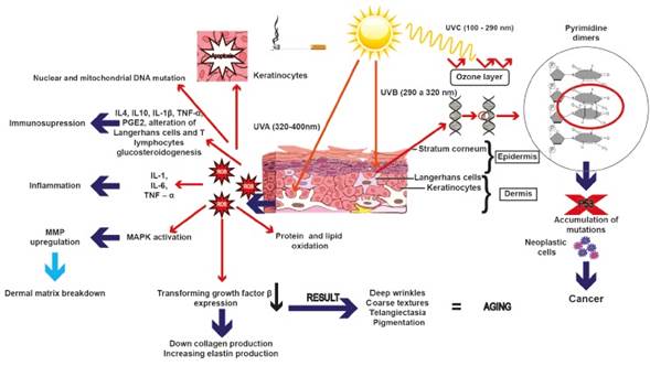
Figure 1 Ultraviolet radiation and cigarette smoking affect the stability of DNA, causing alteration in molecule structure due to the formation of mutations that accumulate and promote cellular malignant transformation. Also, there may be an accumulation of ROS due to p53 inability to remove them, which in turn can lead to inflammation by activating the expression of certain interleukins as well as immunosuppression or the overexpression of metalloproteinases 

MECHANISMS OF SKIN PROTECTION

Human beings have been constantly exposed to sunlight radiation. As the outermost organ, skin acts as the main protective barrier against UV-radiation and other extrinsic agents45)(46. The cutaneous barrier is composed of two primary layers: epidermis and dermis. Each one exhibits unique structural and physiological functions that have important roles against extrinsic factors. Recent advances in the understanding of photodamage mechanisms have enhanced strategy development to prevent alteration of the structure and function of the skin. The first step in order to prevent acute and chronic damage development by UVradiation is photoprotection, which can be understood as a group of measures directed to reduce exposure; this can be accomplished with a set of strategies that involve photoeducation, topical photoprotection (sunscreens), oral photoprotection and photoprotection by devices (clothes, hats, etc.)15.

Regarding oral and topical photoprotection, exogenous nutrients such as α-tocopherol, oral and topical retinoids such as vitamin A and β-carotene (a precursor of vitamin A), lycopene, and lutein all contribute to the formation of a UV-radiation barrier and some of them act as antioxidants47. Indeed, it has been shown that retinoids protect skin from UVradiation because they have the ability to increase the proliferation of epidermal keratinocytes and dermal fibroblasts48, and/or inhibit the expression of matrix metalloproteinases (MMPs), matrix-degrading enzymes, leading to an increase in overall protein and extracellular matrix content30)(32.

On the other hand, vitamins E and D also protect skin from ROS damage. In fact, it has been described that vitamin E inhibits MMP-1 expression, thus avoiding collagen breakdown, and acts as an anti-inflammatory agent, whereas vitamin D has been shown to stimulate innate immunity49)(50)(51.

Previous work has suggested a role of mineral elements such as zinc, copper, and selenium in preventing skin aging. These elements are cofactors of important enzymes that act in protein synthesis of the extracellular matrix, but also have other functions such as radiation absorbance (e.g. zinc)52. Copper is also crucial in this process because it acts as an antioxidant and stimulates collagen and melanin synthesis53. Lastly, selenium has been described to protect skin against oxidative stress54.

Intrinsic skin factors such as the local steroidogenic system also have an important role in cutaneous protection against harmful effects of UV-radiation as it is capable of synthesizing androgens, estradiols, gluco- and mineralo-corticoids from precursors, all of which play anti-cancer roles by getting converted into secosteroids under the influence of UV-radiation55. Additionally, chromophores such as urocanic acid, which is formed from L-histidine in the stratum corneum, is a potent endogenous UV-absorbant56. Nevertheless, melanin, an UV-absorbing pigment that is able to dissipate UV radiation as harmless heat, is probably the most important skin constituent that exerts a defensive function against UV radiation damage. Such pigment is produced by melanocytes and transferred to keratinocytes via long dendritic processes17)(57 and acts as a UV filter that inhibits dimers and 6,4 photoproducts formation or as a ROS scavenger58)(59. In addition, melanin is responsible for skin color which has been suggested is the result of an adaptive response to UVR as skin darkness observed in certain communities relates with the levels of UVR which they are exposed to. It has been suggested that such color changes take place in order to decrease vitamin D synthesis requirements in the skin. Such skin adaptation also seems to prevent UV damage of sweat glands which would have a major impact on thermoregulation, especially in warm areas60)(61. Also, melanin levels have been shown to be inversely correlated with the amount of skin DNA damage induced by UV; therefore, it seems that white Caucasians low pigmentation capacity is responsible for a failure in intrinsic photoprotection mechanisms62)(63.

Another important component in epidermal protection is melatonin, a hormone synthesized by the pineal gland, which is capable of protecting epidermal keratinocytes from oxidative stress by suppressing ROS generation and increasing a reduced glutathione content64)(65. In addition, the skin has natural endogenous enzymatic antioxidants (e.g. superoxide dismutase or catalase) and non-enzymatic antioxidants (e.g. coenzyme Q10, glutathione or vitamin E), which also provide protection against ROS66.

Human skin is also protected from UV radiation damage by the activation of complex molecular repair mechanisms that correct DNA-injuries in order to maintain genomic integrity67. The most important mechanism in this process is nucleotide excision DNA repair (NER) which is in charge of cyclobutane pyrimidine dimers and (6-4) photoproducts removal. Indeed, malfunction of this repair system is related to skin disorders such as xeroderma pigmentosum and photoaging63)(68. Another repairing mechanism is base excision repair (BER) which removes oxidative modifications on a single base in DNA69.

Another mechanism of reparation by NER and BER is via p53 activation by inducing arrest at G2/M checkpoint70. If serious damage occurs, p53-dependent apoptosis takes place in order to get rid of damaged DNA and to prevent cancer occurrence17)(71)(72. Also, it has been demonstrated that upregulation of IL-12 can counteract UVB-induced immunosuppression73.

FUTURE APPROACHES FOR SKIN PROTECTION WITH NATURAL PRODUCTS

In modern society, there is a great increase in the search for eternal youth74. For this reason, new therapeutic options are being explored and many natural active ingredients seem to offer alternatives for skin protection75. Skin protection obtained with some natural products is given by their antioxidant effect, melanin production or activation of transcription factors76)(77)(78)(79. Indeed, the plant Rosmarinus officinalis is a source of rosmarinic acid, which acts as a free radical scavenger and stimulates melanin production76)(77. Recently, it has been described that topical application of forskolin (a product derived from the plant Plectranthus barbatus (Coleus forskolii) up-regulates melanin production via a direct activation of adenylate cyclase that induces the production of cAMP, which results in melanin deposition. This event is melanocortin-1 receptor (MC1R) dependent which is a protein located in the extracellular membranes of epidermal melanocytes80. Also, paeoniflorin (another natural product extracted from the plant Paeonia lactiflora), has been shown to have the ability to repair DNA and probably to reduce facial wrinkles in human skin81.

In various molecules of natural origin (either in the form of plant extracts, fruits and/or tubers), a role has been found in the activation of nuclear factor erythroid 2-like 2 (Nrf2) (a master transcription factor which regulates a battery of genes involved in defense mechanisms against ROS damage). Therefore, compounds that act as activation enhancers of this factor might be useful in prevention of skin damage and/or malignant transformation under various stress conditions78.

In the last decade, some herbal sunscreens have replaced sunscreens containing chemical UV-filters due to the side effects associated with the latter. Many herbal sunscreens are currently available in the market in the forms of creams, lotions, and/or gels formulations which have included a labeled sun protector factor (SPF)82. In addition, plants characterized for protecting themselves from intense heat and UV radiation could be promising in future formulations83. Other innovative options for photoprotection are DNA-oligonucleotides, which enhance DNA-repair and stimulate melanogenesis84.

In addition, antiaging properties have not only been found in plants. Previous studies have described regenerative properties and an antioxidant activity in mollusk Cryptomphalus aspersa85. Also, it has been recently shown that C. aspersa eggs extract promotes migration and prevents cutaneous aging in keratinocytes and dermal fibroblasts in vitro86.

Lastly, different peptides represent an emerging field in cosmeceuticals for photoaging because they have cosmetically interesting activities such as collagen synthesis and chemotaxis stimulation and antistinging effects87)(88.

The majority of publications in the field of natural products and essential oils for potential use in cosmetics are from China and India83)(89)(90)(91)(92. In Latin America, the main publications in this area are form Brazil, Argentina, Mexico and Uruguay, whereas Colombia has published very few studies92)(93)(94. In fact, instead of looking for a direct effect in skin photoaging, most Colombian studies have been focused on the antioxidant or photo-protective activity of some products. One Colombian report has showed the in vitro antioxidant activity of two types of leaves: The Colombian Magnoliaceae and Talauma hernandezii95. In such study, the activity of these magnoliaceae was determined using 2,2′-azino-bis(3- ethylbenzothiazoline-6-sulfonic acid) (ABTS·+) and the stable free radical 2-2-diphenyl-1-picrylhydrazyl (DPPH·), and antioxidant properties obtained were similar to those reported from α-tocopherol, tertbutylated hydroxyanisole (BHA), and ascorbic acid95.

By the use of the free radical α-α-diphenyl- βpicrylhydrazyl (DPPH· ) and the preformed radical monocation 2,2′-azino-bis(3-ethylbenzothiazoline-6- sulphonic acid) (ABTS·+), another Colombian study has proven to have an antioxidant activity comparable to that of α-tocopherol, BHA, ascorbic acid and Troloxin. Some of the products under investigation in this study had antioxidant capabilities in essential oils: from anise (Pimpinella anisum L.), fennel (Foeniculum vulgare Mill.), coriander (Coriandrum sativum L.), rosemary (Rosmarinus officinalis L.), oregano (Origanum vulgare L.), ylang-ylang (Cananga odorata Hook.), verbena (Lippia alba Mill.) and salvia negra (Lepechinia schiedeana (Schlecht) Vatke, syn. Stachys schiedeana Schlecht)96. Another Colombian study has checked the antioxidant and protective abilities of Lobariella pallida and Stereocaulon strictum var. compressum extracts to prevent cell and DNA damage by oxidative stress97. Another study from this same research group has evaluated the antioxidant and photo-protective efficacy of the extract and isolated compounds from the lichen Usnea rocellina Motyka, by evaluating the antioxidant activity through the 2,2-diphenyl-1-picrylhydrazyl (DPPH) assay and ferric reducing power98.

In conclusion, skin aging is a natural process in which different cellular and molecular mechanisms, induced by intrinsic and extrinsic factors, are involved. Currently, increasing attempts are being made to delay skin aging via the ingestion or application of natural products or by formulating new natural sunscreens with higher radiation protective capability. In this respect, plant biodiversity in Latin American countries, and particularly in Colombia, are important sources of potential candidates for topical use. However, anti-aging products innovation requires knowledge of the mechanisms that trigger cell damage due to intrinsic or to extrinsic deleterious factors. More importantly, as the scientific support for the use of the aforementioned products is based on in vitro data, there is a need for a formal evaluation of such products in high quality and well-designed preclinical and phase-1 and -2 clinical trials in order to assess the best dosage and also to determine their real safety and efficacy.

ACKNOWLEDGEMENTS

We are grateful to María Constanza Patiño and to Eduardo García for their technical assistance in language review of the article. Sindy Flórez was sponsored through a grant from the University of Antioquia “Jóvenes Investigadores” program, years 2014-2015. We also thank María Isabel González Gaviria (Facultad de Arquitectura, Universidad Pontificia Bolivariana, Medellín, Colombia), for her assistance in the design of figures.

CONFLICTS OF INTEREST

None to declare

BIBLIOGRAPHIC REFERENCES

1. Nikolakis G, Makrantonaki E, Zouboulis CC. Skin mirrors human aging. Horm Mol Biol Clin Investig. 2013 Dec;16(1):13-28. DOI 10.1515/hmbci-2013-0018. [ Links ]

2. Hashizume H. Skin aging and dry skin. J Dermatol. 2004 Aug;31(8):603-9. [ Links ]

3. Kohl E, Steinbauer J, Landthaler M, Szeimies RM. Skin ageing. J Eur Acad Dermatol Venereol. 2011 Aug;25(8):873-84. DOI 10.1111/j.1468-3083.2010.03963.x. [ Links ]

4. Makrantonaki E, Zouboulis CC, William J. Cunliffe Scientific Awards. Characteristics and pathomechanisms of endogenously aged skin. Dermatology. 2007;214(4):352-60. [ Links ]

5. Poljšak B, Dahmane RG, Godić A. Intrinsic skin aging: the role of oxidative stress. Acta Dermatovenerol Alp Pannonica Adriat. 2012;21(2):33-6. [ Links ]

6. Farage MA, Miller KW, Elsner P, Maibach HI. Functional and physiological characteristics of the aging skin. Aging Clin Exp Res. 2008 Jun;20(3):195-200. [ Links ]

7. Quan T, Shao Y, He T, Voorhees JJ, Fisher GJ. Reduced expression of connective tissue growth factor (CTGF/ CCN2) mediates collagen loss in chronologically aged human skin. J Invest Dermatol. 2010 Feb;130(2):415- 24. DOI 10.1038/jid.2009.224. [ Links ]

8. Ponnappan S, Ponnappan U. Aging and immune function: molecular mechanisms to interventions. Antioxid Redox Signal. 2011 Apr;14(8):1551-85. DOI 10.1089/ars.2010.3228. [ Links ]

9. Bernhard D, Moser C, Backovic A, Wick G. Cigarette smoke--an aging accelerator? Exp Gerontol. 2007 Mar;42(3):160-5. [ Links ]

10. Farage MA, Miller KW, Elsner P, Maibach HI. Intrinsic and extrinsic factors in skin ageing: a review. Int J Cosmet Sci. 2008 Apr;30(2):87-95. DOI 10.1111/j.1468-2494.2007.00415.x. [ Links ]

11. Jenkins G. Molecular mechanisms of skin ageing. Mech Ageing Dev. 2002 Apr;123(7):801-10. [ Links ]

12. Tarbuk A, Grancarić AM, Situm M. Discrepancy of whiteness and UV protection in wet state. Coll Antropol. 2014 Dec;38(4):1099-105. [ Links ]

13. Costa A, Eberlin S, Clerici SP, Abdalla BM. In vitro effects of infrared A radiation on the synthesis of MMP- 1, catalase, superoxide dismutase and GADD45 alpha protein. Inflamm Allergy Drug Targets. 2015;14(1):53-9. [ Links ]

14. Sklar LR, Almutawa F, Lim HW, Hamzavi I. Effects of ultraviolet radiation, visible light, and infrared radiation on erythema and pigmentation: a review. Photochem Photobiol Sci. 2013 Jan;12(1):54-64. DOI 10.1039/c2pp25152c. [ Links ]

15. Schalka S, Steiner D, Ravelli FN, Steiner T, Terena AC, Marçon CR, et al. Brazilian consensus on photoprotection. An Bras Dermatol. 2014 Nov-Dec;89(6 Suppl 1):1-74. DOI 10.1590/abd1806-4841.20143971. [ Links ]

16. McCallion R, Li Wan Po A. Dry and photo-aged skin: manifestations and management. J Clin Pharm Ther. 1993 Feb;18(1):15-32. [ Links ]

17. Maverakis E, Miyamura Y, Bowen MP, Correa G, Ono Y, Goodarzi H. Light, including ultraviolet. J Autoimmun. 2010 May;34(3):J247-57. DOI 10.1016/j.jaut.2009.11.011. [ Links ]

18. Ziegler A, Jonason AS, Leffell DJ, Simon JA, Sharma HW, Kimmelman J, et al. Sunburn and p53 in the onset of skin cancer. Nature. 1994 Dec;372(6508):773-6. [ Links ]

19. Krämer M, Stein B, Mai S, Kunz E, König H, Loferer H, et al. Radiation-induced activation of transcription factors in mammalian cells. Radiat Environ Biophys. 1990;29(4):303-13. [ Links ]

20. Cadet J, Wagner JR. DNA base damage by reactive oxygen species, oxidizing agents, and UV radiation. Cold Spring Harb Perspect Biol. 2013 Feb;5(2). pii:a012559. DOI 10.1101/cshperspect.a012559. [ Links ]

21. Kielbassa C, Roza L, Epe B. Wavelength dependence of oxidative DNA damage induced by UV and visible light. Carcinogenesis. 1997 Apr;18(4):811-6. [ Links ]

22. Chen L, Hu JY, Wang SQ. The role of antioxidants in photoprotection: a critical review. J Am Acad Dermatol. 2012 Nov;67(5):1013-24. DOI 10.1016/j.jaad.2012.02.009. [ Links ]

23. Kulms D, Schwarz T. Molecular mechanisms of UVinduced apoptosis. Photodermatol Photoimmunol Photomed. 2000 Oct;16(5):195-201. [ Links ]

24. Sárdy M. Role of matrix metalloproteinases in skin ageing. Connect Tissue Res. 2009;50(2):132-8. DOI 10.1080/03008200802585622. [ Links ]

25. Morgan MJ, Liu ZG. Crosstalk of reactive oxygen species and NF-κB signaling. Cell Res. 2011 Jan;21(1):103-15. DOI 10.1038/cr.2010.178. [ Links ]

26. Quan T, Qin Z, Xia W, Shao Y, Voorhees JJ, Fisher GJ. Matrix-degrading metalloproteinases in photoaging. J Investig Dermatol Symp Proc. 2009 Aug;14(1):20-4. DOI 10.1038/jidsymp.2009.8. [ Links ]

27. Halliday GM. Inflammation, gene mutation and photoimmunosuppression in response to UVR-induced oxidative damage contributes to photocarcinogenesis. Mutat Res. 2005 Apr;571(1- 2):107-20. [ Links ]

28. Aubin F. Mechanisms involved in ultraviolet lightinduced immunosuppression. Eur J Dermatol. 2003;13(6):515-23. [ Links ]

29. Larsson P, Ollinger K, Rosdahl I. Ultraviolet (UV)Aand UVB-induced redox alterations and activation of nuclear factor-kappaB in human melanocytesprotective effects of alpha-tocopherol. Br J Dermatol. 2006 Aug;155(2):292-300. [ Links ]

30. Fisher GJ, Wang ZQ, Datta SC, Varani J, Kang S, Voorhees JJ. Pathophysiology of premature skin aging induced by ultraviolet light. N Engl J Med. 1997 Nov;337(20):1419-28. [ Links ]

31. Millis AJ, Hoyle M, McCue HM, Martini H. Differential expression of metalloproteinase and tissue inhibitor of metalloproteinase genes in aged human fibroblasts. Exp Cell Res. 1992 Aug;201(2):373-9. [ Links ]

32. Chung JH, Kang S, Varani J, Lin J, Fisher GJ, Voorhees JJ. Decreased extracellular-signal-regulated kinase and increased stress-activated MAP kinase activities in aged human skin in vivo. J Invest Dermatol. 2000 Aug;115(2):177-82. [ Links ]

33. Uitto J. The role of elastin and collagen in cutaneous aging: intrinsic aging versus photoexposure. J Drugs Dermatol. 2008 Feb;7(2 Suppl):s12-6. [ Links ]

34. Krutmann J, Morita A, Chung JH. Sun exposure: what molecular photodermatology tells us about its good and bad sides. J Invest Dermatol. 2012 Mar;132(3 Pt 2):976-84. DOI 10.1038/jid.2011.394. [ Links ]

35. Shreedhar V, Giese T, Sung VW, Ullrich SE. A cytokine cascade including prostaglandin E2, IL-4, and IL- 10 is responsible for UV-induced systemic immune suppression. J Immunol. 1998 Apr;160(8):3783-9. [ Links ]

36. Iwai I, Hatao M, Naganuma M, Kumano Y, Ichihashi M. UVA-induced immune suppression through an oxidative pathway. J Invest Dermatol. 1999 Jan;112(1):19-24. [ Links ]

37. Noonan FP, Bucana C, Sauder DN, De Fabo EC. Mechanism of systemic immune suppression by UV irradiation in vivo. II. The UV effects on number and morphology of epidermal Langerhans cells and the UV-induced suppression of contact hypersensitivity have different wavelength dependencies. J Immunol. 1984 May;132(5):2408-16. [ Links ]

38. Meunier L, Bata-Csorgo Z, Cooper KD. In human dermis, ultraviolet radiation induces expansion of a CD36+ CD11b+ CD1- macrophage subset by infiltration and proliferation; CD1+ Langerhans-like dendritic antigen-presenting cells are concomitantly depleted. J Invest Dermatol. 1995 Dec;105(6):782-8. [ Links ]

39. Taguchi K, Fukunaga A, Ogura K, Nishigori C. The role of epidermal Langerhans cells in NB-UVBinduced immunosuppression. Kobe J Med Sci. 2013 Apr;59(1):E1-9. [ Links ]

40. Slominski AT. Ultraviolet radiation (UVR) activates central neuro-endocrine-immune system. Photodermatol Photoimmunol Photomed. 2015 May;31(3):121-3. DOI 10.1111/phpp.12165. [ Links ]

41. Freiman A, Bird G, Metelitsa AI, Barankin B, Lauzon GJ. Cutaneous effects of smoking. J Cutan Med Surg. 2004 Nov-Dec;8(6):415-23. [ Links ]

42. Metelitsa AI, Lauzon GJ. Tobacco and the skin. Clin Dermatol. 2010 Jul-Aug;28(4):384-90. DOI 10.1016/j.clindermatol.2010.03.021. [ Links ]

43. Lønnberg AS, Skov L, Skytthe A, Kyvik KO, Pedersen OB, Thomsen SF. Smoking and risk for psoriasis: a population-based twin study. Int J Dermatol. 2016 Feb;55(2):e72-8. DOI 10.1111/ijd.13073. [ Links ]

44. Morita A, Torii K, Maeda A, Yamaguchi Y. Molecular basis of tobacco smoke-induced premature skin aging. J Investig Dermatol Symp Proc. 2009 Aug;14(1):53-5. DOI 10.1038/jidsymp.2009.13. [ Links ]

45. Elias PM, Friend DS. The permeability barrier in mammalian epidermis. J Cell Biol. 1975 Apr;65(1):180-91. [ Links ]

46. Bouwstra JA, Ponec M. The skin barrier in healthy and diseased state. Biochim Biophys Acta. 2006 Dec;1758(12):2080-95. [ Links ]

47. Evans JA, Johnson EJ. The role of phytonutrients in skin health. Nutrients. 2010 Aug;2(8):903-28. DOI 10.3390/nu2080903. [ Links ]

48. Varani J, Perone P, Griffiths CE, Inman DR, Fligiel SE, Voorhees JJ. All-trans retinoic acid (RA) stimulates events in organ-cultured human skin that underlie repair. Adult skin from sun-protected and sunexposed sites responds in an identical manner to RA while neonatal foreskin responds differently. J Clin Invest. 1994 Nov;94(5):1747-56. [ Links ]

49. Ricciarelli R, Maroni P, Ozer N, Zingg JM, Azzi A. Agedependent increase of collagenase expression can be reduced by alpha-tocopherol via protein kinase C inhibition. Free Radic Biol Med. 1999 Oct;27(7-8):729- 37. [ Links ]

50. Meydani SN, Barklund MP, Liu S, Meydani M, Miller RA, et al. Vitamin E supplementation enhances cellmediated immunity in healthy elderly subjects. Am J Clin Nutr. 1990 Sep;52(3):557-63. [ Links ]

51. Gombart AF, Borregaard N, Koeffler HP. Human cathelicidin antimicrobial peptide (CAMP) gene is a direct target of the vitamin D receptor and is strongly upregulated in myeloid cells by 1,25-dihydroxyvitamin D3. FASEB J. 2005 Jul;19(9):1067-77. [ Links ]

52. Mitchnick MA, Fairhurst D, Pinnell SR. Microfine zinc oxide (Z-cote) as a photostable UVA/UVB sunblock agent. J Am Acad Dermatol. 1999 Jan;40(1):85-90. [ Links ]

53. Pickart L, Vasquez-Soltero JM, Margolina A. The human tripeptide GHK-Cu in prevention of oxidative stress and degenerative conditions of aging: implications for cognitive health. Oxid Med Cell Longev. 2012;2012:324832. DOI 10.1155/2012/324832. [ Links ]

54. Rafferty TS, McKenzie RC, Hunter JA, Howie AF, Arthur JR, Nicol F, et al. Differential expression of selenoproteins by human skin cells and protection by selenium from UVB-radiation-induced cell death. Biochem J. 1998 May;332( Pt 1):231-6. [ Links ]

55. Slominski AT, Zmijewski MA, Semak I, Zbytek B, Pisarchik A, Li W, et al. Cytochromes p450 and skin cancer: role of local endocrine pathways. Anticancer Agents Med Chem. 2014 Jan;14(1):77-96. [ Links ]

56. Young AR. Chromophores in human skin. Phys Med Biol. 1997 May;42(5):789-802. [ Links ]

57. Godic A, Poljšak B, Adamic M, Dahmane R. The role of antioxidants in skin cancer prevention and treatment. Oxid Med Cell Longev. 2014;2014:860479. DOI 10.1155/2014/860479. [ Links ]

58. Brenner M, Hearing VJ. The protective role of melanin against UV damage in human skin. Photochem Photobiol. 2008 May-Jun;84(3):539-49. DOI 10.1111/j.1751-1097.2007.00226.x. [ Links ]

59. Kobayashi N, Muramatsu T, Yamashina Y, Shirai T, Ohnishi T, Mori T. Melanin reduces ultravioletinduced DNA damage formation and killing rate in cultured human melanoma cells. J Invest Dermatol. 1993 Nov;101(5):685-9. [ Links ]

60. Jeong C, Di Rienzo A. Adaptations to local environments in modern human populations. Curr Opin Genet Dev. 2014 Dec;29:1-8. DOI 10.1016/j.gde.2014.06.011. [ Links ]

61. Sanclemente G, Falabella R, Garcia JJ. Vitiligo. Rev Col Dermatol.2004;12(4):12-22. [ Links ]

62. Yamaguchi Y, Beer JZ, Hearing VJ. Melanin mediated apoptosis of epidermal cells damaged by ultraviolet radiation: factors influencing the incidence of skin cancer. Arch Dermatol Res. 2008 Apr;300 Suppl 1:S43-50. [ Links ]

63. Reichrath J, Rass K. Ultraviolet damage, DNA repair and vitamin D in nonmelanoma skin cancer and in malignant melanoma: an update. Adv Exp Med Biol. 2014;810:208-33. [ Links ]

64. Fischer TW, Zbytek B, Sayre RM, Apostolov EO, Basnakian AG, Sweatman TW, et al. Melatonin increases survival of HaCaT keratinocytes by suppressing UV-induced apoptosis. J Pineal Res. 2006 Jan;40(1):18-26. [ Links ]

65. Janjetovic Z, Nahmias ZP, Hanna S, Jarrett SG, Kim TK, Reiter RJ, et al. Melatonin and its metabolites ameliorate ultraviolet B-induced damage in human epidermal keratinocytes. J Pineal Res. 2014 Aug;57(1):90-102. DOI 10.1111/jpi.12146. [ Links ]

66. Shindo Y, Witt E, Han D, Epstein W, Packer L. Enzymic and non-enzymic antioxidants in epidermis and dermis of human skin. J Invest Dermatol. 1994 Jan;102(1):122-4. [ Links ]

67. Friedberg EC. How nucleotide excision repair protects against cancer. Nat Rev Cancer. 2001 Oct;1(1):22-33. [ Links ]

68. Takahashi Y, Moriwaki S, Sugiyama Y, Endo Y, Yamazaki K, Mori T, et al. Decreased gene expression responsible for post-ultraviolet DNA repair synthesis in aging: a possible mechanism of age-related reduction in DNA repair capacity. J Invest Dermatol. 2005 Feb;124(2):435-42. [ Links ]

69. Melis JP, van Steeg H, Luijten M. Oxidative DNA damage and nucleotide excision repair. Antioxid Redox Signal. 2013 Jun;18(18):2409-19. DOI 10.1089/ars.2012.5036. [ Links ]

70. el-Deiry WS, Tokino T, Velculescu VE, Levy DB, Parsons R, Trent JM, et al. WAF1, a potential mediator of p53 tumor suppression. Cell. 1993 Nov;75(4):817-25. [ Links ]

71. Hill LL, Ouhtit A, Loughlin SM, Kripke ML, Ananthaswamy HN, Owen-Schaub LB. Fas ligand: a sensor for DNA damage critical in skin cancer etiology. Science. 1999 Aug;285(5429):898-900. [ Links ]

72. Brash DE. Cellular proofreading. Nat Med. 1996 May;2(5):525-6. [ Links ]

73. Reeve VE, Tyrrell RM. Heme oxygenase induction mediates the photoimmunoprotective activity of UVA radiation in the mouse. Proc Natl Acad Sci U S A. 1999 Aug;96(16):9317-21. [ Links ]

74. Ramos-e-Silva M, Celem LR, Ramos-e-Silva S, Fucci-daCosta AP. Anti-aging cosmetics: facts and controversies. Clin Dermatol. 2013 Nov-Dec;31(6):750-8. DOI 10.1016/j.clindermatol.2013.05.013. [ Links ]

75. Jadoon S, Karim S, Bin Asad MH, Akram MR, Khan AK, Malik A, et al. Anti-Aging Potential of Phytoextract Loaded-Pharmaceutical Creams for Human Skin Cell Longetivity. Oxid Med Cell Longev. 2015;2015:709628. DOI 10.1155/2015/709628. [ Links ]

76. Leelapornpisid P, Wickett RR, Chansakaow S, Wongwattananukul N. Potential of native Thai aromatic plant extracts in antiwrinkle body creams. J Cosmet Sci. 2015 Jul-Aug;66(4):219-31. [ Links ]

77. Sánchez-Campillo M, Gabaldon JA, Castillo J, Benavente-García O, Del Baño MJ, Alcaraz M, et al. Rosmarinic acid, a photo-protective agent against UV and other ionizing radiations. Food Chem Toxicol. 2009 Feb;47(2):386-92. DOI 10.1016/j.fct.2008.11.026. [ Links ]

78. Schäfer M, Werner S. Nrf2--A regulator of keratinocyte redox signaling. Free Radic Biol Med. 2015 Nov;88(Pt B):243-52. DOI 10.1016/j.freeradbiomed.2015.04.018. [ Links ]

79. Tundis R, Loizzo MR, Bonesi M, Menichini F. Potential role of natural compounds against skin aging. Curr Med Chem. 2015;22(12):1515-38. [ Links ]

80. Amaro-Ortiz A, Yan B, D’Orazio JA. Ultraviolet radiation, aging and the skin: prevention of damage by topical cAMP manipulation. Molecules. 2014 May;19(5):6202-19. DOI 10.3390/molecules19056202. [ Links ]

81. Lee S, Lim JM, Jin MH, Park HK, Lee EJ, Kang S, et al. Partially purified paeoniflorin exerts protective effects on UV-induced DNA damage and reduces facial wrinkles in human skin. J Cosmet Sci. 2006 Jan-Feb;57(1):57-64. [ Links ]

82. Shweta K, Swarnlata S. Efficacy Study of Sunscreens Containing Various Herbs for Protecting Skin from UVA and UVB Sunrays. Pharmacogn Mag. 2009;5(19):238-48. DOI 10.1155/2015/709628. [ Links ]

83. Korać RR, Khambholja KM. Potential of herbs in skin protection from ultraviolet radiation. Pharmacogn Rev. 2011 Jul;5(10):164-73. DOI 10.4103/0973-7847.91114. [ Links ]

84. Arad S, Konnikov N, Goukassian DA, Gilchrest BA. Quantification of inducible SOS-like photoprotective responses in human skin. J Invest Dermatol. 2007 Nov;127(11):2629-36. [ Links ]

85. Brieva A, Philips N, Tejedor R, Guerrero A, Pivel JP, Alonso-Lebrero JL, et al. Molecular basis for the regenerative properties of a secretion of the mollusk Cryptomphalus aspersa. Skin Pharmacol Physiol. 2008;21(1):15-22. [ Links ]

86. Cruz MC, Sanz-Rodríguez F, Zamarrón A, Reyes E, Carrasco E, González S, et al. A secretion of the mollusc Cryptomphalus aspersa promotes proliferation, migration and survival of keratinocytes and dermal fibroblasts in vitro. Int J Cosmet Sci. 2012 Apr;34(2):183-9. DOI 10.1111/j.1468-2494.2011.00699.x. [ Links ]

87. Malerich S, Berson D. Next generation cosmeceuticals: the latest in peptides, growth factors, cytokines, and stem cells. Dermatol Clin. 2014 Jan;32(1):13-21. DOI 10.1016/j.det.2013.09.003. [ Links ]

88. Lintner K, Peschard O. Biologically active peptides: from a laboratory bench curiosity to a functional skin care product. Int J Cosmet Sci. 2000 Jun;22(3):207-18. DOI 10.1046/j.1467-2494.2000.00010.x. [ Links ]

89. Bupesh G, Vijayakumar TS, Manivannan S, Beerammal M, Manikandan E, Shanthi P, et al. Identification of Secondary Metabolites, Antimicrobial and Antioxidant Activity of Grape Fruit (Vitis vinifera) Skin Extract. Dia Obe Int J. 2016;1(1):DOIJ-MS-ID-000102. [ Links ]

90. Kanlayavattanakul M, Lourith N. An update on cutaneous aging treatment using herbs. J Cosmet Laser Ther. 2015;17(6):343-52. DOI 10.3109/14764172.2015.1039036. [ Links ]

91. Rojas J, Londoño C, Ciro Y. The health benefits of natural skin UVA photoprotective compounds found in botanical sources. Int J Pharm Pharm Sci. 2016;8(3):13-23. [ Links ]

92. Centro de Investigación de Excelencia CENIVAM. Aplicaciones cosmecéuticas de los aceites esenciales y compuestos naturales en el cuidado de la piel [Internet]. Bucaramanga: CENIVAM; 2008 [consultado 2026 Ago 5]. Disponible en: http://cenivam.uis.edu.co/cenivam/documentos/libros/2.pdfLinks ]

93. Avila Acevedo JG, Castañeda CM, Benitez FJ, Durán DA, Barroso VR, Martínez CG, et al. Photoprotective activity of Buddleja scordioides. Fitoterapia. 2005 Jun;76(3-4):301-9. [ Links ]

94. da Costa César I, Castro Braga F, Vianna-Soares C, de Aguiar Nunan E, Pianetti G, Moreira-Campos LM. Quantitation of genistein and genistin in soy dry extracts by UV-Visible spectrophotometric method. Quím Nova. 2008;31(8):1933-6. DOI 10.1590/S0100-40422008000800003. [ Links ]

95. Puertas M MA, Mesa V AM, Sáez V JA. In vitro radical scavenging activity of two Columbian Magnoliaceae. Naturwissenschaften. 2005 Aug;92(8):381-4. [ Links ]

96. Puertas-Mejía M, Hillebrand S, Stashenko E, Winterhalter P. In vitro radical scavenging activity of essential oils from Columbian plants and fractions from oregano (Origanum vulgare L.) essential oil. Flavour Frag J. 2002 Sept-Oct;17(5):380-4. DOI 10.1002/ffj.1110. [ Links ]

97. Perico-Franco LS, Rojas JL, Cerbón MA, GonzálezSánchez I, Valencia-Islas NA. Antioxidant Activity and Protective Effect on Cell and DNA Oxidative Damage of Substances isolated from Lichens of Colombian páramo. UK J Pharm Biosci. 2015;3(4):9-17. [ Links ]

98. Rojas JL, Díaz-Santos M, Valencia-Islas NA. Metabolites with antioxidant and photo-protective properties from Usnea roccellina Motyka, a lichen from Colombian Andes. UK J Pharm Biosci. 2015;3(4):18- 26. DOI 10.20510/ukjpb/3/i4/89454. [ Links ]

1Cómo citar: Mesa-Arango AC, Flórez-Muñoz SV, Sanclemente G. Mechanisms of skin aging. Iatreia. 2017 Abr-Jun;30(2):160-170. DOI 10.17533/udea.iatreia.v30n2a05.

Received: April 29, 2016; Accepted: August 25, 2016

*Correspondencia: Ana Cecilia Mesa-Arango; ana.mesa@udea.edu.co

Creative Commons License This is an open-access article distributed under the terms of the Creative Commons Attribution License