SciELO - Scientific Electronic Library Online

 
vol.29 issue54Exploratory Modeling of Software Performance on Message-Oriented MiddlewareLearning about Programming and Epistemic Emotions: A Gendered Analysis author indexsubject indexarticles search
Home Pagealphabetic serial listing  

Services on Demand

Journal

Article

Indicators

Related links

  • On index processCited by Google
  • Have no similar articlesSimilars in SciELO
  • On index processSimilars in Google

Share


Revista Facultad de Ingeniería

Print version ISSN 0121-1129On-line version ISSN 2357-5328

Rev. Fac. ing. vol.29 no.54 Tunja Jan./Mar. 2020  Epub Dec 30, 2020

https://doi.org/10.19053/01211129.v29.n54.2020.11769 

Papers

SOPHIA: System for Ophthalmic Image Acquisition, Transmission, and Intelligent Analysis

SOPHIA: Sistema para adquisición, transmisión, y análisis inteligente de imágenes oftálmicas

SOPHIA: Sistema para aquisição, transmissão e análise inteligente de imagens oftálmicas

Oscar-Julián Perdomo-Charry Ph. D1 
http://orcid.org/0000-0001-9493-2324

Andrés-Daniel Pérez2 
http://orcid.org/0000-0002-2007-3202

Melissa de-la-Pava-Rodríguez3 
http://orcid.org/0000-0001-7357-1080

Hernán-Andrés Ríos-Calixto4 
http://orcid.org/0000-0002-9422-7112

Víctor-Alfonso Arias-Vanegas5 
http://orcid.org/0000-0002-2358-5908

Juan-Sebastián Lara-Ramírez6 
http://orcid.org/0000-0003-3408-003X

Santiago Toledo-Cortés Ph. D. (c)7 
http://orcid.org/0000-0003-4172-9263

Jorge-Eliecer Camargo-Mendoza Ph. D8 
http://orcid.org/0000-0002-3562-4441

Francisco-José Rodríguez-Alvira9 
http://orcid.org/0000-0002-1728-3444

Fabio-Augusto González-Osorio Ph. D10 
http://orcid.org/0000-0001-9009-7288

1Universidad del Rosario (Bogotá-Distrito Capital, Colombia)

2Universidad Nacional de Colombia (Bogotá-Distrito Capital, Colombia)

3Universidad Nacional de Colombia (Bogotá-Distrito Capital, Colombia)

4Fundación Oftalmológica Nacional (Bogotá-Distrito Capital, Colombia).

5Universidad Nacional de Colombia (Bogotá-Distrito Capital, Colombia)

6Universidad Nacional de Colombia (Bogotá-Distrito Capital, Colombia)

7Universidad Nacional de Colombia (Bogotá-Distrito Capital, Colombia)

8Universidad Nacional de Colombia (Bogotá-Distrito Capital, Colombia)

9Fundación Oftalmológica Nacional (Bogotá-Distrito Capital, Colombia)

10Universidad Nacional de Colombia (Bogotá-Distrito Capital, Colombia)


Abstract

Ocular diseases are one of the main causes of irreversible disability in people in productive age. In 2020, approximately 18% of the worldwide population was estimated to suffer of diabetic retinopathy and diabetic macular edema, but, unfortunately, only half of these people were correctly diagnosed. On the other hand, in Colombia, the diabetic population (8% of the country’s total population) presents or has presented some ocular complication that has led to other associated costs and, in some cases, has caused vision limitation or blindness. Eye fundus images are the fastest and most economical source of ocular information that can provide a full clinical assessment of the retinal condition of patients. However, the number of ophthalmologists is insufficient and the clinical settings, as well as the attention of these experts, are limited to urban areas. Also, the analysis of said images by professionals requires extensive training, and even for experienced ones, it is a cumbersome and error-prone process. Deep learning methods have marked important breakthroughs in medical imaging due to outstanding performance in segmentation, detection, and disease classification tasks. This article presents SOPHIA, a deep learning-based system for ophthalmic image acquisition, transmission, intelligent analysis, and clinical decision support for the diagnosis of ocular diseases. The system is under active development in a project that brings together healthcare provider institutions, ophthalmology specialists, and computer scientists. Finally, the preliminary results in the automatic analysis of ocular images using deep learning are presented, as well as future work necessary for the implementation and validation of the system in Colombia.

Keywords clinical decision support; deep learning; intelligent analysis; ocular diseases; ophthalmic image acquisition; telemedicine

Resumen

Las enfermedades oculares son una de las principales causas de incapacidad irreversible en personas en edad productiva. En 2020, la población mundial con retinopatía diabética y edema macular diabético está estimada como el 18% de la población mundial, aproximadamente, desafortunadamente, solo la mitad de estas personas fueron diagnosticadas correctamente. Por otro lado, en Colombia, la población diabética (8% de la población total del país) presenta o ha presentado alguna complicación ocular que ha llevado a otros costos asociados y, en algunos casos, ha provocado limitación de la visión o ceguera. Las imágenes de fondo de ojo son la fuente de información ocular más rápida y económica que puede proveer una valoración clínica del estado de la retina de los pacientes. Sin embargo, el número de oftalmólogos es insuficiente, la atención de estos expertos está limitada a zonas urbanas, y el análisis de dichas imágenes por parte de profesionales requiere una amplia formación; incluso para los más experimentados, es un proceso engorroso y propenso a errores. Los métodos de aprendizaje profundo han marcado avances importantes en imágenes médicas debido al desempeño sobresaliente en tareas de segmentación, detección y clasificación de enfermedades. Este artículo presenta SOPHIA, un sistema basado en el aprendizaje profundo para la adquisición, transmisión, análisis inteligente y soporte de decisiones clínicas para el diagnóstico de enfermedades oculares. El sistema se encuentra en desarrollo activo en un proyecto que reúne a instituciones proveedoras de salud, especialistas en oftalmología e informáticos. Finalmente, los resultados preliminares en el análisis automático de imágenes oculares utilizando el aprendizaje profundo son presentados, y se discute el trabajo futuro necesario para la implementación y validación del sistema en Colombia.

Palabras clave adquisición de imágenes oftálmicas; análisis inteligente; apoyo a la decisión clínica; aprendizaje profundo; enfermedades oculares; telemedicina

Resumo

As enfermidades oculares são uma das principais causas de incapacidade irreversível em pessoas em idade produtiva. Em 2020, a população mundial com retinopatia diabética e edema macular diabético está estimada como 18% da população mundial, aproximadamente, desafortunadamente, só a metade destas pessoas foram diagnosticadas corretamente. Por outro lado, na Colômbia, a população diabética (8% da população total do país) apresenta ou já apresentou alguma complicação ocular que tem levado a outros custos associados e, em alguns casos, tem provocado limitação da visão ou cegueira. As imagens de fundo de olho são a fonte de informação ocular mais rápida e econômica que pode prover uma valoração clínica do estado da retina dos pacientes. Porém, o número de oftalmologistas é insuficiente, a atenção destes expertos está limitada a zonas urbanas, e a análise de tais imagens por parte de profissionais requer uma ampla formação; incluso para os mais experimentados, é um processo complexo e propenso a erros. Os métodos de aprendizagem profunda têm marcado avanços importantes em imagens médicas devido ao desempenho sobressaliente em tarefas de segmentação, detecção e classificação de enfermidades. Este artigo apresenta SOPHIA, um sistema baseado na aprendizagem profunda para a aquisição, transmissão, análise inteligente e suporte de decisões clínicas para o diagnóstico de enfermidades oculares. O sistema encontra-se em desenvolvimento ativo em um projeto que reúne a instituições provedoras de saúde, especialistas em oftalmologia e informáticos. Finalmente, os resultados preliminares na análise automática de imagens oculares utilizando a aprendizagem profunda são apresentados, e discute-se o trabalho futuro necessário para a implementação e validação do sistema na Colômbia.

Palavras-chave aquisição de imagens oftálmicas; análise inteligente; apoio à decisão clínica; aprendizagem profunda; enfermidades oculares; telemedicina

I. Introduction

Ocular imaging has been continuously evolving and constitutes a useful tool in the clinical care of patients with retinal diseases. Over the last few decades, the use of different imaging techniques has provided a very detailed description of several retinal diseases. The different ocular image modalities provide information about the anatomy and functional changes in the retina with high-resolution images [1].

In addition, ocular images are essential for the prognosis, diagnosis, and follow-up of patients with retinal diseases. Currently, some modalities commonly used by ophthalmologists are fundus photography (FP) and optical coherence tomography (OCT). The FP presents a 2D representation of the retinal semitransparent tissues projected in a 3D image plane using reflected light. On the other hand, OCT uses low coherence light interferometry to create a detailed image of retinal and choroidal layers [2]. Both are widely used for the detection and treatment of diabetes-related eye diseases, such as diabetic retinopathy (DR) and diabetic macular edema (DME).

DME and DR are complications responsible for an estimated 37 million cases of blindness worldwide, also, they are a major cause of vision loss among people in working age [3].

Around the world, the dedicated budget to diseases related to vision disorders has increased exponentially in recent years. In addition, the growth in life expectancy requires more eye care services, pushing health care systems to bring adequate care to rural and remote populations [4].

Deep learning-based methods for automatic analysis of eye images have proven to be a valuable tool to support medical decision making [5, 6, 7, 8]. Moreover, the synergy of teleophthalmology and deep learning algorithms is considered a solution that offers an appropriate and efficient alternative, especially in diseases of the retina, where images are useful for diagnosis and follow-up procedures [9]. This article presents the general architecture of a system based on deep learning techniques, called SOPHIA, for the diagnosis of eye diseases. SOPHIA comprises five blocks: acquisition, storage, analysis, access, and presentation. The system supports different types of acquisition devices, in particular portable and low-cost devices based on a conventional smartphone.

The article's structure is organized as follows: Section 2 describes the general architecture of the proposed system. Results are presented in Section 3. Discussion, conclusions, and future work are mentioned in Section 4. Finally, Section 5 contains the acknowledgments and funding of the study.

II. Methodology

SOPHIA is an acronym for System for OPHthalmic image acquisition, transmission, and Intelligent Analysis, which is a deep learning algorithm-based system for managing ophthalmic medical images. Its general architecture corresponds to a picture archiving and communication system (PACS), i.e. a system that provides storage and access to medical images [10]. However, SOPHIA's design is driven by particular design objectives and constraints: the solution must be focused on its low cost, using free software tools, open-source code, and low hardware requirements; the solution must support images from conventional ophthalmic imaging devices and low-cost acquisition devices (3D printing); access to images should support different mechanisms from conventional text search of image metadata to retrieval mechanisms based on visual content, and, finally, an interface that supports different types of users and platforms (both a web-based and a mobile front-end) must be provided.

The overall SOPHIA architecture is depicted in Figure 1. The architecture is organized in five different blocks: acquisition, storage, analysis, access, and presentation.
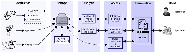
Fig. 1. Block diagram with the SOPHIA structure. 

A. Acquisition

The main static devices used in ophthalmology facilities are the ones from the following brands: Zeiss, Optovue, Canon, Topcom, and Heidelberg. These devices are limited by the high associated cost, non-portability and by the manufacturer's limitations for post-image analysis. On the other hand, some fundus cameras are portable, allow easy acquisition, and subsequent analysis, but are considerably expensive, the most widely used include Volk Pictor plus, Horus Scope, and Welch Allyn RetinaVue 100 Imager [10].

B. Storage

The storage block corresponds to a database in the cloud that stores images and metadata. However, images acquired with mobile devices are evaluated using automatic methods to improve illumination, contrast, and edge enhancement according to clinical criteria.

C. Analysis

This block uses methods for two main purposes: diagnostic support and visual/textual content analysis to improve image retrieval [11, 12, 13, 14]. The intelligent image analysis module includes different models of machine learning and deep learning aimed at specific disease diagnosis. The multimodal indexing module includes models that allow the visual content of the image, and the textual content of the metadata to be analyzed jointly to extract useful patterns for content-based image information retrieval.

D. Access

The access block provides several modules that implement different system functionalities. The intelligent diagnostic support stage provides additional interpretable clinical information for disease prediction. On the other hand, the use of algorithms to retrieve clinically meaningful information from large medical databases supports diagnosis and decision making. Finally, the image annotation and metadata editing stages allow to add new information about clinical data, as well as local and global characteristics to be included in the image dataset.

E. Presentation

This block corresponds to the system's user interface, which is a web-based application that can be accessed from a desktop browser or mobile device.

III. Results

This section presents the main results obtained for quality assessment, DR, DME, and clinical findings detection. The databases, clinical criteria, and results obtained were validated by professionals from the Fundación Oftalmológica Nacional.

A. Automatic Evaluation of Ophthalmic Image Quality

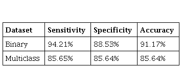
The automatic quality assessment was developed using the eye fundus image set provided by the California Healthcare Foundation and Kaggle for the detection of diabetic retinopathy [15]. Images from this database were classified by experts as images in a binary-class problem with acceptable quality and rejected images [16]. Also, they were classified in a multi-class problem with good, usable, and poor-quality image labels, respectively [17].

The architecture of the proposed model contains a series of 5 blocks of convolutional and max-pooling layers with different filters (16, 32, 64, 64, and 64), and with kernel sizes of 11x11, 9x9, 7x7, 6x6, and 6x6, respectively. Finally, it contains three dense layers with 256 and 64 neurons in the first two layers, while the last layer uses the number of classes, as presented in Figure 2.

Fig. 2. Deep learning model for the evaluation of eye fundus image quality. 

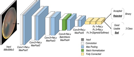
The results obtained for the databases in test sets are presented in Table 1.

Table 1 Performance measures for the fundus image quality evaluation 

B. Models for the Diabetic Retinopathy Detection

Models for diabetic retinopathy detection were developed using two datasets. The Kaggle challenge dataset, consisting of about 88,000 fundus images, and containing the following numerical classification: 0 for healthy individuals and 1-5 for mild, moderate, severe, and proliferative DR, respectively [15]. The second set is Messidor-2 [18], with 1748 eye fundus images from a French research program, and binary labels for referable and non-referable DR.

The InceptionV3 architecture was trained and fine-tuned using the pre-trained weights from the ImageNet dataset [20], as shown in Figure 3.

Fig. 3. Architecture used for automatic DR detection [19]. 

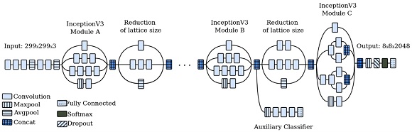
The model uses a low learning rate (LR) to perform fine-tuning with a particular partition of the Kaggle public dataset. Each image was associated with a label of 0 for healthy or non-referable patients, and 1 for a patient with any DR or referable diagnosis. Initially, features are extracted using the InceptionV3 pre-trained with the ImageNet dataset, these features are used as input to the classification model, on which a systematic exploration was performed to determine the best hyperparameters. In our case, the parameters were a LR =1x10⁻³ and the batch size was of 32. Finally, a fine tuning of the pre-trained weights of the last two blocks of the InceptionV3 architecture during 300 epochs was made, using as optimization algorithm the stochastic gradient descent (SGD), with LR =1x10⁻⁵, and a momentum of 0.9; the loss employed was categorical cross-entropy and a batch size of 32. Thus, an AUC of 0.92, a sensitivity of 89.74%, a specificity of 92.44%, and an accuracy of 90.10% were obtained using the Messidor-2 dataset.

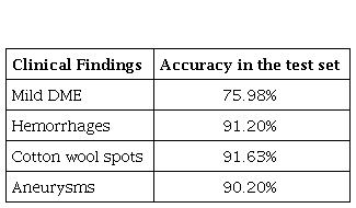
C. Obtained Results in the Detection of DME and Clinical Findings

The proposed model was trained using a VGG model to classify the images with moderate DME, using an Adam adaptive optimizer, with an LR = 1x10⁻⁵ and a batch size of 2. The number of dense layers and nodes per layer for the classifier were explored in a systematic search, using 25 epochs and a binary cross-entropy function to model the loss. The results with 2 dense layers, and with 4096 and 512 units presented the best results in training and validation. The best model was evaluated with the test set, the results are presented in Table 2.

Table 2 Accuracy in the detection of mild DME and findings associated with DR 

IV. Discussion and conclusions

The implemented deep learning methods have achieved good performance for eye fundus image quality assessment or improvement, however, the enhancement of images taken with mobile devices is still a challenge [21]. In addition, deep learning offers the best performance for the description of visual content compared to other methods [22], therefore, its application in ophthalmic information retrieval is a promising research opportunity [23].

Ophthalmological databases have additional information that is not fully exploited, such as clinical data, diagnostic reports, or other data that could be included to improve the models' performance. In this aspect, there is a need for multimodal systems that allow the effective exploitation of joint information between different information sources.

There is great potential for the design and implementation of low-cost systems based on machine and deep learning techniques in low-income and developing countries. The proposed system combines a low-cost image acquisition and computer vision methods with the highest quality clinical requirements to ensure accurate medical diagnosis in remote and hard-to-reach areas. The implementation and future validation of such systems, in support of clinical staff, represent major challenges that could potentially help to reduce the low coverage of public health systems, the lack of specialized services, and the high cost of specialized medical examinations.

Acknowledgments

The authors are grateful for the support of MinCiencias with the research project number 1101-807-63563, named "Detección Temprana de daño ocular en Diabéticos usando un sistema de Inteligencia Artificial en Imágenes de Fondo de Ojo", as well as with the Universidad Nacional de Colombia for the support in the project "Clasificación de retinopatía diabética y edema macular diabético en imágenes de fondo de ojo mediante redes neuronales convolucionales", number 202010029118. This work was partially supported by a Google Research Award.

References

[1] American Diabetes Association, “Classification and diagnosis of diabetes,” Diabetes Care, vol. 39 (1), S13-S22, 2016. https://doi.org/10.2337/dc16-S005Links ]

[2] M. Abràmoff, M. Garvin, and M. Sonka, "Retinal imaging and image analysis,” IEEE reviews in biomedical engineering, vol. 3, pp. 169-208, 2010. https://doi.org/10.1109/RBME.2010.2084567Links ]

[3] J. Köberlein, K. Beifus, C. Schaffert, and R. Finger, “The economic burden of visual impairment and blindness: a systematic review,” BMJ open, vol. 3 (11), e003471, 2013. https://doi.org/10.1136/bmjopen-2013-003471Links ]

[4] G. Labiris, E. Panagiotopoulou, and V. Kozobolis, “A systematic review of teleophthalmological studies in Europe,” International journal of ophthalmology, vol. 11 (2), pp. 314-325, 2018. https://doi.org/10.18240/ijo.2018.02.22Links ]

[5] R. Gargeya, and T. Leng, “Automated identification of diabetic retinopathy using deep learning,” Ophthalmology, vol. 124 (7), pp. 962-969, 2017. https://doi.org/10.1016/j.ophtha.2017.02.008Links ]

[6] S. Otálora, O. Perdomo, F. González, and H. Müller, “Training deep convolutional neural networks with active learning for exudate classification in eye fundus images,” In Intravascular Imaging and Computer Assisted Stenting, and Large-Scale Annotation of Biomedical Data and Expert Label Synthesis, pp. 146-154, 2017. https://doi.org/10.1007/978-3-319-67534-3_16Links ]

[7] C. Lam, C. Yu, L. Huang, and D. Rubin, “Retinal lesion detection with deep learning using image patches,” Investigative ophthalmology & visual science, vol. 59 (1), pp. 590-596, 2018. https://doi.org/10.1167/iovs.17-22721Links ]

[8] O. Perdomo, S. Otálora, F. Rodríguez, J. Arévalo, and F. González, “A novel machine learning model based on exudate localization to detect diabetic macular edema,” in Proceedings of the Ophthalmic Medical Image Analysis Third International Workshop, pp. 137-144, 2016. https://doi.org/10.17077/omia.1057Links ]

[9] B. Host, A. Turner, and J. Muir, “Real‐time teleophthalmology video consultation: an analysis of patient satisfaction in rural Western Australia,” Clinical and Experimental Optometry, vol. 101 (1), pp. 129-134, 2018. https://doi.org/10.1111/cxo.12535Links ]

[10] J. Micheletti, A. Hendrick, F. Khan, D. Ziemer, and F. Pasquel, “Current and next generation portable screening devices for diabetic retinopathy,” Journal of diabetes science and technology, vol. 10 (2), pp. 295-300, 2016. https://doi.org/10.1177/1932296816629158Links ]

[11] W. Alyoubi, W. Shalash, and M. Abulkhair, “Diabetic retinopathy detection through deep learning techniques: A review,” Informatics in Medicine Unlocked, vol. 20, e100377, 2016. https://doi.org/10.1016/j.imu.2020.100377Links ]

[12] K. Stebbins, “Diabetic Retinal Examinations in Frontline Care Using RetinaVue Care Delivery Model,” Point of Care, vol. 18 (1), pp. 37-39, 2019. https://doi.org/10.1097/POC.0000000000000183Links ]

[13] O. Perdomo, J. Arévalo, and F. González, “Convolutional network to detect exudates in eye fundus images of diabetic subjects,” in 12th International Symposium on Medical Information Processing and Analysis, 2017, e101600T. https://doi.org/10.1117/12.2256939Links ]

[14] O. Perdomo, V. Andrearczyk, F. Meriaudeau, H. Müller, and F. González, “Glaucoma diagnosis from eye fundus images based on deep morphometric feature estimation,” inComputational pathology and ophthalmic medical image analysis, pp. 319-327, 2018. https://doi.org/10.1007/978-3-030-00949-6_38Links ]

[15] B. Graham, “Kaggle diabetic retinopathy detection competition report,” Master Thesis, University of Warwick, United Kingdom, 2015. [ Links ]

[16] K. Zhou, Z. Gu, A. Li, J. Cheng, S. Gao, and J. Liu, “Fundus image quality-guided diabetic retinopathy grading,” inComputational Pathology and Ophthalmic Medical Image Analysis, pp. 245-252, 2018. https://doi.org/10.1007/978-3-030-00949-6_29Links ]

[17] H. Fu, B. Wang, J. Shen, S. Cui, Y. Xu, J. Liu, and L. Shao, “Evaluation of retinal image quality assessment networks in different color-spaces,” in International Conference on Medical Image Computing and Computer-Assisted Intervention, pp. 48-56, 2019. https://doi.org/10.1007/978-3-030-32239-7_6Links ]

[18] E. Decencière, X. Zhang, G. Cazuguel, B. Lay, B. Cochener, C. Trone, P. Gain, R. Ordonez, P. Massin, A. Erginay, B. Charton and J-C. Klein, “Feedback on a publicly distributed image database: the Messidor database,” Image Analysis & Stereology, vol. 33 (3), pp. 231-234, 2014. https://doi.org/10.5566/ias.1155Links ]

[19] C. Szegedy, V. Vanhoucke, S. Ioffe, J. Shlens, and Z. Wojna, “Rethinking the inception architecture for computer vision,” in Proceedings of the IEEE conference on computer vision and pattern recognition, pp. 2818-2826, 2016. https://doi.org/10.1109/CVPR.2016.308Links ]

[20] O. Russakovsky, J. Deng, H. Su, J. Krause, S. Satheesh, S. Ma, Z. Huang, A. Karpathy, A. Khosla, M. Bernstein, A. Berg and L. Fei-Fei, “Imagenet large scale visual recognition challenge,” International journal of computer vision, vol. 115 (3), pp. 211-252, 2015. https://doi.org/10.1007/s11263-015-0816-yLinks ]

[21] T. Vu, C. Van Nguyen, T. Pham, T. Luu, and C. Yoo, “Fast and efficient image quality enhancement via desubpixel convolutional neural networks,” inProceedings of the European Conference on Computer Vision (ECCV), 2018. https://doi.org/10.1007/978-3-030-11021-5_16Links ]

[22] J. Wan, D. Wang, S. Hoi, P. Wu, J. Zhu, Y. Zhang, and J. Li, “Deep learning for content-based image retrieval: A comprehensive study,” in Proceedings of the 22nd ACM international conference on Multimedia, pp. 157-166, 2014. https://doi.org/10.1145/2647868.2654948Links ]

[23] Y. Cao, S. Steffey, J. He, D. Xiao, C. Tao, P. Chen, and H. Müller, “Medical image retrieval: a multimodal approach,” Cancer informatics, vol. 13, e14053, 2014. https://doi.org/10.4137/CIN.S14053Links ]

Received: July 23, 2020; Accepted: September 14, 2020

Conflict of interest: The authors state there is no conflict of interest.

Author's contribution: Oscar-Julián Perdomo-Charry; Andrés-Daniel Pérez-Pérez; Melissa de-la-Pava-Rodríguez; Víctor-Alfonso Arias-Vanegas; Juan-Sebastián Lara-Ramírez; Santiago Toledo-Cortés: Formal analysis, Research, Methodology, Writing – original draft. Hernán-Andrés Ríos-Calixto; Francisco-José Rodríguez-Alvira: Conceptualization, Research, Validation. Jorge-Eliecer Camargo-Mendoza; Fabio-Augusto González-Osorio: Conceptualization, Methodology, Supervision, Writing - review and editing.

Creative Commons License This work is under License Creative Commons Attribution 4.0 International.