


Services on Demand
Journal
Article
 text in
  Spanish (pdf)
 Article in xml format
 Article references
 Send this article by e-mail
Indicators
-
 Cited by SciELO
 -
 Access statistics
Related links
-
 Cited by Google
 -
 Similars in
SciELO
Similars in
SciELO -
 Similars in Google
Share

 Permalink
Revista Facultad de Odontología Universidad de Antioquia
Print version ISSN 0121-246X
Rev Fac Odontol Univ Antioq vol.23 no.1 Medellín July/Dec. 2011
CASE REPORT
Mandibular bilateral macrodontia and hyperdontia: a clinical case report
Nathalia Rúa Cardona1, John Jairo Tapias2, Jorge Mario Castaño Henao3
1Dentist, Universidad de Antioquia. Specialist in Pathology and Oral
Medicine UAM-Mexico; Professor instructor at Universidad CES and
Adjunct professor at Universidad de Antioquia, Schools of Dentistry.
E-mail address: nathyrua@hotmail.com
2Dentist, Universidad de Antioquia. Maxillofacial Prosthesis Resident,
School of Dentistry, Universidad Nacional Autónoma de México. E-mail
address: johnjtapias@hotmail.com
3Dentist, CES. Orthodontics Resident, School of Dentistry, Universidad
Cooperativa de Colombia. E-mail address: jorgecastano74@hotmail.com
SUBMITTED: NOVEMBER 30, 2010-ACCEPTED: SEPTEMBER 13, 2011
CORRESPONDING AUTHORSNathalia Rúa Cardona
E-mail address: nathyrua@hotmail.com
John Jairo Tapias
E-mail address:johnjtapias@hotmail.com
Rúa N, Tapias JJ, Castaño JM. Mandibular bilateral macrodontia and hyperdontia: a clinical case report. Rev Fac Odontol Univ Antioq 2011; 23(1): 174-181.
ABSTRACT
Macrodontia is a dental size anomaly of unknown etiology, characterized by abnormal tooth size when compared to the rest of the dental formula. Hyperdontia is the presence of an increased number of teeth in the dental formula. Although it is rare, in some cases both anomalies have been reported simultaneously, and they might be associated or not with a syndrome. A 12-year-old male patient who was scheduled for a routine dental exam revealed at the radiographic examination impaction of teeth 34, 35 and 45 with macrodontia of the same teeth and multiple hyperdontia (three extra teeth, located occlusal to 34, 35 and 45); no other relevant pathology was found. Multiple macrodontias and hyperdontias are rare dental anomalies that may cause pathologies and alterations to adjacent permanent teeth.
Key words: dental anomalies, hyperdontia, macrodontia.
INTRODUCTION
Macrodontia (megalodontia, megadontia) is a rare dental anomaly.1 The term refers exclusively to teeth that are physically bigger than usual and must not be used to refer to teeth that are altered by either fusion or germination.2 In contrast, the so-called relative macrodontia refers to teeth of normal size crowded inside a small maxilla.
Hyperdontia (supernumerary teeth) is defined as the excess of teeth in a normal dental formula, regardless of their localization and morphology.3-4
It is an uncommon anomaly, with less than 1% prevalence among the world’s population.5-6 It is then a relatively rare condition, which has often been associated to certain syndromes, although some other authors classify it and have reported it as “multiple supernumerary teeth with no syndrome”, which means that its presence is not connected to a syndrome in particular.5
Hyperdontia may be considered multiple when having one or more supernumerary teeth in two or more dental groups. These hyperdontias may be associated to some syndromes, such as the Gardner, Fabry-Anderson, or Ehler Danlos syndromes, as well as facial fissures or cleidocranial dysplasia, among others. In absence of these complex syndromes, hyperdontia is rare. These forms of hyperdontia seem to happen only in permanent dentition, and they usually involve the coexistence of supernumerary teeth in the anterior section or premolar area.3
Their etiology is still unknown,3, 6, 7 although numerous genetic theories have been suggested, such as: phylogenetic atavism or reversion, tooth bud dichotomy, dental lamina hyperactivity, autosomal dominant,3-6 autosomal recessive, and chromosome X-linked heredities, and multifactorial6 genetic patterns. Besides these, a combination of genetic and environmental factors has been mentioned.7
Depending on their buccal topographic location, they are classified into: mesiodens: Supernumerary tooth in the maxilla anterior region (incisors). Distomolar or distodens: an extra forth molar. Paramolar: A posterior supernumerary situated lingual or buccal to the molar.2 Parapremolar: a doubled premolar tooth.
According to their form, supernumerary teeth may be: Supplemental (eumorphic): a tooth with normal shape and size. Rudimentary (dysmorphic): a tooth with smaller shape and size; this includes conical, tuberculate and molariform teeth.5
Macrodontias are not very frequent, while hyperdontias are more common; but finding both anomalies in a single patient occurs only in isolated cases.
Due to their uncommon occurrence, this report presents a clinical case with developmental alterations in both dental number and size.
CLINICAL CASE
A 12-year old male patient requested private consultation for routine dental examination. The patient had above average body build in both height and weight (weight: 58 kg, height: 5’5”); he did not report any relevant personal or family medical record at the time of consultation. Intraoral examination revealed deciduous molar 74 yet to be shed but with degree 3 mobility and almost complete root resorption; it also displayed mild generalized enamel hypoplasia.
Treatment began with dental and periodontal preparation, and extraction of deciduous tooth 74 was performed. The standard panoramic radiograph image revealed that premolars 34, 35 and 45 were retained in the mandibular bone, with bigger size and abnormal morphology. Also impacted and located over the occlusal surfaces of each of these teeth, three in-formation supernumerary dysmorphic dental buds were observed (figure 1).
The Cone beam image of (figure 2) more clearly shows the panoramic radiograph images described.
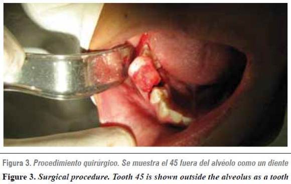
The procedures included simple exodontia of teeth 75 and 85, as well as surgical exodontia of the supernumerary buds and tooth 45 (figure 3), because their permanence in the oral cavity was considered unviable due to their location and size and to the risk of affecting adjacent permanent teeth with other pathologies, such as resorpions and cysts.
As part of treatment, teeth 34 and 35 were kept, in order to position them later by means of orthodontics. In the same surgical work, a direct cementation pin was placed in tooth 35 for traction. It was not possible to do the same with 34 as it was impossible to stick the resin due to difficult access. Right after the extractions, supernumerary dental buds, tooth 45, and their respective follicles were sent to histopathology analysis (figure 4). This analysis reported normal dental tissue and a dentigerous cyst associated to 45 (figure 5).
Although the patient does not present physical, psychological or physiological alterations, it is best to go on with diagnostic aids and medical referrals in order to discard the presence of a syndrome. But due to economic restrictions it is difficult to perform a more exhaustive study.
DISCUSSION
Isolated macrodontia is more commonly found in incisors and canines, but it has also been reported as involving second and third molars; teeth affected by macrodontia tend to appear bilaterally.1 Although macrodontias are more often found in the anterior region, the one in this clinical case is bilateral in the second mandibular premolars, agreeing with other data reported in the literature.1
Supernumerary teeth may erupt or remain impacted inside the maxillaries. About 75% of all the impacted supernumerary teeth are asymptomatic. Consequently, many of these teeth are accidentally found during routine X-ray studies.3 This agrees with the clinical case described here as the patient was asymptomatic and the impacted supernumerary as well as the macrodontic permanent teeth were found in a radiograph image during routine dental procedures.
Supernumerary teeth are often correlated with macrodontia. Some studies have shown that the teeth of patients with hyperdontia are bigger than normal, especially in terms of mesiodistal crown dimensions.2-8 These studies suggest that abnormalities of teeth size and number have similar etiology. Hyperdontia may or may not be accompanied with other developmental disorders.8 In our clinical case we found three supernumerary teeth associated to three macrodontic teeth with enlargement in all their dimensions, not just in the mesiodistal one; also, the patient did not show signs or symptoms that may suggest developmental alterations or association with syndromes related to these anomalies, such as: with hyperdontia: Apert, angioosteohypertrophy, cleidocranial dysplasia, craniometaphyseal displasia, Crouzon, Curtius, Down, Ehlers-Danlos, Ellis van Creveld, Fabry-Anderson, Fucosidosis, Gardner, Hallermann-Streiff, Incontinentia pigmenti, Klippel-Trénaynay-Weber, Laband, Leopardo, Nance Horan, type I and type III oral-facial- digital, Sturge-Weber, tricho-rhino-phalangeal.9
Syndromes associated with macrodontia: pituitary gigantism, otodental syndrome, XYY men, pineal hyperplasia with hyperinsulinism.2-9 Nevertheless, it is necessary to resort to medical referrals and the pertinent diagnostics aids in order to discard more precisely the presence of some of the syndromes previously mentioned.
Whether supernumerary teeth are erupted or impacted, they may induce to or be associated with different disorders. For example, delay in the eruption or no eruption of permanent teeth, late displacement or rotation, adjacent teeth’s root resorption, malformation of secondary roots due to the pressure applied by supernumerary teeth, and cyst formation. The ones more commonly reported are eruption delay, no eruption of permanent teeth, and malformations in neighboring teeth.3 In the previously reported case, none of these disorders was found but they were taken into account at the time of designing the treatment plan, which included histopathology study, oral surgery and orthodontics.
In spite of all the possible origins of hyperdontia, there is nothing conclusive. The closest theory to the possible cause of multiple supernumerary teeth indicates that this anomaly has a hereditary multifactorial pattern, possibly originated in alterations of the dental lamina.5 Several authors have reported recurrence of supernumerary premolars after surgical extraction.10 This is why, besides surgical, pathological, and orthodontic treatment performed on the patient of this study, it is necessary to make radiographic follow up in order to evaluate evolution, possible complications due to periodontal defects and possible emergence of other supernumerary teeth, although this rarely occurs (figure 6).
1. Dugmore CR. Bilateral macrodontia of mandibular second premolars: a case report. Int J Paediatr Dent 2001; 11: 69-73. [ Links ]
2. Neville BW, Damm DD, Allen CM, Bouquot JE. Abnormalities of teeth. En: Oral & Maxillofacial Pathology. 2.ª ed. Philadelphia: WB. Saunders; 2002. p. 49-106. [ Links ]
3. Yagüe-García J, Berini-Aytés L, Gay-Escoda C. Multiple supernumerary teeth not associated with complex syndromes: A retrospective study. Med Oral Patol Oral Cir Bucal 2009; 14(7): E331-E336. [ Links ]
4. Fernández-Montenegro P, Valmaseda-Castellón E, Berini- Aytés L, Gay-Escoda C. Retrospective study of 145 supernumerary teeth. Med Oral Patol Oral Cir Bucal 2006; 11: E 339-344. [ Links ]
5. Blanco-Ballesteros G. Dientes múltiples supernumerarios no relacionados a un síndrome: reporte de un caso. Rev Estomat 2005; 13(1): 13-19. [ Links ]
6. Proff P, Fanghanel J, Allegrini Jr. S, Bayerlein T, Gedrangen T. Problems of supernumerary teeth, hyperdontia or dentes supernumerii. Ann Anat 2006; 188: 163-169. [ Links ]
7. Radi JN, Álvarez GJ. Dientes supernumerarios: reporte de 170 casos y revisión de la literatura. Rev Fac Odontol Univ Antioq 2002; 3(2): 57-67. [ Links ]
8. Dos Santos APP, Ammari MM, Moliterno LFM, Capelli JJ. First report of bilateral supernumerary teeth associated with both primary and permanent maxillary canines. J Oral Sci 2009; 51(1): 145-150. [ Links ]
9. Gorlin RJ, Cohen MM, Hennekam RCM. Syndromes of head and neck. 4.ª ed. Oxford University Press; 2001. [ Links ]
10. Solares R, Romero MI. Supernumerary premolars: A literature review. Pediatr Dent 2004; 26(5): 450-458. [ Links ]

