


Serviços Personalizados
Journal
Artigo
 texto em
  Espanhol (pdf)
 Artigo em XML
 Referências do artigo
 Enviar este artigo por email
Indicadores
-
 Citado por SciELO
 -
 Acessos
Links relacionados
-
 Citado por Google
 -
 Similares em
SciELO
Similares em
SciELO -
 Similares em Google
Compartilhar

 Permalink
Revista Facultad de Odontología Universidad de Antioquia
versão impressa ISSN 0121-246X
Rev Fac Odontol Univ Antioq vol.23 no.2 Medellín jan./jun. 2012
ORIGINAL ARTICLES DERIVED FROM RESEARCH
Nonlinear evaluation of two different posts1
Patricia Lopera Builes2; Federico Latorre Correa3; Junes Abdul Villarraga Ossa4
1 Article derived from a research project conducted by the Research
Group Biomaterials in Dentistry, School of Dentistry, and the Mechanical
Design Group, School of Mechanical Engineering, Universidad de
Antioquia.
2 Dentist, Specialist in Adult Comprehensive Dentistry with Emphasis in
Prosthodontics, School of Dentistry, Universidad de Antioquia.
3 Dentist, Specialist in Adult Comprehensive Dentistry with Emphasis in
Prosthodontics, Associate Professor, School of Dentistry, Universidad
de Antioquia. Email address: flatorre@une.net.co
4 Mechanical Engineer, Universidad Nacional de Colombia at Medellín,
Msc Mechanical Engineering, Universidad Simón Bolívar, Caracas-
Venezuela. Associate Professor, School of Mechanical Engineering,
Universidad de Antioquia. Email address: junes@udea.edu.co
Lopera P, Latorre F, Villaraga JA. Evaluación no lineal de dos postes diferentes. Rev Fac Odontol Univ Antioq 2012; 23(2): 240-255.
ABSTRACT
Introduction: endodontically treated teeth that will be restored with crowns and which have lost part of their dental structure
need a cast post or a prefabricated post to improve final restoration retention and therefore to be able to regain their functionality and
aesthetics. The literature suggests when to use a cast or prefabricated post, but it is not conclusive in terms of which one has a better
stress distribution, which post material or shape is better, or whether their modulus of elasticity is related to load distribution along the
tooth's axis.
Methods: by using Solid Works, two models of a maxillary central incisor with all its supporting structures were made, one
with a cast post and another one with a prefabricated post plus an all-ceramic crown. By means of finite element analysis both teeth were
subjected to loads of 200, 400, 600 and 800 N up to reaching their plasticity.
Results: at 200 and 400 N, both posts showed different stress
distribution patterns without altering the restoration or the tooth's root.
Conclusions: at normal masticatory loads, both posts are likely
to restore a tooth that needs addition of a post.
Key words: cast post, prefabricated post, stress distribution, finite elements method.
INTRODUCTION
Restorative dentistry is daily challenged by the problem of restoring endodontically treated teeth whose coronal and radicular areas are also structurally weakened. An issue under discussion has been the prognosis of these teeth, which require intraradicular elements when adhesive dentistry is not possible, due to significant structure loss and thus achieve restoration retention,1 depending on several factors: the number of adjacent teeth, occlusal contacts, location of the tooth in the dental arch, apical state of the root, radicular collagen degradation, intermolecular relations at the radicular dentin, the amount of hard tissue lost, thickness of residual coronal dentin wall, the type of final restoration, presence of at least 1,5 to 2 mm of residual dentin so that it produces a ferrule effect, the type of post, and the material used for the core.1, 2
Some studies suggest that posts do not strengthen root structure nor they prevent future fractures, as this greatly depends on dental remainders, and the tooth's strength rests on proper application of the diverse post-endodontic procedures.3, 4
The literature specifies when to use a cast post, which is usually recommended when more than 50% of dental structure has been lost and a minimum of 2 mm residual dentin is needed in order to achieve ferrule effect and to thwart lateral stresses, which otherwise would produce instability of the post. Similarly, it states that dentin structures larger than 50% with a minimum remainder of 2 mm could also be treated with prefabricated posts.5, 6
Although there is theoretical clarity, there are no conclusive studies on which of the two types of posts better distributes masticatory forces along the root so that the dental structure receives masticatory stresses in an adequate manner, without altering dental structure or threatening the future restoration.7, 8
The studies do not explain which would be the best material, shape or mechanical properties of the posts,9, 10 and there exists some disagreement, among the diverse reports, as some of them show evidence that favor rigid posts while others suggest that they would be disastrous to dental structure.11
in vitro studies using finite element analysis have been conducted in 2D and 3D, but they haven't been able to provide a clear explanation due to computational limitations and because they have been linear studies (therefore not showing the whole reality of the system) using only static loads.12 Similarly, clinical studies fail to demonstrate or explain superiority of one system over the other due to intrinsic biological variables which differ from one person to another, and because they haven't been able to assess how stresses are distributed;13, 14and in vitro studies applying loads with Instron devices usually analyze teeth subjected to loads that do not usually happen in the oral cavity and therefore portraying a different reality, as usually the tooth is not completely restored.15, 16
In spite of this controversy, the studies agree on the importance of preserving as much dental structure as possible in order to reduce the risks of tooth fracture.17, 18
The finite element method (FEM) involves a series of computational procedures, and it is vastly used in dentistry because it allows finding simple solutions to complex problems, by dividing a structure in a limited or finite number of elements, which are interconnected by means of nodes that behave individually under certain loads. Therefore, this method does not regard the structure as a whole but as an assembly with multiple parts that allow identifying displacements, deformations, stresses and values of fracture resistance. This method is useful to determine the biomechanical properties of human tissues that otherwise would be very difficult to measurein vivo.19
Among the finite elements there are static linear models, in which a direct stress/deformation relation occurs, and non-linear models that take into account the material's non-linearity, as well as the model's big displacements, or sliding contact conditions. For example, in the case of periodontal ligament behavior, non-linear studies are more accurate, since linear studies would show an exaggerated deformation under big loads.20, 21
This is why non-linear analysis has become a powerful tool for calculating stresses in models that involve some linearity. These studies require more computation time and higher costs, and therefore are less common, but they are more accurate at explaining the whole model's behavior.21
The purpose of this study was to assess the nonlinear behavior of two posts subjected to different loads by means of simulation using finite element analysis, in order to verify whether one of the restoration systems is more advantageous than the other.
METHODS
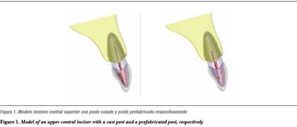
Two models were designed in SolidWorks (Dassault systémes, Suresnes, France), representing the dental structure of an upper central incisor with its supporting tissues. One of the models included a fiberglass prefabricated post, Parapost (Coltene, Whaledent International, Altstatten, Switzerland), and the other one had a metal based cast post (4all, Ivoclar Vivadent, Shaan, Liechtenstein) as well as an all-ceramic crown (emax press, emax ceram Ivoclar Vivadent) (figure 1).
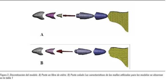
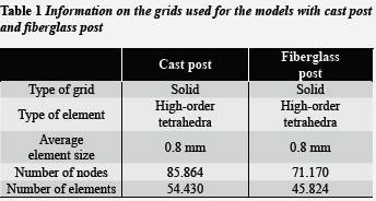
A non-linear model was constructed.21 Tetrahedral elements of the highest order (ten nodes per element) were used in the posts materials, in order to obtain a better approximation to the geometry of the parts (figure 2).
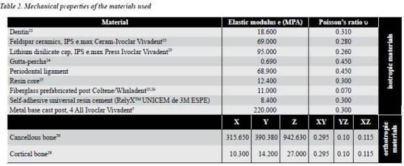
The mechanical properties of the elements that are considered to be linear in this model were obtained from previous studies reported in the literature.22-26 The following elements of the model had isotropic properties: feldspar ceramic, lithium disilicate cap, resin core, gutta-percha, dentin, periodontal fiber, and cement. The non-linear properties of both posts were taken as the structure's non-linear properties, as the literature does not report non-linear values for the rest of the structures,5, 27as well as orthotropic properties for both cortical bone and cancellous bone 28 (table 2).
The load applied to the models was directed at 45° and it was evenly distributed along the palatal surface. The loads ranged from 200 to 800 N with intervals of 200 N, in order to bring the model to its plastic deformation and to observe in which rank it was achieved.
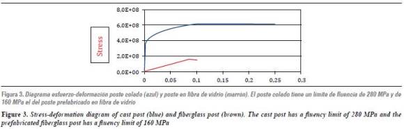
For this purpose, a stress-deformation diagram was outlined (figure 3)in order to observe the difference between the metal cast post and the fiberglass post fluency limits. Convergence of the model was obtained by means of an adaptive solution using the h method, with 98% precision.
Geometry
The upper central incisor's root was modeled with a core of 2 mm of remaining coronal structure, bringing a 1 mm bevel to its periphery in order to achieve ferrule effect, according to the literature, both in the cast post and the prefabricated post.
The root was given a conic shape, with 5 mm in diameter in both models to avoid altering the results due to the great anatomic and root dimensions variability existing from one individual to another.
Modeling of the root canal was performed taking into account that the pulp chamber width would not be bigger than a third of the root width in its narrower area, having at least 1 mm of healthy dentin around it, especially by the apical region.
The root canal's 4 apical mm were modeled with properties of gutta-percha (endodontic filling material) thus retaining the minimum amount of remaining obturation described in the literature to avoid leakage. A post for the remaining root canal and its coronal portion was modeled with the different kinds of materials to be studied. The post was modeled with a coronal diameter of 1.6 mm and an apical diameter of 1.1 mm, thus adjusting to the internal anatomy of the root canal, with its apical portion rounded.
The resin core configuration kept the proportions of an all-ceramic crown preparation for a left upper central incisor, which requires axial reduction of 1.8 mm. This ensured adequate space for modeling the ceramic crown as follows: porcelain thickness was between 1.5 and 1.8mm, except at the incisal portion which was 2.0 mm. All these parameters allow the restoration meeting to meet the aesthetic and thickness requirements recommended for material strength and thus avoid overcontouring.28
Periodontal ligament with root periphery thickness of 0.2 mm was designed taking into account isotropic properties, and it was placed 1.5 mm from the cement-enamel junction.28
The design of the alveolar bone included cancellous bone, which forms the inner part of the maxillary body, and cortical bone, that surrounds both the maxilla and the alveolus.
Both structures were considered to have ortothropic properties (materials with different characteristics in the x, y and z axes).Bone height was 18 mm from the alveolar crest up to the maxillary base.
The cortical bone was 1 mm thick in the peripheral zone from the basal region and 0.5 mm towards the alveolus' internal region. The teeth had a total length of 23.6 mm; the crown being 10.8 mm and the root 12,8 mm. Considering that the anatomy of human beings is variable, average data were used, just as other studies have done it.29
In order to calculate stress and displacement with the model, elastic constants were used (Young's modulus of elasticity: a property of the materials equivalent to the slope of stress curve against the elastic deformation range and Poisson's ratio, which is an elastic constant of materials indicating the relation between axial and lateral deformation) for each of the elements to be analyzed.
RESULTS
Once the tridimensional mathematic model of the two upper endodontically treated and restored central incisors with two different posts was completed, SolidWorks simulation (Dassault systémes, Suresnes, France, 2009) was performed obtaining the following results: in the model with a cast post, the stresses concentrated mainly at the post and the ceramic substructure, as shown in figures 4A and 4B.
Figure 5 shows stress distribution and deformation under each load. In the figures 5A and 5B, the stresses were 200 and 400 N, respectively, and stress concentration occurred at the same spots as in the previous figure (figure 4). As fluency limit was not exceeded, elastic deformation of the post occurred. For loads of 600 and 800 N (figures 5C and 5D), the stresses concentrated at the same spots, but with plastic deformation of the post because the material's fluency limit was exceeded.
Figure 6 displays stress concentration and deformation of the prefabricated post under each of the loads. Notice that the stresses concentrated at the ceramic substructure and the resin core, the prefabricated post suffered deformation within the elastic limit, while the ceramic substructure and the resin presented elastic deformation as their fluency limit was exceeded.
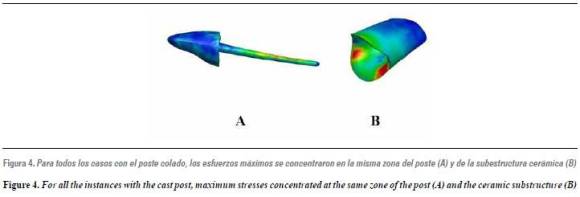
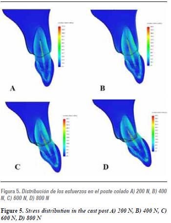
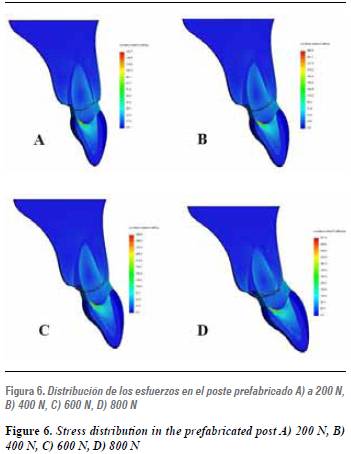
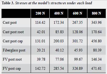
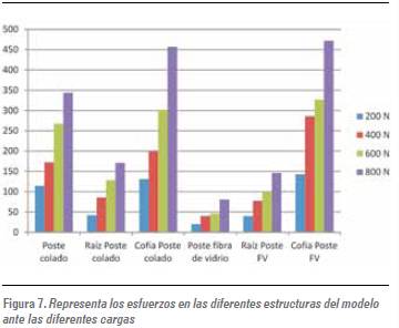
Table 3 and figure 7 show the von Mises stresses of the two types of post under study, which were subjected to four stresses, from 200 N up to 800 N, with differences of 200 N in between, in order to assess stress at the root, the post and the disilicate cap.
DISCUSSION
Finite element analysis is used in dentistry because it allows assessing the mechanical behavior of teeth and the structures with which they are restored. Modeling a tooth with all its structures, just as it is usually restored, by means of a tridimensional model, and conducting a non-linear analysis, allow obtaining more accurate assessment and greater clinical contributions.
The non-linear finite analysis used in this study proved stress distribution differences between two endodontically treated teeth restored with either prefabricated (fiberglass) or cast (metal base) posts; however, each model should be assessed in a different way because each one has different geometry and materials.
This study found out that when the tooth was subjected to loads that are normally found in the oral cavity (between 200 and 400 N), the behavior of both models was similar, indicating that fracture risk should not be considered for concentration of greater or smaller stress in the post. An important characteristic of stress distribution in both models, that was simulated in this study, is that as stresses are greater, they increase in the periodontal ligament, which absorbs part of the stress thus reducing negative effects on the tooth and its structures; therefore, studies that do not simulate the periodontal ligament and its behavior will experience a significant variable error.
This analysis allowed concluding that the most rigid material of each model absorbs the greater force; therefore, in the model with a cast post, the ceramic cap absorbs most of the force, followed by the cast post pole and the root. In the fiberglass post, the ceramic cup absorbs the greatest force, followed by the root and finally by the post pole, which receives the smallest force. This study found out that under physiological masticatory loads of 200 N and 400 N, posts with a modulus of elasticity similar to the dentin's present lower stress concentrations in the posts, and distribute them differently than posts with more rigidity.
This result disagrees with several studies, such as the one by Zarone et al in 2006 that assesses stress distribution patters in a healthy tooth compared to restored teeth with different material configurations, stating that the restoration materials must be similar to the tooth's tissues in order to allow the whole restoring system imitating the mechanical behavior of a natural tooth. Among the limitations of that study is the fact of being linear, which means that some data are lacking to provide a more real conclusion from the system.30
In 2002, Pegoretti et al, by means of a finite element analysis in 2D, examined the mechanical behavior of a fiberglass post in comparison to a carbon fiber post and a gold post cast, having a natural tooth as control variable. Their study favored fiberglass posts; however, it is not a tridimensional study, it is a linear one, and it does not analyze the periodontal ligament,so their conclusions may have a considerable margin of error, as a linear analysis that disregards all the variables would not allow concluding that a rigid post may have fracture effects or system damage.31
Studies such as the one by Mikako et al in 2008 and Lippo et al in 2004 conclude that posts with modulus of elasticity similar to that of the dentin present better behavior under stresses and may be recoverable if fractured, as their fracture patters are less disastrous than rigid posts.32, 33 In 2005, Seefeld et al suggested that using posts with biomechanical properties similar to that of the dentin may be useful as they reduce the risk of tooth fracture;27 similarly, Aquaviva et al, in a 2002 literature review, recommend using posts with a low modulus of elasticity in order to obtain greater adherence and less stress absorption to dental structure, as well as greater protection under masticatory loads.34
Other studies present similarities with our own, such as the one by Naumman et al in 2007, which analyzed the impact of a rigid material (a titanium post) compared to a more "flexible" one (a fiberglass post), finding out that using a post with a modulus of elasticity lower or close to that of the dentin does not imply a better behavior for the system–and that would be a debatable concept–. Their study suggests that when a tooth with different components is subjected to loads, the rigid component has the ability of resisting greater stresses without distorting.35
In 2006, Sorrentino et al conducted a finite element study with the intention of assessing stress distribution in a system composed by a post, a core, and a crown, pointing out the importance of using materials with a higher modulus for the crown's material, as it protects the whole system and avoids greater stresses in the entire tooth. This is also concluded in our study, in which greater stresses initially occur at the ceramic.36
Another in vitro study, by Qing et al in 2007, analyzed the fracture strength of anterior teeth that had been endodontically treated and restored with fiberglass posts and zirconia posts, having teeth restored with cast post as control group.
They found out fracture strength differences between the two groups, thus suggesting that rigid posts may improve deflection resistance. They also verified that the cast post and the core were more resistant to deflection forces, suffering failures under very high loads; therefore, they state that their results do not allow concluding that clinical use of posts is not convenient.37
Schmitteret at, in an interesting study in 2010, combining in vitro analysis and finite elements modeling, concluded that the most important part for system protection is the crown-core junction, and that the fiberglass post effect would not be advantageous for the system.38
In an experiment with lots of coincidences with the present study, in terms of research problem formulation, methodology, tridimensional approach, and use of non-linear analysis, Dejak et al in 2011 obtained very similar results and conclusions, since under physiological loads the posts made of metal or fiberglass do not produce tooth damage, and cast posts apply less stress on the dentin, due to its mechanical properties, as a higher modulus of elasticity.29
Debates among the aforementioned studies demonstrate that previous research has not solved the controversy on which post system provides teeth with greater resistance and permanence.
Based on the results of the present study, no conclusions can be drawn as of one type of post being better than the other, since stresses on roots under normal loads, it is, at 200 and 400 N, behave in a similar manner and, even under efforts that could be disastrous, such as concentrations of 600 and 800 N, no significant differences are found in the roots. The answer as to why fractures happen in any of the elements (post, root or core) when subjected to stresses that hardly occur in the mouth, such as 600 or 800 N, lies on the mechanical properties of each material or tissue–and not because a given material supplies the roots with more stress concentration.
In this study, stresses concentrated differently at the post, the root, or the ceramic cap in both kinds of posts, thus agreeing with the study by Dejak et al.29
This study intentionally omitted the cement because including it would require a greater number of elements, thus increasing computation time and costs, and it has been demonstrated that, due to its mechanical properties (similar to the dentin) it would not be relevant as part of the structure, as it has been proven that cement does not receive the applied loads directly but rather transfer them to the post and root.39
When stresses were increased, the results of this study showed that the cast post, the ceramic cap, and the post pole received the greatest stress, and the cast post pole suffered plastic deformation at 600 MPa, while the prefabricated post displayed greater elastic deformations. This would demonstrate differences between clinical studies and in vitro studies, in which, with a cast post, fractures occur in the post pole and towards cervical, while with a prefabricated post they occur at the core or at cervical, but almost never in the post pole. These findings are also confirmed by Varvara et al in a 2007 in vitro study, conducted with the purpose of evaluating fracture strength of endodontically treated teeth with different levels of residual dentin, concluding that under heavy loads the posts with a high elastic modulus were more resistant than those with more flexibility, but the latter were more susceptible to failures than the former. Nevertheless, the authors conclude that these loads with disastrous failures in the cast posts occur at forces that are not usually found in the oral cavity, agreeing with the findings of our study.8
This study demonstrates that stresses at both roots are very similar, which explains why in presence of roots with a good structure, fracture risks do not occur in any of the two systems.
Concerning the specifications of this study, it is important to point out that it was conducted with static loads; it was non-linear and tridimensional, and subjected teeth to physiological and non-physiological forces. It is recommendable to advance in studies with dynamic forces that totally simulate real clinical conditions in order to complement clinical studies on this field.
CONCLUSIONS
Based on the specifications of this study, the following conclusions may be drawn:
When masticatory forces are within the physiological ranks (200 to 400 N) and the root meets the requirements of minimum space for the posts, without debilitating dental structure, both posts are likely to restore an endodontically treated tooth.
It cannot be stated that one of the systems is better than the other, as they both behave in totally different ways.
Knowing how stress concentrations occur under different loads, and in different ways in each model, would help the dental professional make the right decision according to the chosen system.
CORRESPONDING AUTHOR
Federico Latorre Correa
Facultad de Odontología, Universidad de Antioquia
Calle 64 N.o 52-59
Medellín, Colombia
Correo electrónico:
flatorre@une.net.co
REFERENCES
1. Schwartz R. Post placement and restoration of endodontically treated teeth: A literature review. J Endod 2004; 30(5): 289-301. [ Links ]
2. Naumann M, Kiessling S. Treatment concepts for restoration of endodontically treated teeth: A nationwide survey of dentist in Germany. J Prosthet Dent 2006; 96(5): 332-338. [ Links ]
3. Mikako H, Yutaka T, Satoshi I. Fracture resistance of pulpless teeth restored with post-cores and crowns. Dent Mater 2006; 22: 477-485. [ Links ]
4. Plotino G, Grande N. Flexural properties of endodontic posts and human root dentin. Dent Mater 2007; 23(9): 1129-1135. [ Links ]
5. Torbjorner A. A literature review on the prosthetic treatment of structurally compromised teeth. Intern J Prostho 2004; 17(3): 369-372. [ Links ]
6. Cheung W. A review of the management of endodontically treated teeth. Post, core and the final restoration. Am Dent Assoc 2005; 136 (5): 611-619. [ Links ]
7. Fernandez A, Shetty S, Countinho I. Factors determining post selection: a literature review. J Prosthet Dent 2003; 90: 556-562. [ Links ]
8. Varvara G, Perinneti G, Dilorio D, Murmura G, Caputi S. In vitro evaluation of fracture resistance and failure mode of internally restored endodontically treated maxillary incisors with differing heights of residual dentin. J Prosthet Dent 2007; 98: 365-332. [ Links ]
9. Bëgum A. An in vitro study evaluating the effect of ferrule length on fracture resistance of endodontically treated with fiber-reinforced and zirconia dowel systems. J Prosthet Dent 2004; 92: 155-162. [ Links ]
10. Barjau A, Sancho J, Forner L, Rodriguez P, Pérez A, Sánchez F. Influence of prefabricated post material on restored teeth: Fracture strength and stress distribution. Oper Dent 2006; 31(1): 47-54. [ Links ]
11. Sorrentino R, Aversa R, Ferro V, Auriemma T, Zarone F, Ferrari M et al. Three dimensional finite elements analysis of strain and stress distribution in endodontically treated maxillary central incisors restored with different post, core and crown materials. Dent Mater 2007; 23: 983-993. [ Links ]
12. Sahafi A, Peutzfeld A, Asmussen E. Retention and failure morphology of prefabricated posts. Int J Prosthodont 2004; 17(3): 307-312. [ Links ]
13. Boderau F, Besone E, Baina L. Estudio comparativo de la adaptación de tres sistemas de postes. Rev Intern Prot Estom 2004; 6(3): 183-189. [ Links ]
14. Lippo V, Lassila J, Tanner J. Flexural properties of fiber reinforced root canal posts. Dent Mater 2004; 20(1): 29-36. [ Links ]
15. Kremeier K, Fasen L, Klaiber B. Influence of endodontic post type and luting material on push out bond strength to dentin in vivo. Dent Mater 2008; 24: 660-666. [ Links ]
16. Hew Y, Purton G, Love M. Evaluation of prefabricated root canals posts. J Oral Rehab 2001; 28: 207-211. [ Links ]
17. Fitzpatrick B. Evidence-based dentistry-it subdivided: accepted truths, once divided, may lack validity. Intern J Prosth 2008; 21(4): 358-363. [ Links ]
18. Wiskott A, Meyer M, Perriard J. Rotational fatigue-resistance of seven post types anchored on natural teeth. Den Mater 2007; 23: 1412-1419. [ Links ]
19. Reddy JN. Boundary element method. En: Reddy JN. An introduction to the finite element method. 3.ª ed. New York: McGraw-Hill; 2005. p. 442-443. [ Links ]
20. Geng PJ, Tan KBC, Liu GR. Application of finite element analysis in implant dentistry: A review of the literature. J Prosthet Dent 2001; 85(6): 585-598. [ Links ]
21. Wakabayashi N, Ona M, Suzuki T, Igarashi Y. Nonlinear finite element analysis: advances and challenges in dental applications. J Dent 2008; 36(7): 463-471. [ Links ]
22. Toparli M. Stress analysis in a post restored tooth utilizing the finite elements method. J Oral Rehab 2003; 30: 470-476. [ Links ]
23. Ivoclar Vivadent. Ficha técnica IPS e.max Press [Internet] [fecha de acceso octubre 16 de 2011]; URL disponible en http:// www.ivoclarvivadent.com/es/todos/productos/ ceramica-sin-metal/ips-emax-system-tecnico-dental/ipse_ max-press. [ Links ]
24. Asmussen E, Peutzfeldt A, Sahafi A. Finite element analysis of stress in endodontically treated, dowel-restored teeth. J Prosthet Dent 2005; 94(4): 321-329. [ Links ]
25. Maceri F, Martignoni M, Vairo G. Mechanical behavior of endodontic restoration with multiple prefabricated posts: a finite elements approach. J Biomech 2006; 40(11): 2386-2398. [ Links ]
26. 3M ESPE. Ficha técnica Rely XTM Unicem. Aplicap TM/ Maxicap TM [internet]. [fecha de acceso octubre 16 de 2011]; URL disponible en: http://www.ddsltdlab.com/pdf/ CementationTechniques.pdf. [ Links ]
27. Seefeld F, Wenz HJ, Ludwig K, Kern M. Resistance to fracture and structural characteristics of different fiber reinforced post systems. Dent Mater 2007; 23(3): 265-71. [ Links ]
28. Heoung-Jae Chun, Ha-Shik Shin, Chong-Hyun Han, Soo- Hong Lee. Influence of implant abutment type on stress distribution in bone under various loading conditions using finite element analysis. Int J Oral Maxillofac Implants 2006; 21: 195-202. [ Links ]
29. Dejak B, Andrzej M. Finite element analysis of strength and adhesion of cast posts compared to glass fiber-reinforced composite resin posts in anterior teeth. J Prosthet Dent 2011; 105: 115-126. [ Links ]
30. Zarone F, Sorrentino R, Apicella D, Valentino B, Ferrari M, Aversa R et al. Evaluation of the biomechanical behaviour of maxillary central incisors restored by means of endocrowns compared to a natural tooth: a 3D static linear finite element analysis. Dent Mater 2006; 22: 1035-1044. [ Links ]
31. Pegoretti A, Fambri L, Zappini G, Biancheti M. Finite element analysis of a glass fibre reinforced composite endodontic post. Biomaterials 2002; 23: 2667-2682. [ Links ]
32. Mikako H, Atsushi S, Yutaka T, Satoshi I. Static and fatigue fracture resistances of pulpless teeth restored with postcores. Dent Mater 2008; 24(9): 1178-1186. [ Links ]
33. Lippo V, Lassila J, Tanner J. Flexural properties of fiber reinforced root canal posts. Dent Mater 2004; 20(1): 29-36. [ Links ]
34. Aquaviva S, Fernandes, Sharat S. Factores que influyen en la resistencia a la fractura de los dientes reconstruidos con postes y muñones: Revisión. Rev Intern Prot Estom 2002; 4(1): 23-31. [ Links ]
35. Naumman M, Preuss A, Frankenberger R. Reinforcement effect of adhesively luted fiber reinforced composite versus titanium post. Dent Mater 2007; 23(2):138-144. [ Links ]
36. Sorrentino R, Aversa R, Ferro V, Auriemma T, Zarone F, Ferrari M et al. Three-dimensional finite element analysis of strain and stress distributions in endodontically treated maxillary central incisors restored with different post, core, and crown materials. Dent Mater 2006; 23(8): 983-993. [ Links ]
37. Qing H, Zhu Z, Chao Y, Zhang WQ. In vitro evaluation of the fracture resistance of anterior endodontically treated teeth restored with glass fiber and zircon posts. J Prosthet Dent 2007; 97(2):93-8. [ Links ]
38. Schmitter M, Rammelsberg P, Lenz J, Scheuber S, Schweizerhof K, Rues S. Teeth restored using fiberreinforced posts: In vitro fracture tests and finite element analysis. Acta Biomaterialia 2010; 6: 3747-3754. [ Links ]
39. Orozco M, Villarraga J, Latorre F, Escobar JC. Influencia de los materiales de cementación en la distribución de los esfuerzos en un incisivo central superior rehabilitado con poste. Análisis de elementos finitos. Rev Fac Odontol Univ Antioq 2011; 23(1): 56-75. [ Links ]
