


Services on Demand
Journal
Article
 text in
  Spanish (pdf)
 Article in xml format
 Article references
 Send this article by e-mail
Indicators
-
 Cited by SciELO
 -
 Access statistics
Related links
-
 Cited by Google
 -
 Similars in
SciELO
Similars in
SciELO -
 Similars in Google
Share

 Permalink
Revista Facultad de Odontología Universidad de Antioquia
Print version ISSN 0121-246X
Rev Fac Odontol Univ Antioq vol.25 no.2 Medellín Jan./June 2014
ORIGINAL ARTICLES DERIVED FROM RESEARCH
A COMPARISON OF ANGLE MEASURE REPRODUCIBILITY BETWEEN MANUAL AND COMPUTERIZED TRACING
Margarita María Bonilla Londoño1, Judith Patricia Barrera Chaparro2, Ángela Patricia Arroyave Godoy3, Mónica Eliana Díaz Roa4
1 Dentist, Specialist in Orthodontics, Fundación Universitaria San Martín, Bogotá, Colombia
2 Dentist, Specialist in Epidemiology. Research Professor, Graduate School of Dentistry, Fundación Universitaria San Martín, Bogotá, Colombia. Email address: barrerajudith@gmail.com
3 Dentist, Specialist in Orthodontics, Fundación Universitaria San Martín, Bogotá, Colombia
4 Dentist, Specialist in Orthodontics, Fundación Universitaria San Martín, Bogotá, Colombia
SUBMITTED: MAY 8/2012-ACCEPTED: JULY 30/2013
Bonilla MM, Barrera JP, Arroyave ÁP, Díaz ME. A comparison of angle measure reproducibility between manual and computerized tracing. Rev Fac Odontol Univ Antioq 2014; 25 (2):.
ABSTRACT
INTRODUCTION: digital cephalometry allows handling errors produced during manual tracing; the purpose of this study was therefore to evaluate the reproducibility and precision of angle measures between manual tracing and that obtained with Cephapoint in digital radiography. METHODS: 11 direct digital radiographs taken to orthodontics students were introduced in the Cephapoint computer program. 9 angles were measured in both hand-tracing digital radiography and Cephapoint. All measurements were made by 3 operators with 1-week interval. We calculated the average interobserver error to find the reproducibility of each angle measure, and the average intra-observer error to determine the accuracy of each observer. RESULTS: : the FH/N/Pg angle showed the smallest interobserver error difference (0.10°) in both methods, favoring manual tracing. On the other hand, the angles with the smallest inter-observer error difference in computerized tracing were LI-NB (0.11°) and N-A/Pg (0.11°). Intraobserver reproducibility showed excellent Intraclass Correlation Coefficient (ICC) in both methods. CONCLUSIONS: reproducibility of angular measurements did not show significant differences between manual and computerized tracing. According to the findings of this study, the methods under evaluation offer equal diagnostic validity.
Key words: cephalometry, reproducibility of results, radiography, radiographic imaging by dual photon emission.
INTRODUCTION
In orthodontics, a great deal of treatment success and patient satisfaction depend on aspects such as diagnosis, treatment options, and the operator's skills. Timely and appropriate diagnosis is essential to establish treatment goals, with various means and tools available, including cephalometric tracing, which has been used since 1930 to evaluate anthropometric data.1 This technique consists of taking measurements on a radiograph of the patient's skull, where points are located, planes are traced, and angles are measured. The results are compared with measures previously established by several studies.2-6 These studies have achieved the standardization of methods of analysis so that cephalometric tracing is now considered a reliable diagnosis technique7
Tracings can be made manually or digitally, with computer programs such as Quick Ceph,8 Dolphin Imaging,9-12 or VistaDent,13 which provide accurate diagnosis besides offering ways to storage patients' images.14
Proper manual tracing provides results comparable to those obtained with cephalometric analysis performed with computerized methods, so it is considered a reliable method with high clinical validity.15, 16
Cephalometric analysis errors can be systematic or random; the latter include errors of location and identification of reference points, as well as measurement errors.14 Digital cephalometry can eliminate the systematic errors produced during the manual method while tracing lines between points of reference and taking measurement with a protractor.14
Recently, Bonilla et al17 conducted a study to determine the reproducibility of 14 cephalometric points in hard tissue. For this study, they created and used Cephapoint, a software that allows landmark identification on a computer monitor directly with the cursor. They used a sample of 22 films, 11 digital and 11 conventional radiographs, each pair taken on the same patient and with the same equipment; they took the digital radiograph first followed by the conventional one. They found out that all the points present similar reproducibility in both types of radiograph, with the least inter-observer error in direct digital imaging. The authors recommend conducting further research to assess angular measurements in direct digital radiography and to compare it to other methods,
The objective of this study was to assess angle measure reproducibility between manual tracing and that obtained with Cephapoint on a digital image.
METHODS
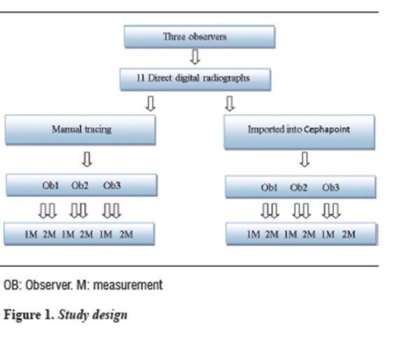
This was a concordance study on 11 profile direct digital radiographs taken by orthodontic students and used in a previous study by Bonilla et al.17
The radiographs were taken in the natural position of the head by a trained operator. Each participant was taken one direct phosphoactivated digital radiographic image with an FCR CAPSULA X® equipment, which immediately transfers the image to the computer monitor. Each participant's radiographic image was exported to Cephapoint, a computer program designed in a previous study.17
A selection of angle measures was performed, and three observers clearly determined their locations, as follows: position of the maxilla with respect to the base of the skull, which is measured by the angle formed by the planes going from the Sella Turcica to Nasion and from Nasion to point A (SNA); position of the mandible with respect to the base of the skull, an angle taken at the intersection of the Sella Turcica to Nasion and from Nasion to point B (SNB); upper incisor inclination, an angle formed by the longitudinal axis of the most vestibular upper incisor and the palatal plane (U1-PP); Lande's angle, formed by the intersection of the Frankfort plane and the line that goes from Nasion to point A (FH/NA); facial depth, an angle formed by the Frankfort plane and the line from point A to Pogonion (FH/N/Pg); angle of hard tissue convexity, formed by the intersection of the planes going from Nasion to point A and from point A to Pogonion (N-A/Pg); inclination of the lower incisor with the NB angle formed by the longitudinal axis of the most vestibular lower incisor and the plane going from Nasion to point B (LI/NB); nasolabial angle, formed by the intersection of the planes going from Columella to Subnasale and from Subnasale to Labrale superius (Cm/Sn/Ls); and the soft tissue convexity angle taken at the intersection of the plane between the Glabella of soft tissue and Subnasale, and the plane that goes from Subnasale to the Pogonion of soft tissue (G'/Sn-Pg').
All these manual tracings were made using digital radiography printing on radiographic paper, with a DRY PIX 2000 printer (FUJI FILM USA®), a corrected 1:1 scale, and a negatoscope under artificial light. A same operator made the profilograph for each digital radiograph, which was made 3 times in order to avoid differences greater than 1 mm among the traced anatomical structures. We also used cephalometric paper (Ortho Organizers®) 0.76 mm thick (0.03 inches), and a HB 0.5 mm lead (Faber Castell®). A millimeter ruler (Faber Castell®) was used for plane tracing, and a Bimler appliance was used for measurements. The three observers made the manual tracing of angles for each digital radiograph.
In this study, the average position for each angle measure identified by the three observers was defined as the "benchmark". This benchmark was used to determine interobserver errors in manual and digital tracing. The average differences in degrees between the benchmark and the measurements made by the observers were defined as interobserver error, and this in turn was used as the variable that determines reproducibility for each angle measure. As a result, reproducibility in the identification of angle measures for each method (manual and digital) could be defined as the differences in magnitude of these distances from the average between the two types of tracing.
According to each observer's measurements, intra-observer precision or error is defined as the level that indicates how close are the angular measurements obtained during the first and second observations with weekly intervals. It is obtained by comparing the measures of each operator and is calculated by means of intraclass correlation coefficient (ICC). Figure 1 shows the study design.
STATISTICAL ANALYSIS
The data were collected in Microsoft Excel 2007, and analyzed in the statistical program for Social Sciences SPSS, version 15.0. The quantitative variables were analyzed through averages and standard deviations. To compare manual and computerized tracing, we used Student's- t test for independent groups and the Levene test for equality of variances. We used a significance level of α = 0.05 for all the tests, and confidence intervals with 95% reliability.
RESULTS
In the measures taken in both the manual digital radiograph tracing and the digital radiograph image imported to Cephapoint, the interobserver error and standard deviation displayed in table 1 shows levels above 7.9° in these two angles: Cm/Sn/Ls and U1-PP in both methods (manual and digital tracing).
En el trazado manual, los ángulos que presentaron mayor diferencia en el promedio de error interobservador, comparado con el trazo digital, fueron: G'/Sn-Pg' (0,32°), II-NB (0,11°) y N-A/Pg (0,11°). En el trazado computarizado, los ángulos que presentaron mayor diferencia en el error interobservador, fueron SNA (0,52°), U1-PP (0,52°), Cm/Sn/Ls (0,47°), SNB (0,21°), FH/NA (0,20°) y FH/N/Pg (0,10°). Al comparar el promedio del error interobservador entre el trazo manual y el computarizado, no se encontraron diferencias significativas. (p ≥ 0,05).
In most of the angles, the level of inter-observer error dispersion was lower in manual tracing, as the standard deviation data indicate (table 1).
When comparing both tracing methods in terms of their standard deviations, the greatest differences were found in the manual method for these angles: FH/NA, G'/Sn-Pg', N-A/Pg, and SNA, and for the computerized method in Cm/Sn/Ls, FH/N/Pg, LI-NB, SNB, and U1-PP, with no significant statistical differences.
The FH/N/Pg angle had the smallest interobserver error difference (0.10°) in both methods, favoring the manual tracing; also, the angles with the smallest inter-observer error difference in computerized tracing were LI-NB (0.11°) and N-A/Pg (0.11°). While SNA (0.52°) and U1-PP (0.52°) were the angles with the greatest inter-observer error difference for the manual method, and G'/Sn-Pg' (0.32°) was the angle with the greatest inter-observer error difference for the computerized method.
As for the evaluation of intra-observer accuracy or error, the manual method showed an excellent intraclass correlation coefficient (ICC), being over 0.9 for all the measurements, except for FH/NA, which was 0,847 (Observer 2) (table 2).
In the computerized method, the ICC was above 0,844, with the exception of angle N/FH/Pg, which was 0,784 (Observer 2), and N-A/ Pg, which was 0,793 (Observer 1) (table 3).
DISCUSSION
The present study showed that inter-observer error averages of manual versus computerized tracing did not present significant differences between both methods; However, there were higher values for the SNA and U1-PP angles in computerized tracing, and for G'/Sn-Pg' in manual tracing.
Concerning the SNA angle, some authors1, 12-18 maintain that the computerized method shows a decrease in cephalometric measurement differences, and is therefore a more accurate method due to different software characteristics such as pixels, contrast and brightness. These factors make it a more reliable method, especially when benchmark location must be done in a contour with bone depth, such as A, B and N. The location of these benchmarks is important when determining the magnitude of horizontal discrepancy in the maxilla, in an angular measurement like SNA, so that benchmark errors along the horizontal axis would be more significant than errors along the vertical axis.1
In this sense, any change in the horizontal position of point A means a significant change in SNA results. Selecting a benchmark in cephalometric analysis is important for successful diagnosis and treatment planning.1 Accordingly, Lim et al18 found out that anatomical benchmarks with low radiodensity, such as point A, tend to be less reliable to identify in computed radiography.
The results obtained for the U1-PP angle in the present study are similar to those obtained by Collins et al,19 who suggest that the maxillary and mandibular planes in a radiograph are marked between two points that are difficult to locate. In addition, they claim that the increase in measure variability occurs because it is necessary to digitize four points to measure certain angles (U1-PP), and 3 points for other angles (SNA and SNB). Other reasons for this variation are root superposition, which makes it difficult to accurately locate apices,12, 20 and the lack of contrast in this area,12 which makes the measures related to the root apices of the incisors located in the benchmarks less reproducible.21, 22 Similarly, Bonilla et al17 found differences in the X and Y axis for ENA and ENP, both in the conventional image and the digital one; this alters the reproducibility of these points and affects the correct tracing of the palatal plane.
The G'/Sn-Pg' angle presented greater inter-observer error difference in the manual tracing, since the Subnasale point has a higher average error on the X axis; also, it is hard to locate the Pogonion on a curve, as reported by Chen.20
In this study, the FH/N/Pg angle showed less inter-observer error, i.e., high reproducibility in both methods, contrasting the report by Sayinsu et al11 for whom all the parameters with lower correlations were measures related to the Frankfort plane, which goes through the Porion and the Orbital. Similarly, other authors12, 20, 24 observed that the Porion is located in complex radiopaque structures that overlap to each other, and the Orbital point is more inexact, probably due to the narrow vertical alignment on the left and right sides of the orbits.9 Likewise, Bonilla et al17 found out that the Porion presented greater standard deviation on the Y axis, and the Infra-orbital point on the X axis in conventional imaging. This is consistent with Geelen et al,21 who found extensive error distribution dispersion in both axes, indicating an inexact point. Similarly, McClure et al9 reported that the Pogonion is located on the Menton contour curve, and therefore this point may be difficult to identify.
In this study, the Cm/Sn/Ls angle was within the highest inter-observer error averages in computerized tracing. Concerning this measure, some authors24 report that the Nasolabial angle is a measure with great clinical relevance in soft tissue analysis, requiring the construction of two lines along the bottom contour of nose and lip. However, there are large variations among the tracing methods, as other studies indicate.3, 25-27 On the other hand, Hwang et al24 found significant differences and low reproducibility for the Nasolabial angle when using the tangent line tracing method, a result that was attributed to the anatomical shape ("S" shape) of the lower part of the nose, and not to the lack of coherence in tangent construction. They also concluded that variability in the elaboration of a tangent on the upper lip contributes to the low reproducibility of the Nasolabial angle. In another study, Swennen et al12 found no statistically significant differences between the methods of analysis and the image format, with the exception of the Nasolabial angle, which exceeded the clinical significance. The difficulty in constructing this angle is therefore evident.
The values seen in table 1, with respect to inter-observer error averages, correspond to the average of angles obtained for manual and computerized tracing. The malocclusion type was used as a sample inclusion criterion, and therefore the values in the table reflect this variability among the angles measured; this is why the analysis of results was based on the differences between the methods.
All the intraclass correlation coefficients (ICC) indicate high levels of precision in both methods; however, a lower correlation for the FH/NA angle was found in manual tracing, and for FH/N/Pg, N-A/Pg angles in the computerized method.
According to the results of the present study, the differences in these methods are not clinically relevant, so the application of either analysis does not affect diagnosis. Regardless of the method used, the clinician must be trained and calibrated for it.11 Therefore, the choice of the method of analysis depends on the orthodontist's criterion in terms of advantages, disadvantages, cost, time, accessibility, and comfort.
CONCLUSIONS
Angle measure reproducibility between manual and computerized tracing methods did not show significant differences, suggesting that both methods offer equal diagnostic validity.
REFERENCES
1. De Araújo P, Nascimento J, Mesquita F, Nery E. A comparative study of manual vs. computerized cephalometric analysis. Dental Press J Orthod 2010; 15(2): 44-51. [ Links ]
2. Steiner C. Cephalometrics for you and me. Am J Orthod 1953; 39(10): 729-755. [ Links ]
3. Legan HL, Burstone CJ. Soft tissue cephalometric analysis for orthognathic surgery. J Oral Surg 1980; 38(10): 744-751. [ Links ]
4. Burstone CJ, James RB, Legan H, Murphy GA, Norton LA. Cephalometrics for orthognathic surgery. J Oral Surg 1978; 36(4): 269-277. [ Links ]
5. McNamara JA Jr. A method of cephalometric evaluation. Am J Orthod 1984; 86(6): 449-469. [ Links ]
6. Sassouni V. A classification of skeletal facial types. Am J Orthod 1969; 55(2): 109-123. [ Links ]
7. Dana JM, Goldstein M, Burch JG, Hartigan PC. Comparative study of manual and computerized cephalometric analyses. J Clin Orthod 2004; 38(5): 293-296. [ Links ]
8. Roden-Johnson D, English J, Gallerano R. Comparison of hand-traced and computerized cephalograms: landmark identification, measurement, and superimposition accuracy. Am J Orthod Dentofacial Orthop 2008; 133(4): 556-564. [ Links ]
9. McClure SR, Sadowsky LP, Ferreira A, Jacobson A. Reliability of digital versus conventional cephalometric radiology: a comparative evaluation of landmark identification error. Semin Orthod 2005; 11(2): 98-110. [ Links ]
10. Bruntz LQ, Palomo JM, Baden S, Hans MG. A comparison of scanned lateral cephalograms with corresponding original radiographs. Am J Orthod Dentofacial Orthop 2006; 130(3): 340-348. [ Links ]
11. Sayinsu K, Isik F, Trakyali G, Arun T. An evaluation of the errors in cephalometric measurements on scanned cephalometric images and conventional tracings. Eur J Orthod 2007; 29(1): 105-108. [ Links ]
12. Swennen GR, Grimaldi H, Berten JL, Kramer FJ, Dempf R, Schwestka-Polly R et al.. Reliability and validity of a modified lateral cephalometric analysis for evaluation of craniofacial morphology and growth in patients with clefts. J Craniofac Surg 2004; 15(3): 399-412. [ Links ]
13. Polat-Ozsoy O, Gokcelik A, Toygar Memikoglu TU. Differences in cephalometric measurements: a comparison of digital versus hand-tracing methods. Eur J Orthod 2009; 31(3): 254-259. [ Links ]
14. Chen YJ, Chen SK, Yao JC, Chang HF. The effects of differences in landmark identification on the cephalometric measurements in traditional versus digitized cephalometry. Angle Orthod 2004; 74(2): 155-161. [ Links ]
15. Richardson A. A comparison of traditional and computerized methods of cephalometric analysis. Eur J Orthod 1981; 3(1): 15-20. [ Links ]
16. Sandler PJ. Reproducibility of cephalometric measurements. Br J Orthod 1988; 15(2): 105-110. [ Links ]
17. Bonilla M, Barrera J, Gutiérrez D, Paredes M, Puentes J. Comparación del error en la ubicación de puntos cefalométricos entre una imagen digital directa y una convencional. Punto de Contacto 2011; 18(17): 63-71. [ Links ]
18. Lim KF, Foong KW. Phosphor-stimulated computed cephalometry: reliability of landmark identification. Br J Orthod 1997; 24(4): 301-308. [ Links ]
19. Collins J, Shah A, McCarthy C, Sandler J. Comparison of measurements from photographed lateral cephalograms and scanned cephalograms. Am J Orthod Dentofacial Orthop 2007; 132(6): 830-833. [ Links ]
20. Chen YJ, Chen SK, Chang HF, Chen KC. Comparison of landmark identification in traditional versus computer-aided digital cephalometry Angle Orthod 2000; 70(5): 387-392. [ Links ]
21. Geelen W, Wenzel A, Gotfredsen E, Kruger M, Hansson LG. Reproducibility of cephalometric landmarks on conventional film, hardcopy, and monitor-displayed images obtained by the storage phosphor technique. Eur J Orthod 1998; 20(3): 331-340. [ Links ]
22. Baumrind S, Frantz RC. The reliability of head film measurements. 1. Landmark identification. Am J Orthod 1971; 60(2): 111-127. [ Links ]
23. Yu SH, Nahm DS, Baek SH. Reliability of landmark identification on monitor-displayed lateral cephalometric images. Am J Orthod Dentofacial Orthop 2008; 133(6): 790. [ Links ]
24. Hwang HS, Kim WS, McNamara JA Jr. A comparative study of two methods of quantifying the soft tissue profile. Angle Orthod 2000; 70(3): 200-207. [ Links ]
25. Park YC, Burstone CJ. Soft-tissue profile -fallacies of hard-tissue standards in treatment planning. Am J Orthod Dentofacial Orthop 1986; 90(1): 52-62. [ Links ]
26. Nanda RS, Meng H, Kapila S, Goorhuis J. Growth changes in the soft tissue facial profile. Angle Orthod 1990; 60(3): 177-190. [ Links ]
27. Formby WA, Nanda RS, Currier GF. Longitudinal changes in the adult facial profile. Am J Orthod Dentofacial Orthop 1994; 105(5): 464-476. [ Links ]
7. Cobankara FK, Unlun N, Altinoz HC, Fusun O. Effect of home bleaching agents on the roughness and surface morphology of human enamel and dentine. Int Dent J 2004; 54(4): 211-218. [ Links ]
8. Pinto CF, Oliveira R, Cavalli V, Giannini M. Peroxide bleaching agent effects on enamel surface microhardness, roughness and morphology. Braz Oral Res 2004; 18(4): 306-311. [ Links ]
9. Gotz H, Duschner H, White DJ, Klukowska A, Malgorzata A. Effects of elevated hydrogen peroxide 'strip' bleaching on surface and subsurface enamel including subsurface histomorphology, micro-chemical composition and fluorescence changes. J Dent 2007; 35: 457-466. [ Links ]
10. Claus PE. Effects of hydrogen peroxide-containing bleaching agents on the morphology of human enamel. Quintessence Int 1996; 27(1): 53-56. [ Links ]
11. Moreira de Freitas PM, Turssi CP, Hara AT, Serra MC. Dentin microhardness during and after whitening treatments. Quintessence Int 2004; 35(5): 411-417. [ Links ]
12. Romero AD, Bueno GJ. Radicales libres del oxígeno y antioxidantes en medicina. Rev Clin Española 1998; 184(7): 345-346. [ Links ]
13. Joiner E. The bleaching of teeth: A review of the literature. J Dent 2006; 34(7): 412-419. [ Links ]
14. Goldstein RE, Garber DA. Complete dental bleaching. Chicago: Quintessence Publishing; 1995. [ Links ]
15. Domínguez MN, González LS, Menéndez NM. Study of the diffusion ways in the white spot enamel lesion. RCOE 2002; 7(5): 469-476. [ Links ]
16. Price RBT, Sedarous M, Hiltz GS. The pH of tooth-whitening products. J Can Dent Assoc 2000; 66: 421-426. [ Links ]
17. Cheesman KH, Slater TF. Free radicals in medicine. Br Med Bull 1998; 49: 118-121. [ Links ]
18. Floyd RA. The effect of peroxides and free radicals on body tissues. J Am Dent Assoc 1997; 128: 37-44. [ Links ]
19. Venereo JR. Daño oxidativo, radicales libres y antioxidantes. Rev Cub Med Mil 2002; 31(2): 126-133 [ Links ]
20. Mc Guckin RS, Thurmond BA, Osovitz S. Enamel shear bond strengths after vital bleaching. Am J Dent 1992; 5: 216-222. [ Links ]
21. Titley KC, Torneck CD, Smith DC, Chernecky R, Adibfar A. Scanning electron microscopy observation on the penetration and structure of resin "tags" in bleached and unbleached bovine enamel. J Endod 1991; 17: 72-75. [ Links ]
22. Carvalli V, Giannini M, Carvalho R. Effect of carbamide peroxide bleaching agents on tensile strength of human enamel. Dent Mat 2004; 20: 733-739. [ Links ]
23. Rotstein I. Role of catalase in the elimination of residual hydrogen peroxide following tooth bleaching. J Endod 1993; 19: 567-569. [ Links ]
24. Shinohara MS, Peris AR, Pimenta LA, Ambrosano GM. Shear bond strength evaluation of composite resin on enamel and dentin after nonvital bleaching. J Esthet Restor Dent 2005; 17(1): 22-29. [ Links ]
25. Sung EC, Chan SM, Mito R, Caputo AA. Effect of carbamide peroxide bleaching on the shear bond strength of composite to dental bonding agent enhanced enamel. J Prosthet Dent 1999; 82(5): 595-599. [ Links ]
26. Cadenaro M, Breschi L, Antoniolli F, Mazzoni A, Di Lenarda R. Influence of whitening on the degree of conversion of dental adhesives on dentin. Eur J Oral Sci. 2006; 114(3): 257-262. [ Links ]
27. Ben-Amar A, Liberman R, Gorfil C, Bernstein Y. Effect of night guard bleaching on enamel surface. Am J Dent 1995; 8: 29-32. [ Links ]
28. Josey AL, Meyers IA, Romaniuk K, Symons AL. The effect of vital bleaching technique on enamel surface morphology and the bonding of composite resin to enamel. J Oral Rehabil 1996; 23: 244-250. [ Links ]
29. Baldión PA, Arcos LC, Mora MA. Efecto de los fluoruros en la composición química del esmalte dental posblanqueamiento. Univ Odontol 2011; 30(65): 41-49. [ Links ]
30. Manzini JL. Declaración de Helsinki: Principios éticos para la investigación médica sobre sujetos humanos. Acta Bioethica 2000; 6(2): 321-334. [ Links ]
31. Colombia Ministerio de Salud. Resolución N.° 8430 de 1993, octubre 4, por la cual se establecen las normas científicas, técnicas y administrativas para la investigación en salud. Título II. De la investigación en seres humanos. Capítulo 1. De los aspectos éticos de la investigación en seres humanos: Artículos 4 al 16. Bogotá: El Ministerio; 1993. [ Links ]
32. Comité Materiales Odontológicos. NTC 4882. Métodos de ensayo para la evaluación de la unión adhesiva entre los materiales odontológicos y la estructura dental. Instituto Colombiano de Normas Técnicas y Certificación, ICONTEC. Sector: 11-Tecnología del cuidado de la salud. Fecha de ratificación: 25/10/2000. Actualización: Ninguna. [ Links ]
33. Tschoppe P, Zandim DL, Martus P, Kielbassa AM. Enamel and dentine remineralization by nano-hydroxyapatite toothpastes. J Den 2011; 39(6): 430-437. [ Links ]
34. Tschoppe P, Meyer-Lueckel H. Effects of regular and highly fluoridated toothpastes in combination with saliva substitutes on artificial enamel caries lesions differing in mineral content. Arch Oral Biol 2012; 57(7): 931-939. [ Links ]
35. Tschoppe P, Kielbassa AM, Meyer-Lueckel H. Evaluation of the remineralising capacities of modified saliva substitutes in vitro. Arch Oral Biol 2009; 54(9): 810-816. [ Links ]
36. 3M ESPE Casa comercial. Perfil técnico del producto: Adpter® Single Bond. Sistema adhesivo dental. [en línea] [fecha de acceso 15 de octubre de 2012 ]; URL disponible en: http://multimedia.3m.com/mws/mediawebserver? UUUUUUC04ehUnx7UGx7UUUPJtEtttttS- [ Links ]
37. Torneck CD, Titley KC, Smith DO, Adibfar A. Effect of water leaching on the adhesion of composite resin to bleached and unbleached enamel. J Endodon 1991; 17: 156-160. [ Links ]
38. Gokce B, Comlekoglu ME, Ozpinar B, Turkun M, Demirbas AK. Effect of antioxidant treatment on bond strength of a luting resin to bleached enamel. J Dent 2008; 36: 780- 785. [ Links ]
39. Kimyai S, Oskoee SS, Rafighi A, Valizadeh H, Ajami AA, Helali ZZ. Comparison of the effect of hydrogel and solution forms of sodium ascorbate on orthodontic bracket- enamel shear bond strength immediately after bleaching: An in vitro study. Indian J Dent Res 2010; 21: 54-58. [ Links ]
40. Suelieman M, Addy M, Macdonald E, Ress J. The leaching depth of a 35% hydrogen peroxide based in-office product: a study in vitro. J Dent 2005; 33: 33-40. [ Links ]
41. Titley KC, Torneck CD, Ruse ND. The effect of carbamide- peroxide gel on the shear bond strength of a microfilm resin to bovine enamel. J Dent Res 1992; 71: 20-24. [ Links ]
42. Miranda AM, Bermejo GN, Bazan JE, Saravia MA. Efectos de un blanqueamiento dental con ozono y otro con peróxido de carbamida al 22% sobre la fuerza de adhesión al esmalte en diferentes intervalos de tiempo. Acta Odontol Venez 2009; 47(4): 69-77. [ Links ]
43. Cavalli V, Reis AF, Giannini M, Ambrosano GM. The effect of elapsed time following bleaching on enamel bond strength of resin composite. Oper Dent 2001; 26: 597-602. [ Links ]
44. Gómez ME, Campos A. Esmalte. En: Histología y embriología bucodental. 2ed. Madrid: Médica Panamericana; 2003. p. 273-316. [ Links ]
45. Whittaker DK. Structural variations in the surface zone of human tooth enamel observed by scanning electron microscopy. Archs Oral Biol 1982; 27(5): 383-392. [ Links ]
46. Shi XC, Ma H, Zhou JL, Li W. The effect of cold-light-activated bleaching treatment on enamel surfaces in vitro. Int J Oral Sci 2012; 4: 208-213. [ Links ]
47. Hegedüs C, Bistey T, Flóra-Nagy E, Keszthelyi G, Jenei A. An atomic force microscopy study on the effect of bleaching agents on enamel surface. J Dent 1999; 27: 509-515. [ Links ]
48. Pérez LF, Díaz AM, Aguirre M, Alcántara CM, Aguilar RE, Acedo JE et al. Efecto del peróxido de carbamida sobre el esmalte dentario a diferentes concentraciones y tiempos de exposición (estudio in vitro). Odontol Sanmarquina 2004; 8(1): 25-29. [ Links ]
49. Rodrígues JA, Oliveira GP, Amaral CM. Effect of thickener agents on dental enamel microhardness submitted to at-home bleaching. Braz Oral Res 2007; 21(2): 170-175. [ Links ]
50. Chen HP, Chang CH, Liu JK, Chuang SF, Yang JY. Effect of fluoride containing bleaching agents on enamel surface properties. J Dent 2008; 36(9): 718-725. [ Links ]
51. Dominguez JA, Bittencourt B, Michel M, Sabino N, Gomes JC, Gomes OM. Ultrastructural evaluation of enamel after dental bleaching associated with fluoride. Microsc Res Tech 2012; 75(8): 1093-1098. [ Links ]
52. Adebayo OA, Burrow MF, Tyas MJ. Effects of conditioners on microshear bond strength to enamel after carbamide peroxide bleaching and/or casein phosphopeptide-amorphous calcium phosphate (CPP-ACP) treatment. J Dent 2007; 35(11): 862-870. [ Links ]
53. Zhao J, Liu Y, Sun WB, Zhang H. Amorphous calcium phosphate and its application in dentistry. Chem Cent J 2011; 5: 40. [ Links ]
54. Goswami M, Saha S, Chaitra TR. Latest developments in non-fluoridated remineralizing technologies. J Indian Soc Pedod Prev Dent 2012, 30(1): 2-6. [ Links ]
55. Cvitko E, Deheny GE, Swift Jr EJ, Pires JA. Bond strength of composite resin to enamel bleached with carbamide peroxide. J Esthet Dent 1991; 3: 100-102. [ Links ]
56. Rotstein I, Dankner E, Goldman A, Heling I, Stabholz A, ZalkindM. Histochemical analysis of dental hard tissues following bleaching. J Endod 1996; 22: 23-26.
57. Baldión PA, Viteri LN, Lozano E. Efecto de la peroxidasa sobre la resistencia de unión de una resina compuesta al esmalte dental posblanqueamiento. Rev Fac Odontol Univ Antioq 2012; 24(1): 8-21. [ Links ]
58. Kaya AD, Türkün M. Reversal of dentin bonding to bleached teeth. Oper Dent 2003; 28(6): 825-829. [ Links ]
59. Türkün M, Kaya AD. Effect of 10% sodium ascorbate on the shear bond strength of composite resin to bleached bovine enamel. J Oral Rehab 2004; 31: 1184-1191. [ Links ]
60. Barghi N, Godwin JM. Reducing the adverse effect of bleaching on composite-enamel bond. J Esthet Dent 1994; 6: 157-161. [ Links ]
61. García EJ, Oldoni TL, Alencar SM, Reis A, Loguercio AD, Grande RH. Antioxidant activity by DPPH assay of potential solutions to be applied on bleached teeth. Braz Dent J 2012; 23: 22-27. [ Links ]
62. Da Costa DB, Mazur RF. Effects of the new formulas of bleaching gel and fluoride application on enamel microhardness: An in vitro study. Oper Dent 2007; 32: 589-594. [ Links ]
63. Barkvoll P. Effect of sodium lauryl sulfate on the uptake of fluoride from NaF and MFP by etched enamel in vitro. J Biol Buccale 1991; 19(3): 235-239. [ Links ]
64. Kennedy DO, Scholey AB. The psychopharmacology of European herbs with cognition-enhancing properties. Curr Pharm Des 2006; 12(35): 4613-4623. [ Links ]
65. Dedio I. Value of Calendula officinalis as a tannin source. Herba Pol 1983; 29(3-4): 211-216. [ Links ]
66. Brasseur T, Abgenot L, Pricemail J, Debev C. Free radical formation inhibiting and antioxidant properties of flavonoids. J Exptl Biol Med 1986; 37: 533-548. [ Links ]
67. Groppo FC, de Cássia BC, Cogo K, Franz-Montan M, Lopes RH, Dias AE. Use of phytotherapy in dentistry. Phytother Res 2008; 22(8): 993-1133. [ Links ]
68. Lu Y, Foo LY. Flavonoid and phenolic glycosides from Salvia officinalis. Phytochemistry 2000; 55(3): 263-267. [ Links ]
69. Halliwell B. Rafter J, Jenner A. Health promotion by flavonoids, tocopherols, tocotrienols, and other phenols: direct or indirect effects? Antioxidant or not? Am J Clin Nutr 2005. 81(suppl): 268-276. [ Links ]
70. Roginsky V. Chain-breaking antioxidant activity of natural polyphenols as determined during the chain oxidation of methyl linoleate in Triton X-100 micelles. Arch Biochem Biophys 2003; 414: 261-270. [ Links ]
71. Freire A, Souza E, Biazzetto D, Ribeiro E, Cynthia C, Marins R et al. Reaction kinetics of sodium ascorbate and dental bleaching gel. J Dent 2009; 37: 932-936. [ Links ]
72. Lai S, Mak Y, Cheung G, Osorio R, Toledano M. Reversal of compromised bonding to oxidized etched dentin. J Dent Res 2001, 80: 1919-1924. [ Links ]
73. Tezel H, Ertas OS, Ozata F, Dalgar H, Korkut ZO. Effect of bleaching agents on calcium loss from the enamel surface. Quintessence Int 2007; 38(4): 339-347. [ Links ]
74. Attin T, Kielbassa AM, Schwanenberg M, Hellwig E. Effect of fluoride treatment on remineralization of bleached enamel. J Oral Rehabil 1997; 24: 282-286. [ Links ]
75. Attin T, Hanning G, Wiegand A, Attin R. Effect of bleaching on restorative materials and restorations. A systematic review. Dent Mater 2004; 20: 852-861. [ Links ]
76. Cochrane NJ, Cai F, Huq NL, Burrow MF, Reynolds EC. New approaches to enhanced remineralization of tooth enamel. J Dent Res 2010; 89: 1187-1197. [ Links ]
77. Lewinstein I. Effect of different peroxide bleaching regimens and subsequent fluoridation on the hardness of human enamel and dentin. J Prosthet Dent 2004; 92: 337-342. [ Links ]
78. Cavalli V, Rodrigues LK, Paes-Leme AF, Brancalion ML, Arruda MA, Berger SB, et al. Effects of bleaching agents containing fluoride and calcium on human enamel. Quintessence Int 2010; 41: 157-165. [ Links ]
79. Tschoppe P, Neumann K, Mueller J, Kielbassa AM. Effect of fluoridate bleaching gels on the remineralization of predemineralized bovine enamel in vitro. J Dent 2009: 37(2): 156-162. [ Links ]
