


Servicios Personalizados
Revista
Articulo
 texto en
  Español (pdf)
 Articulo en XML
 Referencias del artículo
 Enviar articulo por email
Indicadores
-
 Citado por SciELO
 -
 Accesos
Links relacionados
-
 Citado por Google
 -
 Similares en
SciELO
Similares en
SciELO -
 Similares en Google
Compartir

 Permalink
Revista Facultad de Odontología Universidad de Antioquia
versión impresa ISSN 0121-246X
Rev Fac Odontol Univ Antioq vol.26 no.1 Medellín jul./dic. 2014
ORIGINAL ARTICLES DERIVED FROM RESEARCH
THE EFFECT OF SILVER DIAMINE FLUORIDE ON CARIES INDUCED IN WISTAR RATS DISILICATE
Susana Vanegas1, Astrid Godoy1, Leonidas Urdaneta2, Daniela Olávez3, Karla Padrón3, Eduvigis Solórzano4
1 DMD. Universidad de Los Andes, Mérida, Venezuela. Dentist in private
practice
2 BA in Bioanalysis. Magister Scientiae in Clinical Microbiology,
Biopatology Research/Comprehensive Laboratory of Cellular and
Molecular Biology Research Group. Adjunct Microbiology Professor,
School of Dentistry, Universidad de Los Andes. Mérida, Venezuela
3 DMD. Biopatology Research/Comprehensive Laboratory of Cellular
and Molecular Biology Research Group. Adjunct Histology Professor,
School of Dentistry, Universidad de Los Andes. Mérida, Venezuela
4 DMD. PhD in Biologic Anthropology. Biopatology Research/
Comprehensive Laboratory of Cellular and Molecular Biology
Research Group. Adjunct Histology Professor, School of Dentistry,
Universidad de Los Andes. Mérida, Venezuela. eMAIL ADDRESS: eduvigis@ula.ve
SUBMITTED: AUGUST 13/2013-ACCEPTED: APRIL 22/2014
Vanegas S, Godoy A, Urdaneta L, Olávez D, Padrón K, Solórzano E. The effect of Silver Diamine Fluoride on caries induced in Wistar rats. Rev Fac Odontol Univ Antioq 2014; 26(1): 76-88.
ABSTRACT
INTRODUCTION: dental caries is considered a public health problem. Various biomaterials have been suggested for treating it, including
fluride+silver nitrate mix, known as silver diamine fluoride (SDF) which contains cariostatic, remineralizing, and bactericide properties and is
used on primary teeth. Only a few studies have assessed the response of dental tissue when this substance is applied; the objective of this study
was therefore to determine the effect of SDF on caries induced in Wistar rats.
METHODS: dental caries was induced in Wistar rats' molars by
inoculating them with S. mutans CVCM656 and feeding them a sucrose-rich diet for 12 weeks. Once caries were diagnosed and sorted out into
control groups (C1, C2) and experimental groups (E1, E2), softened dentine was removed from molars; only the molars from experimental groups
received a topical application of 3.8% SDF. Finally, clinical assessment was performed and samples were collected for histological study 7 and
13 weeks after treatment.
RESULTS: the control groups experienced an increased number of carious lesions and progression in severity, while the
same parameters in the experimental groups remained unchanged, showing hard dark dentine surfaces. The histological report showed increased
predentine thickness in molars from the experimental groups only.
CONCLUSIONS: the protocol of caries induction in Wistar rats was successful.
Treatment with SDF deactivated the carious processes, arresting not only severity progression but also the increase in number of caries; these
findings may be extrapolated to humans.
Key words: dental caries, primary teeth, Silver Diamine Fluoride (SDF), animal model.
INTRODUCTION
As pathology of the oral cavity, dental caries is considered a public health problem. Although it does not represent a potential life hazard, it brings about considerable aesthetic and functional consequences, as well as pain, infections, and eventually, tooth loss if not treated on time. Development of tooth decay, according to Keyes' triad of modified etiology, involves three factors maintained over time: a susceptible host, cariogenic microbiota located on dental biofilm, and a suitable substrate provided by the diet, which serves as a source of energy to microorganisms;1 in addition, caries is related to environmental, socio-economic, cultural, and genetic factors, to name just a few.
Traditionally, tooth decay treatment has included not- too-conservative therapies, following the principles of extension by prevention designed by Black, which implied greater destruction of healthy dental tissue in order to increase the restorative material's mechanical retention. Minimally invasive treatment has been proposed as alternative therapy, which has evolved in parallel with the development of new biomaterials and is being used in primary cavitated teeth in order to maintain pulp vitality, enabling space conservation and allowing the natural process of exfoliation and tooth replacement. 2
Among the materials used for tooth decay prevention are fluorides, whose main mechanism of action is their post-eruptive effect, which includes demineralization inhibition, re-mineralization stimulation, and antibacterial activity.3 They are mostly applied topically via rinses, gels, varnishes, toothpaste, mouthwash, dental floss, prophylactic toothpaste, and chewing gums that contain fluoride in different concentrations.3 Other forms of massive administration are fluoridation of public drinking water and programs of fluoridation of salt for consumption.4
Taking into account the characteristics of fluorides, a mixture of fluoride and silver nitrate, known as silver diamine fluoride (SDF), has recently been suggested to control caries in primary teeth.5 SDF is a topical cariogenic solution with re-mineralizing and bactericidal properties that has been used in the dental practice of some American, African and European countries to treat children's enamel and dentin caries in a non-invasive manner; its most effective concentration is 38%.6
Among the most important properties attributed to SDF are: increase resistance of tooth enamel, inhibit dental biofilm formation, decrease acid production of microorganisms in carious dentin, reduce proliferation of S. mutans, and obliterate the exposed dentinal tubules.6, 7 Therefore, it produces a cariogenic action by blocking decay progression; in addition, it has been reported that the SDF solution has an antimicrobial effect, so it is used for root canals asepsis, and in vitro studies have shown that it prevents collagen degradation in demineralized dentin.8 Due to these properties and its relatively low cost, SDF is ideal for massive application, 5-9 since it allows non-traumatic tooth decay treatment, especially in children populations in rural or suburban areas, where access to dental care is limited. Also, some studies have reported the cariogenic effect of 38% SDF in primary teeth, from a clinical and radiographic perspective;9-11 however, few studies have structurally assessed dental tissues response after applying this biomaterial.12 Therefore, the objective of the present study was to determine the effect of SDF on caries induced in Wistar rats. This experimental model was used because its histological features are similar to those of humans,13 and this allowed to clinically and histologically assess the reaction of dental tissues when exposed to SDF.
METHODS
The sample of this descriptive experimental study consisted of 40 decayed teeth after inducing carious process in 108 molars of 9 Wistar male rats, weighing between 200 and 250 g. The rats were provided by the Universidad de Los Andes Animal Laboratory (BIOULA) and maintained in its facilities during the course of the experiment, which followed the ethical guidelines established for the management of experimental animals, 14 and was endorsed by the Ethics Committee of BIOULA.
Induction of dental caries in animal models
After sedating 9 Wistar rats by intraperitoneally applying 0.5 ml of ketamine (LabMalgene) with doses of 5 mg/kg, the oral cavity of each animal was clinically assessed to verify the absence of carious processes. Next, the occlusal surfaces of upper and lower molars were cleansed, brushes were used to take cytological samples, and each molar was inoculated with colonies of S. mutans CVCM 656 strain, collected with a sterile brush, making an application at a time and re-inoculating with the same strain 24 hours later. Additionally, the animals were fed a sucrose-rich diet, adding 15% sugar in 500 ml of water for daily consumption during the twelve weeks of the caries induction process.
During this caries induction phase, animals that showed symptoms of pain, such as bristly fur, lack of cleanliness, any kind of eye pigmentation, or that were not consuming food or water, were applied 1.0 ml Meloxicam (Vivax) subcutaneously once a day for 3 days. Similarly, two blood tests were taken to verify the absence of any systemic conditions due to the sugar supply during this stage. To do this, after a 12-hour overnight fasting, a sample of blood from tip tails was obtained to determine blood sugar level (normal values = 126 mg / dL).
Clinical evaluation following dental caries induction
Twelve weeks after induction of the carious process, the oral cavities of Wistar rats were assessed again to determine the number of decayed molars in every animal and to classify them according to the International Caries Detection and Assessment System (ICDAS);15 the obtained data were recorded in a form specifically designed for this purpose. According to the obtained caries in enamel and dentin, the 9 animals were intentionally distributed in four study groups, two control groups (C1 with 2 animals and C2 with 3 animals) and two experimental groups (E1 and E2 with 2 animals each), so that each group had 10 decayed molars with varying degrees of severity.
Treatment phase
A commercial presentation of 38% SDF (NAF laboratories) was diluted in distilled water 1:10 and used at a concentration of 3.8% because of the report by Gotjamanos and Maque, who mention dental enamel lesions in Sprague-Dawley rats consistent with generalized fluorosis six weeks after treatment with 4% SDF.6 Following the sedation protocol, the softened dentin from all decayed molars of the four study groups was removed using a probe and then the samples received a topical application of 3.8% SDF for 3 minutes, using a sterile micro-applicator and a sterile swab soaked with distilled water. This procedure was performed on the molars of animals from the experimental groups (E1 and E2). Animals in the control groups were not treated. All animals were fed ad libitum and the supply of 15% sugar in water was suspended in order to remove the substrate of the induction process.
Animals in groups C1 and E1 were clinically evaluated again seven weeks after treatment and slaughtered using 100% Halothane BP (Omicron) inhaled in a glass chamber for 3 to 5 minutes. On the other hand, animals from groups C2 and E2 were evaluated thirteen weeks after treatment and sacrificed in a similar way, following the bioethics rules for handling experimental animals.
Histological study
For histological processing, the samples' jaws were dissected using a scalpel blade #15 and a portable micromotor (Thumb) with diamond edge discs, obtaining four fragments of each quadrant by animal, which were inserted in 10% formalin in amber bottles properly labeled for 48 hours. Then they were demineralized with 8% nitric acid and processed according to a routine histological technique, making transverse and longitudinal cuts of 0.5 to 2 µm in thickness in mesial-distal direction. The samples were colored with hematoxylin-eosin and observed on an optical trinocular microscope (LEICA®, model DNR-CH) with a built-in camera.
Statistical analysis
The data obtained from the clinical observation of caries were collected on forms and analyzed with the statistical program SPSS v. 17, applying descriptive statistics and X2 test (Chi square) to determine the association of the study's variables and to establish statistical significance. The histological findings were described according to the observations.
RESULTS
Twelve weeks after the caries induction process, 40 molars of the 108 that were inoculated with the S. mutans CVCM 656 strain were diagnosed with carious lesions, representing 37% of the total number of treated teeth. The identified caries were classified considering the degree of severity, following the ICDAS parameters as shown in table 1.

Groups C1 and E1 were clinically evaluated seven weeks after treatment, noting that in group C1 (cavities not treated with SDF) the caries that had initially been classified 3 according to ICDAS increased to 4 in severity, while in group E1 (cavities treated with SDF) there was no increase in the number of existing lesions nor progression in their severity (table 2). Also, presence of softened dentin was observed in samples from group C1, unlike the decayed molars from group E1, which showed a hard dark surface.
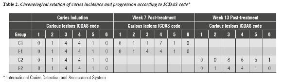
The clinical evaluation of groups C2 and E2 was done by week 13, noting that group C2 presented 10 new carious lesions, 8 of which were on enamel (severity 3) and 2 on dentin (severity 4 and 5). In addition, 5 caries initially reported as 2, 3, 4 and 5 progressed to 4, 5 and 6 in severity with presence of softened dentine (figure 1). Group E2 remained unchanged in number of carious lesions and in progression of existing cavities, showing hard dark dentine surfaces (table 2). The Chi-square test (X2) showed statistical significance in terms of SDF effectiveness-carious process association in both clinical evaluations, with a p-value = 0.000 by week seven and a p-value = 0.003 by week thirteen.

From the histological point of view, there were no differences between control and experimental groups in terms of hard tissue; however, it is important to mention the reaction of pulp tissue in the studied samples of both groups as it showed increased cellularity (plasmocytes in particular), prevalence of chronic inflammatory infiltrate (polymorphonuclear), fibrosis and angiogenesis. It is also important to highlight that groups E1 and E2 showed a thicker predentine layer compared to that of molars in control groups (figure 2), which was evident in both the first and the second histological evaluations, while in the cavitated molars not treated with SDF, predentine thickness remained unchanged, at least until 13 weeks after removing the carious process.

DISCUSSION
In this study, we performed a caries induction protocol in Wistar rats by inoculating a portion of the S. mutans CVCM 656 strain in the occlusal side of each animal's molars plus an ad libitum diet and 15% sugar in daily drinking water for twelve continuous weeks, obtaining 37% of caries in various degrees of severity according to the ICDAS classification, in both jaws. These results are satisfactory, considering that the protocol is quite simple and was conducted in a relatively short time, compared with those proposed by other authors. 16-18
The protocol used in this study did not produce soft or periodontal tissue pathologies and it demonstrated that the sugar supply was adequate, since it did not produce any systemic change, such as diabetes mellitus. It is also important to highlight that during the treatment phase of the control group, the caries progressed in number and severity regardless of diet, which supports the assumption that the cariogenic substrate is not essential after inducing the carious process.
Tooth decay is still the most important problem in dentistry; different therapies have been proposed over time seeking little loss of healthy dental tissue, especially by using minimally invasive operative movements and by applying cariostatic substances such as 38% SDF, which has been used to minimize premature loss of temporary teeth affected by carious lesions, close to their stage of normal exfoliation.
The use of SDF has been controversial among authors, especially if used in primary dentition. There are those who support and encourage it, 3, 6-8 while others reject it due to its toxicity and soft tissue irritation, among other adverse factors.6 In this direction, it is important to mention the study by Llodra et al,9 who analyzed the effectiveness of 38% SDF, showing an average decrease in new lesions per surface in temporary dentition and permanent first molars in the group treated with topical SDF. By using an animal model, the present study showed that all the molars treated with SDF experienced hardened dental tissue, at least during the 13 weeks of the experiment, and no new carious lesions were reported, while molars in the control groups showed increased lesion severity and new carious processes. These results demonstrate the effectiveness of SDF as a cariostatic agent and support the authors who promote its use.
From the histological point of view, 7 and 13 weeks after initiating the study we observed increased cellularity in pulp tissue, prevalence of polymorphonuclear inflammatory infiltrate, fibrosis and angiogenesis in both the experimental and control samples, which means an inflammatory reaction as a defensive response of the pulp to the lesion produced by the carious process; 19 it is therefore clear that this finding is not attributed to the use of SDF.
Also, the histological study reported a significant increase in the average thickness of the predentine, which could not be quantified due to technical limitations but that was only evident in the samples treated with SDF, which suggests a possible deposit of reparative dentin in less time than in untreated molars not reported in the literature. However, the study by Elizondo et al,10 who clinically and radiographically compared the effects of calcium hydroxide and 38% SDF on caries in primary teeth, showed increased dentin tissue thickness and density three and six months after treatment, with no statistical significant differences in the group treated with calcium hydroxide, but this may support the findings of our study. In any case, we recommend making additional studies on this issue. Finally, both groups showed chronic inflammatory infiltrate which suggests a normal tissue response to noxa (caries).
CONCLUSIONS
In conclusion, the standardization of the protocol of dental caries induction in Wistar rats was successful under the methodological conditions used in this study. In addition, this study showed that treatment with SDF inactivated the induced carious processes, stopped progression in lesion severity and blocked increase in number of cavities, at least 13 weeks after its application; these findings can be extrapolated to humans, as it minimizes the formation of new carious lesions and the progression of existing cavities. We suggest its use as a strategy to control dental caries in cavitated primary teeth (involving enamel and dentine), in order to keep pulp vitality until its natural replacement.
CONFLICTS OF INTEREST
The authors declare having no conflicts of interest.
REFERENCIAS
1. Liébana J. Microbiología oral. 2.a ed. Granada: Mc Graw Hill Interamericana; 2002. [ Links ]
2. Rojas F. Algunas consideraciones sobre caries dental, fluoruros, su metabolismo y mecanismos de acción. Act Odont Ven 2008; 46(4): 1-11. [ Links ]
3. Swan E. Fluoride supplements and dietary sources of fluoride. J Can Dent Assoc [Internet]. 2000; 66: 362- 363 [Consultado 2012 Jun 2] Disponible en: http://www. fluoridation.com/cda-fluoride.htm [ Links ]
4. Organización Mundial de la Salud. Publicación de una revisión de guías para la calidad del agua potable con el fin de prevenir brotes epidémicos y enfermedades relacionadas con el agua. Centro de prensa [Internet] [Consultado 2012 Jun 23]. Disponible en: http://www.who.int/mediacentre/ news/releases/2004/pr67/es/index,html [ Links ]
5. Gomes R, Vasconcellos M, Rastelli M, Cziusniak G, Wambier D. Diaminofluoreto de prata: umarevisão de literatura. Ci Biol Saúde 2006; 12(2): 45-52. [ Links ]
6. Gotjamanos T, Ma P. Potential of 4 per cent Silver Fluoride to induce fluorosis in rats: clinical implications. Aust Dent J 2000; 45(3): 187-192. [ Links ]
7. Mei ML, Chu CH, Low KH, Che CM, Lo EC. Caries arresting effect of silver diamine fluoride on dentine carious lesion with S. mutans and L. acidophilus dualspecies cariogenic biofilm. Med Oral Patol Oral Cir Bucal 2013; 18(6): e824-e831. [ Links ]
8. Mei ML, Ito L, Cao Y, Li QL, Lo EC, Chu CH. Inhibitory effect of silver diamine fluoride on dentine demineralisation and collagen degradation. J Dent 2013; 41(9): 809-817. [ Links ]
9. Llodra J, Rodriguez A, Ferrer B, Menardia V, Ramos T, Morato M. Efficacy of silver diamine fluoride for caries reduction in primary teeth and first permanent molars of schoolchildren: 36-month clinical trial. J Dent Res 2005; 84(8): 721-724. [ Links ]
10. Elizondo M, Lucas G, Rosa G. Estudio preliminar del efecto del hidróxido de calcio y del fluoruro diamino de plata al 38% en el tratamiento de las caries dentinarias profundas en molares primarios. Comunicaciones Científicas y Tecnológicas Univ Nac Nordeste. 2004 [Internet] [Consultado 2012 Jun 9]. Disponible en: www.unne.edu. ar/unnevieja/Web/cyt/com2004/3-Medicina/M-054.pdf [ Links ]
11. Yee R, Holmgren C, Mulder J, Lama D, Walker D, Van W. Efficacy of silver diamine fluoride for arresting caries treatment. J Dent Res 2009; 88(7): 644-647. [ Links ]
12. Knight G, McIntyre J, Craig G, Mulyani, Zilm P, Gully N. An in vitro model to measure the effect of a silver fluoride and potassium iodide treatment on the permeability of desmineralized dentine to Streptococcus mutans. J Aust Dent 2005; 50(4): 242-245. [ Links ]
13. Ramalho SA, Daruge E, De La Cruz B, Francesquini Jr L, Francesquini, MA, Daruge Jr E et al. La importancia del peritaje en el estudio comparativo histomorfológico del esmalte, dentina y cemento de dientes humanos y otros animales. Acta Odontol Venez. 2006. 44(1). [Internet] [Consultado 2012 Jun 17] Disponible en: http:// www.actaodontologica.com/ediciones/2006/1/peritaje_ histomorfologico_esmalte_dentina_cemento.asp [ Links ]
14. Ministerio del poder popular para ciencia, tecnología e industrias intermedias. Fondo nacional de ciencia, tecnología e innovación. Código de ética y bioseguridad. 3ª ed. Ediciones del Fondo Nacional de Ciencia, Tecnología e Innovación: Caracas; 2012 [Internet]: [Consultado 2013 May 24]. Disponible en: http://www.ciens.ucv. ve:8080/generador/sites/biolanimlab/archivos/codigo_ fonacit_2008.pdf [ Links ]
15. Braga M, Mendes F, Ekstrand K. Detection activity assessment and diagnosis of dental caries lesions. Dent Clin North Am 2010; 54(3): 479-493. [ Links ]
16. Ooshima T, Sumi N, Izumitani A, Sobue S. Maternal transmission and dental caries induction in Sprague- Dawley rats infected with Streptococcus mutans. Microbiol Immunol 1988; 32(8): 785-794. [ Links ]
17. Ooshima T, Yoshida T, Aono W, Takei T, Izumitani A, Sobue S et al. Changes with time in the oral microflora and dental caries induction in hyposalivated rats fed on sucrose diet. Microbiol Immunol 1992; 36(12): 1223-1231. [ Links ]
18. Tanzer JM, Grant LP, Mcmahon T, Clinton D, Eanes ED. Simultaneous caries induction and calculus formation in rats. J Dent Res 1993; 72(5): 858-864. [ Links ]
19. Gómez M, Campos A. Histología y embriología bucodental. 2.a ed. Madrid: Panamericana; 2007. [ Links ]
