SciELO - Scientific Electronic Library Online

 
vol.27 issue1ORIGIN, FREQUENCY, AND VARIABILITY OF PROTOSTYLID IN HUMAN POPULATIONS FROM SOUTHWESTERN COLOMBIA. A SYSTEMATIC LITERATURE REVIEWTHE ROLE OF ENAMELYSIN (MMP-20) IN TOOTH DEVELOPMENT. SYSTEMATIC REVIEW author indexsubject indexarticles search
Home Pagealphabetic serial listing  

Services on Demand

Journal

Article

Indicators

Related links

  • On index processCited by Google
  • Have no similar articlesSimilars in SciELO
  • On index processSimilars in Google

Share


Revista Facultad de Odontología Universidad de Antioquia

Print version ISSN 0121-246X

Rev Fac Odontol Univ Antioq vol.27 no.1 Medellín July/Dec. 2015

https://doi.org/10.17533/udea.rfo.v27n1a7 

REVISIÓN DE LITERATURA

INCREASED LONGEVITY OF RESINS BASED COMPOSITE RESTORATIONS AND THEIR ADHESIVE BOND. A LITERATURE REVIEW1

Gustavo Moncada1  * 

Patricio Vildósola2 

Eduardo Fernandez3 

Juan Estay4 

Osmir B. de Oliveira Junior5 

Javier Martin6 

1 DDS, Cariology, School of Dentistry, Universidad Mayor, Santiago, Chile.

2 DDS, Operative Dentistry, School of Dentistry, Universidad de Chile, Chile. Email: fmorenog@javerianacali.edu.co

3 DDS, Operative Dentistry, School of Dentistry, Universidad de Chile, Chile.

4 DDS, Operative Dentistry, School of Dentistry, Universidad de Chile, Chile.

5 DDS, Ph.D. School of Dentistry, Department of Restorative Dentistry, Universidade Estatal de São Paulo, Araraquara, Brasil.

6 DDS, Operative Dentistry, School of Dentistry, Universidad de Chile, Chile.


ABSTRACT.

Introduction:

the goal of this literature review is to analyze the findings included in the literature concerning different alternatives to increase longevity of resin-based composite restorations and their adhesive bond.

Methods:

bibliographies in the EBSCO database (Elton B Stephens Company) were reviewed using the following key words: "composite repair bond strength"; "restorations sealing"; composite restorations longevity"; "restorations refurbishment"; "composite bond strategy"; "dental adhesive collagen cross linking"; "proanthocyanidin dentin bond strength"; "multiple layer dentin bond strength", and "dentin adhesive evaporation bond strength". Clinical and laboratory results were analyzed in terms of repairing, sealing, and refurbishing defective restorations, in addition to improvements in impregnation of adhesive surfaces, resistance of adhesive polymers, and the progress recently made concerning resistance to hydrolytic collagen degradation.

Results:

repairing, sealing, or refurbishing defective restorations allow keeping tooth structure healthy, reducing potential damage to dental pulp, as well as operatory pain, often caused without anesthetics. New bonding management techniques provide adhesive procedures with increased longevity.

Conclusions:

repairing, refurbishing, or sealing defective composite resins increase the longevity of restorations and restored teeth by using minimally invasive dental techniques. Other methods, such as improving impregnation of adhesive surfaces, increasing adhesive polymers strength, and hydrolytic degradation of collagen are promising advances that modify the management of bonding techniques, providing patients with restorative treatments of increased longevity.

Key words: resin-based composites; restorations longevity; restorations repair; failed restorations; operative dentistry

RESUMEN.

Introducción:

el objetivo de esta revisión es analizar los resultados de las diferentes alternativas que presenta la literatura para incrementar la longevidad de las restauraciones con base a resinas compuestas.

Métodos:

fueron revisadas las bibliografías en la base de datos EBSCO (Elton B Stephens Company), en idioma inglés bajo los siguientes acrónimos " composite repair bond strengh"; "restorations sealing"; composite restorations longevity; "restorations refurbishment"; "composite bond estrategy"; "dental adhesive collagen cross linking"; "proanthocyanidin dentin bond strength";" multiple layer dentin bond strength"; and "dentin adhesive evaporation bond strength". Se examinan los resultados clínicos y de laboratorio de reparación, sellado y remodelado de restauraciones defectuosas, además de las mejoras en la impregnación de las superficies adhesivas, la resistencia de los polímeros adhesivos y los avances en el aumento de la resistencia a la degradación hidrolítica del colágeno.

Resultados:

la reparación, sellado y remodelado de restauraciones defectuosas permite la preservación de estructura dentaria sana, reducción del potencial daño a la pulpa dental y reducción del dolor operatorio, la mayoría de las veces efectuado sin uso de anestésicos. Nuevas técnicas para el manejo de la adhesión proporcionan incremento en la longevidad de los procedimientos adhesivos.

Conclusiones:

la reparación, remodelado y sellado de resinas compuestas defectuosas, incrementa la longevidad de las restauraciones y de los dientes restaurados con la aplicación de técnicas de odontología mínimamente invasiva. Adicionalmente otras metodologías, tales como mejoras en la impregnación de las superficies adhesivas, aumento de la resistencia de los polímeros adhesivos e incremento de la resistencia a la degradación hidrolítica del colágeno constituyen promisorios avances que modifican el manejo de las técnicas adhesivas, que permitirá ofrecer tratamientos restauradores de mayor longevidad a la población.

Palabras clave: resinas compuestas; longevidad; reparación de restauraciones; fracaso de restauraciones; operatoria dental

INTRODUCTION

Resin-based composite restorations (CRs) have a limited lifespan especially due to the presence of carious lesions in their margins; other causes of failure include fracture of teeth or restorations, marginal damage, dental sensibilities, loss of relationship contacts, and stains or color changes, just to name a few.1)(2)(3)(4)(5)(6)(7)(8

The time a restoration lasts is an important aspect in clinical decision-making. Clinical evidence shows that clinicians spend over 60% of time (ranging from 50% to 75%) in replacing failed restorations.9 In addition, most patients are unaware of their restorations life cycles, and dental services usually lack a comprehensive consideration of restorations lifespan as a budgeting parameter.

Reports on the life expectancy of restorations generally disagree with one other due to factors such as varying study designs, diverse criteria for case selection, different ways to determine success or failure, and survival estimates. Concerning design, these studies rely on prospective and retrospective analysis, and only some of them mention clinicians′ calibration. Prospective studies usually present fewer distortions because they collect data from controlled design studies and consistently observe variables over time; however, these studies require many years to achieve sufficient clinical validation, and there might be bias related to either operator or patient. Retrospective studies are conveniently completed in a short period of time and require lower costs but might imply higher risks of inaccuracies due to omissions. On the other hand, the survival analysis of prospective studies usually presents greater accuracy and the risk of inaccuracies can be counterbalanced by including control cases in which failure time cannot be intervened.6)(10)(11)(12)(13 A number of methodologies have been used to evaluate the quality of restorations but the one adopted by the U.S. Public Health System (USPHS), originally designed by Ryge et al, is the most widely accepted standard nowadays.14)(15)(16)(17)(18

A wide range of factors converge when dentists try to decide whether or not to perform a restoration, ranging from scientific evidence to their personal experiences to patient′s preferences, as well as associated risk factors, costs, and aesthetic factors; particularly important is longevity, as it allows learning about treatment predictability.19)(20)(21)(22)(23 It is well known that longevity is lower in patients at high risk of developing carious lesions because of the presence of secondary caries.1)(4)(7)(12)(24)(25)(26 Some studies show that the highest failure rates are associated with larger restorations and the diverse positions of teeth on the arch, being smaller restorations the ones that enjoy greater longevity,12)(27)(28)(29)(30 while other studies argue that no matter the size of the surfaces to be treated,31 the combination of large restorations and patients with high risk of caries presents high failure rates.32

Variables associated to operators may also affect longevity, as some operators are more efficient than others in achieving high longevity rates. Other variables such as type of restoration and material may also affect longevity; glass ionomer cements are the materials with the lowest longevity. Moreover, patients who change dentists have higher rates of restoration replacements, which affect longevity.2)(4)(27)(33)(34

The average failure rate of CRs is considered to be 2.2% per year,28 being secondary caries and fractures the most common reasons for it;1)(28)(35 replacement has been the traditional treatment but it implies loss of healthy tooth tissue-even in areas far from the failure-and risks of tooth weakening.9)(36 Repairing restorations that have localized defects is an alternative treatment to increase longevity;9)(10)(11)(12)(13)(14)(15)(16)(17)(18)(19)(20)(21)(22)(23)(24)(25)(26)(27)(28)(29)(30)(31)(32)(33)(34)(35)(36)(37)(38)(39 however, these techniques have not been confirmed by Cochrane reviews due to lack of randomized clinical trials.40

Nearly half of all restorations performed by general dentists are replaced because of defects or failures after 10 years.41)(42 The reasons for this replacement are divided into three broad categories:7 factors related to the clinician, properties of materials, and patient-related factors. Regardless of the reasons, it is often difficult to identify which factor was the most determinant for failure. It is sometimes a combination of factors but clinicians rarely record more than one reason for replacement. Most failures occur gradually but they may also occur all of a sudden, as in the case of fractures, where the defect does not necessarily coincide with restoration failure and replacement is immediately indicated. In general, as failures develop gradually, they provide an opportunity to indicate minimally invasive treatments.

During recent years, the literature has included abundant information on treatments to increase longevity through alternative methods such as repairing, sealing, or refurbishing restorations, although this information is scarce in comparison to other methods to overcome technical, chemical, or physical problems in order to enable increased longevity without additional interventions.43)(44)(45

The goal of this literature review is to analyze the findings of different alternatives suggested in the literature to increase longevity of compositeresin restorations and their adhesive bonds.

1. ALTERNATIVE TREATMENTS

1a. CR repair

Repair techniques have appeared in the literature since the 1970s.46)(47)(48 The repair criteria have been established over the past years48)(49)(50 and currently most dental schools include repairing restorations as part of their undergraduate teaching (Japan 95% - USA and Europe 71%).8)(51 The results of published studies deal with repairing marginal damage, secondary caries, or anatomical defects. Gordan et al (after two and seven years) and Martin et al (after four and five years) reported that repairing, sealing, and refurbishing show high survival rates, similar to those of total replacement, improving the quality of restorations with minimally invasive interventions.9)(36)(48)(52)(53)(54)(55)(56

Repair is defined as the removal of part of the restoration adjacent to the defect, as an exploratory cavity, allowing adequate diagnosis and evaluation of extension as well as elimination of the defect; it is then recommended to perform mechanical retentions in the inside of pre-existing restorations, as well as complete isolation with a rubber dam.17

The advantages of repairing have been summarized by Blum et al as the lowest loss and the greatest preservation of healthy tooth structure, reduction of potential damage to dental pulp, pain reduction (usually performed without anesthesia), and less iatrogenic damage to neighboring teeth; in addition, reduction in treatment time and costs, good patient acceptance, and increased longevity of restorations.45

In its 2008 General Assembly, the Scientific Committee of the International Dental Federation (FDI for its French initials) defined the criteria for repair indication, which were confirmed in 2010. These criteria were classified into three groups with subgroups and aesthetic, functional, and biological parameters; each criterion is expressed in five levels: three are acceptable and two are unacceptable (one for repair and one for replacement) (Table 1). The following failures are repairable: defects located in marginal gaps, "splintering" margins, staining margins, fractures of specific portions of the restoration, and secondary caries or wear; size and accessibility should also be considered when repairing.57)(58

Table 1 Criteria defined by the FDI for repair indication57  

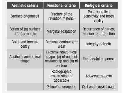
In all three groups, the following levels are used for evaluation:

1. Clinically excellent / very good.

2. Clinically good.

3. Clinically sufficient / satisfactory.

4. Clinically unsatisfactory.

5. Clinically poor.

Laboratory studies have provided information on CR repair techniques; for example, in the case of silorane-based composite resins, pre-treatment with sandpaper disks to roughen the surface provides the same results as for methacrylate-based CRs.59 Other recommendations include: using diamond stones,60 sandblasting with aluminum oxide,60 coating with silica,60)(61 and cleansing with phosphoric acid and other bonding agents.59)(60)(61 The use of hydrofluoric acid or silorane primer proved to be less efficient,60)(61 as well as using ozone or cleaning with acetone or ethanol, which did not affect the reparation′s bonding.62)(63)(64 The best results in repairing silorane-based CRs were obtained by combining silane and bonding agent, similar to what happens in BisGMA-based CRs (Bisphenol-a-glycidyl Methacrylate).59)(62)(65)(66)(67 Compatibility of silorane-based CRs and BisGMA-based CRs increase bond strength when silanes and adhesive phosphor-dimethacrylates

are used in the repairing process.59)(60)(68 In general, all aged CR surfaces that were repaired in labs showed reduced traction strength values ranging from 47 to 73% of the cohesive bonding of nonaged resins; in vitro, CRs repaired with nanohybrid fillings show the greatest traction strength values, and the lowest values occur when nanohybrid CRs are repaired with microfilled CRs.(69

One critical topic is defining treatment criteria and selecting patients for repair indication. It is necessary to point out that repair studies in recent years have included localized failures stating that the reminder restoration must be in good conditions; they basically include margin defects, gaps, marginal staining, localized secondary caries and wear, and repair is not indicated in the presence of secondary caries that is inaccessible through a small cavity or when it affects the restoration′s resistance to functional forces because it may compromise deep areas or reduce support for the remaining restoration. Neither is it indicated when the restoration has overall defects, or when the patient rejects alternative treatments or has a history of repair failure; also in cases with two concomitant failures, such as restoration fracture plus marginal defects, in which full replacement is deemed more reasonable.33)(45 Repair indications must be approached flexibly, as clinical history conditions may appear during the repair process, suggesting a change in indication-for example, in the presence of a deeper or more extensive carious lesion than initially observed-. It is also especially recommended to search for a clear cause of restoration failure, whether this variable will affect the repair process, or if it is possible to modify that variable, as in the case of very convex proximal walls with broken restorative material, in which the repair will probably experience the same problem.70 Full replacement remains a necessary indication, since usually not all restorations defects are assessed if they are small, while they often affect a greater extension of the restoration. However, repairs due to caries failures normally have better prognosis compared with restorations failing by fracture.5 Repairing extends the longevity of restorations, reduces the destructive effect of invasive procedures, reduces the probability of switching indications by indirect restorations, and significantly reduces pulp compromise, which is convenient for patients.4)(71

1b. Sealing

Sealing defective margins is another minimally invasive procedure with the ability to greatly increase the longevity of restorations with interventions that are simple, fast, and well tolerated by patients.8)(36)(38)(44)(45)(48)(53)(55)(72)(73)(74)(75 In addition to being a conservative technique, it is rather cost-effective as it requires short times and effectively eliminates marginal defects that could lead to secondary caries.42)(76)(77)(78)(79)(80)(81)(82)(83)(84 Currently there are no publications about its contraindications or recommendations regarding maximum magnitude and the effects of gap location and extension to indicate sealing.

Technically, sealing consists on using sealants or flowable resins52 to eliminate marginal gaps in defective restorations by means of absolute isolation, acid etching, and adhesive procedures, producing clinical results that are similar to those of replacement of restorations after being controlled during five years-the maximum reported period of clinical performance of sealed defective gaps-a time when it should be evaluated whether it is possible to seal the gap again-if the original conditions remain stable-or if it is necessary to apply another restoring technique.37

1c. Refurbishing

Refurbishing implies re-carving anatomic shape defects, eliminating extra contours, and improving surfaces by carving and polishing;39)(82 it has proven to be capable of recovering the morphologic, functional, and aesthetic characteristics of CR restorations with minimal interventions,55)(82)(85)(86 thus increasing longevity. It consists on re-carving occlusal anatomies using carbide or steel burs with multiple blades, discs, and sandpaper bands.55)(86 Limitations of refurbishing include very superficial restorations or risk of permanent damage.

1 d. Reduction of adhesive interface degradation

It is well known that performance and longevity of CR restorations are closely related to quality of the adhesive interface. Preventing or reducing interface degradation has been suggested as a new way to increase the longevity of CR restorations. We′ll analyze studies that show that using active adhesive systems, applying multiple layers of adhesive material, using additional hydrophobic layer, and increasing polymerization times are all techniques that promote resistance to adhesive interface degradation, with clinical consequences such as lower margin staining or prevalence of secondary caries. Furthermore, applying warm air to evaporate the solvent, using a wet technique with ethanol and eventually improving collagen strength through matrix metalloproteinase inhibitors (MMPIs) and collagen protectors, minimize adhesive interface degradation, contributing to increased longevity of CRs.

2. IMPROVING ADHESIVE SURFACE IMPREGNATION

2a. Active application

It has been traditionally considered that the two critical elements to achieve proper resin adhesion to dentin are a) dentin moistening by components of the adhesive material and B9 micro-mechanic assembly through resin penetration in the collagen fibers exposed by acid etching.87)(88

It has been shown that the adhesive agent suffers lower bonding degradation if demineralized dentin is vigorously rubbed for 10 s with two layers of adhesives and manual pressure of about 34 kg/cm,2 followed by evaporation of solvent for 10 s with the tip of the air syringe positioned at a distance of 20 cm plus light curing for 30 s in order to improve impregnation of the active surface, as demonstrated in laboratory tests and clinical results;89)(90 this may happen immediately or in the long term in both dry and wet demineralized dentin. This has prompted debate about generalized techniques in recent years, as dentin must remain hydrated to prevent the collagen mesh from collapsing, avoiding gaps that would block adequate dentin infiltration; in addition, active application provides higher adhesive values. One study shows that the difficulties in keeping dentin moisture and its failures could be overcome by vigorously rubbing the demineralized dentin surface.89

Vigorous application was already known in the case of dentinal adhesives containing the 10 MPD poly-functional monomer (10-metacril-oxo decylphosphate- dihydrogenated), which improves biodegradation resistance of the adhesive interface, resulting in the formation of multiple nano-layers of calcium salts that are attached to the 10 MPD molecule of 3.5 nm thick on each layer of dentin and protect collagen fibers from interphase biodegradation by hydrolysis.90 These nano-layers may explain the high stability of the bonding as well as its physical strength, proven in clinical and laboratory studies,91)(92 thus improving longevity. Interaction with hydroxyapatite occurs with acids of low pH but higher than the traditional ones, so it is necessary to recommend prior selective etching of enamel. The Ca-10-MDP bonding occurs clinically after rubbing the tooth surface for 20 to 30 s.92)(93)(94 This new formulation leaves the traditional considerations of total etching behind and incorporates adhesion to dentin by chemical bonding. As in all new developments, there are still unanswered questions regarding the biochemical secrets of these processes.93)(95)(96)(97)(98

Further studies on the chemical interactions of adhesive interfaces are needed to improve understanding of the activity of Ca-10-MDP nanolayers. The bonding of nano-layers to the dental substrate has been studied and is now understood, but there is not available information on nanolayers bonding in the transition from ultra-adhesive structure to restorative or cementing resin. So far, it has not been explained whether nano-layers are evenly spread over the entire tooth surface or if they are sets of bonding spots in nano-layers; if so, how are the gaps in between nano-layers formed and how they behave, or how they are related to the rest of the hybrid zone?

Theoretically, the size of a molecule of 10-MDP has been established as 1.95 nm; thus, the size of a Ca-10-MDP compound, consisting of two molecules of 10-MDP, would be 3.9 nm.94)(99 It is also known that Ca is 4 nm in diameter. These combined dimensions (7.9 nm) compared to the thickness of each nano-coating of Ca-10-MDP salt (3.5 nm), in both ClearFil SE (CFSE) and Single Bond Universal (SBU), are currently difficult to explain. These dimensional asymmetries could be explained by polymerization shrinkage phenomena in the new Ca-MDP formation; however, this evidence has not been built yet. It has been established that the dimensions of Ca-10-MDP nano-layers are interpreted as a fingerprint that could reveal the functional monomers that make part of the adhesive, but it does not explain the combined dimensions of its components.

It would be especially interesting to have additional knowledge on the interaction with some adhesive components, such as copolymers (from polialkenoic acid) of glass ionomers in adhesives (such as SBU), which could contest for the same HA calcium molecules (hydroxyapatite) used by 10-MDP to form nano-layers. Another important aspect would be to know if the presence of HEMA (hidroetil methacrylate acid) significantly affects the chemical interaction of MDP, preventing the formation of bonds, salts, and nano-layers by reducing the rate of surface demineralization-a pre-requirement for the formation of Ca-MDP salt in its chemical interaction with HA100-and whether such chemical interactions affect the longevity of CRs.

Another original contribution is the recent publication by Mena-Serrano et al, who demonstrated that sonic application of the adhesive to a 170 Hz oscillation frequency can improve the µTBS of dentin/resin bonding, reduce nano-leakage, and slow down the degradation of adhesive bonding.101

2b. Application of multi-layer

Laboratory studies have shown that application of multi-layer adhesives can increase adhesion in the presence of leakage by increasing adhesives values. A clinically-controlled, randomized, prospective study on the behavior of CR restorations made with self-etching and etch-and-rinse adhesives in non-carious cervical lesions (Loguercio and Reis) concluded that the multi-layer adhesive technique significantly improves retention rates in both adhesive techniques 12 and 18 months after application; however, it does not reach the level required by the ADA (American Dental Association′s Guideline) for bonding between dentin and adhesive materials. It is worth noting that the greater the amount of adhesive layers the lower the amount of failures in Class V adhesive restorations,102 making the application of a double layer of adhesive a clinical standard-but accompanied by just one light-curing process-. This is an original contribution easy to follow, presented by only one publication, and therefore more reports should be expected before considering its incorporation in daily clinical use.

2c. Application of an additional hydrophobic layer

The clinical performance of adhesives has significantly improved in recent times, achieving current adhesive restorations with higher levels of predictability in terms of clinical success. Modern adhesive systems are better than the early ones in terms of retention.103)(104 However, the chemistry of self-etching adhesive systems remains a great challenge, as it incorporates hydrophilic and hydrophobic monomers, resulting in highly hydrophilic systems, which produce semi-permeable membranes with water diffusion from dentin through the adhesive layer105-a process that decreases the mechanical properties of polymers in the resinous matrix-.106 Retention of residual water because of incomplete evaporation of the adhesive agent or due to moisture from underlying dentin as a result of high osmolarity of adhesives produces channels filled with water inside the adhesive material. (105 As a contribution to the solution to this problem, it has been suggested incorporating a hydrophobic layer on the self-etching adhesive. Experimental and clinical tests107)(108 have demonstrated that including an extra hydrophobic layer in addition to the self-etching adhesive procedure improves the clinical behavior observed 6, 12, and 24 months after application, mainly in terms of retention of CR restorations performed on Class V non-carious cervical lesions, as a result of lower degradation of adhesive bond.90)(109)(110 However, longer-term clinical studies are required to complement this information.

Recently, observations by Muñoz et al suggested that reducing nano-leaking by applying an additional hydrophobic layer is dependent on the composition of the adhesive agent rather than on the adhesive strategy itself.111

2d. Late polymerization or passive evaporation of solvent

Dentin adhesives contain monomers of hydrophilic resins, which are dissolved in organic solvents such as acetone and ethanol. Using these solvents favors the movement of water from dentin surface and facilitates monomer penetration to the microporosities exposed by acid etching.112)(113 Including a solvent has increased adhesive bond values and its presence along with water is considered essential for the plastic behavior of collagen bundles and for managing their collapsing.114)(115 Ideally, solvents must be completely removed before light-curing the adhesive in order to prevent unwanted effects to monomers,116 such as reduction of their mechanical properties, poor polymerization, formation of cracks on adhesive agent, and premature failure.117)(118)(119)(120

A lower degradation of adhesive bonding has been observed when slowing down the polymerization of adhesive monomers; in addition, under this conditions, the micro-traction strength values increase if waiting 300 s before light-curing.121 As early as 2002, one published study on the Scotch-Bond adhesive system claimed that increasing the waiting time to 30 s before light-curing significantly increased shear strength.121 The explanation to this phenomenon may be related to the fact of giving the adhesive agent more time to evaporate its alcohol solvents and water, especially since it has been well known that adding water to adhesive systems promotes the formation of water channels through the hybrid zone, when it is associated to alcohols; this happens because adding water to both comonomers and ethanol increases the retention of both ethanol and water in the system, because the two solvents can join the hydrogens of monomers.122 Increasing exposure time for evaporation of the adhesive system after its application to even more time than that recommended by manufacturers does not prevent its degradation but can increase bond strength immediately and for up to six months, perhaps as a result of the amount of polymer chains;110 unfortunately, the literature lacks additional studies on higher times.

3. IMPROVEMENTS TO POLYMERS STRENGTH

3a. Using hot air to evaporate solvents

Initially, water absorption in resins was considered favorable as it compensated the effect of shrinkage by polymerization;123)(124 however, water absorption is currently associated with internal resin weaknesses, which facilitate the extraction of free monomers or residual materials from the polymerization of CR. Water molecules can form clusters that induce softening and deformation of the matrix that surrounds it, decreasing the restoration rigidity.125)(126

Degradation and the shorter longevity of RC restorations are related to deleterious phenomena at the hybrid zone, with degradation of collagen fibers inadequately protected.105)(127)(128 Several clinical approaches have been developed in search for solutions to this conflict, one of them being the incorporation of a flow of hot pressurized air to promote forced evaporation of the adhesive agent′s solvents, concluding that this method can improve the bonding interface over time (six months), especially in water/ethanol-based adhesive systems.129)(130 This is based on the improved quality of the hybrid layer considering its ability to produce lower nano-leakage and to reduce the number of pores, apparently without modifying the resins′ conversion degree.131 A recent study shows that using hot air to evaporate solvents was not efficient in reducing water absorption or adhesive agents solubility, keeping the increase in adhesive bond values.127 The truth is that evaporation of solvents remains and unsolved issue, since the evaporation times recommended by manufacturers for acetoneor ethanol-based adhesive agents retain 5% to 10% after having been blown for 120 s122-a period more than 10 times higher than recommended-, but when the solvent is mixed with 30% of water it can retain up to 41%.122

From the perspective of pulp biology, it is well known that several factors affect the temperature increase in pulp (for instance, in terms of residual dentin thickness), as dentin has a low thermal conductivity and acts as a shield against thermal action;132 however, there are no studies showing the safety of this approach, which is at least deemed unfavorable by histological studies on vital units; studies on endodontically-treated teeth are then suggested

3b. The ethanol-wet technique

The studies by Sadek et al show that another antidegradation strategy for adhesive bonding is the application of ethanol on dentin surface prior to the adhesive technique. The suggested mechanism of action is saturation of surface by ethanol and water movement, preventing integrity of the hybrid zone by absence of water, according to laboratory observations after 9 and 18 months along with TEM analysis (Transmission Electron Microscopy), in a study conducted with reproduction of dentin hydraulic pressure.133)(134 However, despite showing no degradation of the hybrid layer, from the clinical point of view it will be difficult to implement for the time being, given the undemonstrated biological effects of ethanol application on dentin pulp.

4. IMPROVING THE STRENGTH OF COLLAGEN FIBERS

4a. MMP inhibitors

It is well known that the dentin matrix contains matrix metalloproteinases (MMPs)135)(136)(137 and that some of these MMPs attack collagen (MMP 8 and 20) and gelatin (MMP 2 and 9).136)(138 Dentin contains MMP 2 and 20)(138)(139 and the activity of MMPs derived from host enzymes degrade the hybrid layer in vivo.140)(141

Pretreating dentin with 0.2% chlorhexidine (CHX) for 60 s prevents the collagenolytic activity to levels close to zero142 through inhibition of the activity of MMPs in the hybrid layer, increasing its longevity; unfortunately these studies have been conducted in vivo for the maximum period of 14 months,143 showing that it does not decrease adhesive values in Class V restorations. Besides CHX, other compounds such as EDTA (ethylenediaminetetraacetic acid) produce an inhibitory action on MMPs in collagen degradation in demineralized dentins. MMPs degradation was reduced in resin-infiltrated dentin, and the presence of another element such as zinc produced an additional protective effect.144

Pashley et al have named these new compounds "therapeutic adhesive systems" because they have antibacterial and anti-MMP activity as they contain CHX and benzalkonium chloride.145

Massoni et al claim that using non-toxic MMP inhibitors such as CHX is an additional step in adhesive systems in order to increase the longevity of adhesive restorations.146

Although this scheme has detractors, recently Luhrs et al conducted laboratory analysis noting that the action of MMP inhibitors fails to prevent the decreased micro-traction strength after their samples had been aged; in addition, it does not improve longevity when used in cementing composite resins.147

These results are promising but in vivo randomized trials are needed in the long term in order to assess the exact dimension of these investigations.

4b. Collagen protectors - proanthocyanidin use

It is known that durability of resin-dentin bond requires stable collagen fibers in the hybrid layer; however, using simplified adhesives to demineralize dentin activates the endogenous matrix metalloproteinases, resulting in progressive loss of unprotected collagen fibers.146)(148 Conversely, involvement of endogenous MMPs in the degradation process is minimal, when the self-etching adhesives are softer.90)(149)(150 Modifying the demineralized collagen matrix with external crosslinking agents plays an important role in improving the biomechanical properties of dentin.151 Agents of collagen fibers reinforced by crosslinking show that it decreases the enzymatic degradation, and are therefore considered critical to increase the hybrid layer stability as well as durability of the restoration adhesion. Examples of agents often used as external crosslinkers in dentistry are glutaraldehyde, genipin, carbodiimide, and proanthocyanidin (PA).

PA is a polyphenolic compound classified as a flavonoid plant, which is part of the tannins group; it is found in the bark of pine trees and elm trees, and in grape seeds;1)(52 it is also available in vegetables and fruits but in lower concentrations. PA is a powerful antioxidant and a crosslinking agent of low toxicity. It has been shown that PA from grape seeds extracts improve the traction strength and the rigidity of dentin collagen,151)(153)(154 as well as the long-term stability of dentin collagen matrix.155)(156 In addition to its crosslinking effect, the PA obtained from elm and blueberry extracts has also shown the ability to inhibit the production of MMPs 1, 3, 7, 8, 9, and 13 in macrophages, as well as the catalytic activity of MMPs 1 and 9.157)(158)(159)(160

Dos Santos et al reported that experimentally pretreating demineralized dentin with PA for one hour before applying the adhesive agent significantly improves the nano-mechanical properties (nanohardness and modulus of elasticity) of the resin/dentin interface,158)(159 as well as bond strength.161 Similar results have been observed in studies on dentine affected by carious lesions;162 however, because of the time of application, it is impossible to use this protocol in clinical conditions, and that is why the inclusion of PA in the dental adhesive has been considered, as it allows the PA to act for a long period of time, increasing the crosslinking degree with collagen and resistance to biodegradation.163 Adding PA in an adhesion experiment showed that it had no adverse effects on adhesive bond strength when used in concentrations of up to 2% besides significantly reducing nano-leakage.150 Other observations suggest that using ethanol as adhesive solvent promotes PA/collagen interaction by decreasing the dielectric constant of the adhesive agent and enhances the stability of hydrogen bonding.164 The mechanism of action of PA preserves the triple helix collagen structure and induces the addition of microfibers by displacing water and creating a new hydrogen/ collagen bond.165 Promising experimental results may become available for clinical use in the coming years.

Recently a group of researchers has drawn attention by observing that crosslinking agents are effective in reducing MMPs activity by mixing 0.5% carbodiimide (EDC) and 35% HEMA without affecting the roles of each component.166

CONCLUSIONS

Using minimally invasive techniques such as repair, sealing, and refurbishing defective composite resin restorations show longitudinal clinical evidence in randomized clinical trials which increase longevity in restored teeth.

In addition, other innovative methodologies with clinical evidence still in progress, such as improving impregnation of adhesive surfaces, increasing the strength of adhesive polymers and increasing the strength to collagen hydrolytic degradation, are promising steps that either together or separately modify the management of adhesive techniques and will offer alternative restorative treatments to the population demanding aesthetic solutions of composite resins with longer longevity.

CONFLICT OF INTEREST

The authors of this article certify that they have no property or personal interest of any kind on the products discussed throughout the article, and therefore declare having no conflict of interest

REFERENCES

1. Opdam NJ, Bronkhorst EM, Loomans BA, Huysmans MC. 12-year survival of composite vs. amalgam restorations. J Dent Res 2010; 89(10): 1063-1067. [ Links ]

2. Da Rosa Rodolpho PA, Donassollo TA, Cenci MS, Loguércio AD, Moraes RR, Bronkhorst EM et al. 22-Year clinical evaluation of the performance of two posterior composites with different filler characteristics. Dent Mater 2011; 27(10): 955-963. [ Links ]

3. Opdam NJ, Bronkhorst EM, Cenci MS, Huysmans MC, Wilson NH. Age of failed restorations: a deceptive longevity parameter. J Dent 2011; 39(3): 225-230. [ Links ]

4. Demarco FF, Correa MB, Cenci MS, Moraes RR, Opdam NJ. Longevity of posterior composite restorations: not only a matter of materials. Dent Mater 2012; 28(1): 87-101. [ Links ]

5. Opdam NJ, Bronkhorst EM, Loomans BA, Huysmans MC. Longevity of repaired restorations: a practice based study. J Dent 2012; 40(10): 829-835. [ Links ]

6. Manhart J, Chen H, Hamm G, Hickel R. Buonocore memorial lecture. Review of the clinical survival of direct and indirect restorations in posterior teeth of the permanent dentition. Oper Dent 2004; 29(5): 481-508. [ Links ]

7. Hickel R, Manhart J. Longevity of restorations in posterior teeth and reasons for failure. J Adhes Dent 2001; 3(1): 45-64. [ Links ]

8. Gordan VV, Mjör IA, Blum IR, Wilson N. Teaching students the repair of resin-based composite restorations: a survey of North American dental schools. J Am Dent Assoc 2003; 134(3): 317-323; quiz 38-39. [ Links ]

9. Mjör IA, Reep RL, Kubilis PS, Mondragón BE. Change in size of replaced amalgam restorations: a methodological study. Oper Dent 1998; 23(5): 272-277. [ Links ]

10. Onal B, Pamir T. The two-year clinical performance of esthetic restorative materials in noncarious cervical lesions. J Am Dent Assoc 2005; 136(11): 1547-1555. [ Links ]

11. Chadwick B, Treasure E, Dummer P, Dunstan F, Gilmour A, Jones R et al. Challenges with studies investigating longevity of dental restorations--a critique of a systematic review. J Dent 2001; 29(3): 155-161. [ Links ]

12. Opdam NJ, Bronkhorst EM, Roeters JM, Loomans BA. A retrospective clinical study on longevity of posterior composite and amalgam restorations. Dent Mater 2007; 23(1): 2-8. [ Links ]

13. Kubo S, Kawasaki A, Hayashi Y. Factors associated with the longevity of resin composite restorations. Dent Mater J 2011; 30(3): 374-383. [ Links ]

14. Arhun N, Celik C, Yamanel K. Clinical evaluation of resin-based composites in posterior restorations: twoyear results. Oper Dent 2010; 35(4): 397-404. [ Links ]

15. Goldberg AJ, Rydinge E, Santucci EA, Racz WB. Clinical evaluation methods for posterior composite restorations. J Dent Res 1984; 63(12): 1387-1391. [ Links ]

16. Alves dos Santos MP, Luiz RR, Maia LC. Randomised trial of resin-based restorations in Class I and Class II beveled preparations in primary molars: 48-month results. J Dent 2010; 38(6): 451-459. [ Links ]

17. Moncada G, Martin J, Fernandez E, Hempel MC, Mjor IA, Gordan VV. Sealing, refurbishment and repair of Class I and Class II defective restorations: a three-year clinical trial. J Am Dent Assoc 2009; 140(4): 425-432. [ Links ]

18. Ryge G, Jendresen MD, Glantz PO, Mjör I. Standardization of clinical investigators for studies of restorative materials. Swed Dent J 1981; 5(5-6): 235-239. [ Links ]

19. Forss H, Widström E. Factors influencing the selection of restorative materials in dental care in Finland. J Dent 1996; 24(4): 257-262. [ Links ]

20. Mjör IA, Moorhead JE, Dahl JE. Selection of restorative materials in permanent teeth in general dental practice. Acta Odontol Scand 1999; 57(5): 257-262. [ Links ]

21. Forss H, Widström E. From amalgam to composite: selection of restorative materials and restoration longevity in Finland. Acta Odontol Scand 2001; 59(2): 57-62. [ Links ]

22. Woods N, Considine J, Lucey S, Whelton H, Nyhan T. The influence of economic incentives on treatment patterns in a third-party funded dental service. Community Dent Health 2010; 27(1): 18-22. [ Links ]

23. De Souza JP, Nozawa SR, Honda RT. Improper waste disposal of silver-mercury amalgam. Bull Environ Contam Toxicol 2012; 88(5): 797-801. [ Links ]

24. Goldstein GR. The longevity of direct and indirect posterior restorations is uncertain and may be affected by a number of dentist-, patient-, and material-related factors. J Evid Based Dent Pract 2010; 10(1): 30-31. [ Links ]

25. Lessa FC, Nogueira I, Huck C, Hebling J, Costa CA. Transdentinal cytotoxic effects of different concentrations of chlorhexidine gel applied on acid-conditioned dentin substrate. J Biomed Mater Res B Appl Biomater 2010; 92(1): 40-47. [ Links ]

26. Lindberg A, van Dijken JW, Lindberg M. Nine-year evaluation of a polyacid-modified resin composite/resin composite open sandwich technique in Class II cavities. J Dent 2007; 35(2): 124-129. [ Links ]

27. Kim KL, Namgung C, Cho BH. The effect of clinical performance on the survival estimates of direct restorations. Restor Dent Endod 2013; 38(1): 11-20. [ Links ]

28. Manhart J, Chen H, Hamm G, Hickel R. Buonocore Memorial Lecture. Review of the clinical survival of direct and indirect restorations in posterior teeth of the permanent dentition. Oper Dent 2004; 29(5): 481-508. [ Links ]

29. Mjör IA, Shen C, Eliasson ST, Richter S. Placement and replacement of restorations in general dental practice in Iceland. Oper Dent 2002; 27(2): 117-123. [ Links ]

30. Brunthaler A, König F, Lucas T, Sperr W, Schedle A. Longevity of direct resin composite restorations in posterior teeth. Clin Oral Investig 2003; 7(2): 63-70. [ Links ]

31. Bernardo M, Luis H, Martin MD, Leroux BG, Rue T, Leitao J et al. Survival and reasons for failure of amalgam versus composite posterior. J Am Dent Assoc 2007; 138(6): 775-783. [ Links ]

32. Da Costa J. Summary of: the survival of Class V restorations in general dental practice: part 3, five-year survival. Br Dent J 2012; 212(9): 440-441. [ Links ]

33. Hickel R, Brüshaver K, Ilie N. Repair of restorations-- criteria for decision making and clinical recommendations. Dent Mater 2013; 29(1): 28-50. [ Links ]

34. Burke FJ, Cheung SW, Mjör IA, Wilson NH. Restoration longevity and analysis of reasons for the placement and replacement of restorations provided by vocational dental practitioners and their trainers in the United Kingdom. Quintessence Int 1999; 30(4): 234-242. [ Links ]

35. Forss H, Widström E. Reasons for restorative therapy and the longevity of restorations in adults. Acta Odontol Scand 2004; 62(2): 82-86. [ Links ]

36. Gordan VV, Mondragon E, Shen C. Replacement of resin-based composite: evaluation of cavity design, cavity depth, and shade matching. Quintessence Int 2002; 33(4): 273-278. [ Links ]

37. Martin J, Fernandez E, Estay J, Gordan VV, Mjor IA, Moncada G. Minimal invasive treatment for defective restorations: five-year results using sealants. Oper Dent 2013; 38(2): 125-133. [ Links ]

38. Mjör IA, Gordan VV. Failure, repair, refurbishing and longevity of restorations. Oper Dent 2002; 27(5): 528- 534. [ Links ]

39. Martin J, Fernandez E, Estay J, Gordan VV, Mjör IA, Moncada G. Management of Class I and Class II Amalgam Restorations with localized defects: five-year results. Int J Dent 2013; 2013: 450260. [ Links ]

40. Sharif MO, Catleugh M, Merry A, Tickle M, Dunne SM, Brunton P et al. Replacement versus repair of defective restorations in adults: resin composite. Cochrane Database Syst Rev 2010; (2): CD005971. [ Links ]

41. Jokstad A, Mjör IA, Nilner K, Kaping S. Clinical performance of three anterior restorative materials over 10 years. Quintessence Int 1994; 25(2): 101-108. [ Links ]

42. Mjör IA, Moorhead JE, Dahl JE. Reasons for replacement of restorations in permanent teeth in general dental practice. Int Dent J 2000; 50(6): 361-366. [ Links ]

43. Heintze SD, Rousson V. Clinical effectiveness of direct class II Restorations - a meta-analysis. J Adhes Dent 2012; 14(5): 407-431. [ Links ]

44. Gordan VV, Garvan CW, Blaser PK, Mondragon E, Mjör IA. A long-term evaluation of alternative treatments to replacement of resin-based composite restorations: results of a seven-year study. J Am Dent Assoc 2009; 140(12): 1476-1484. [ Links ]

45. Blum IR, Jagger DC, Wilson NH. Defective dental restorations: to repair or not to repair? Part 1: direct composite restorations. Dent Update 2011; 38(2): 78-80, 82-84. [ Links ]

46. Inoue T. Repair of restoration materials: repair of amalgam and composite resin used in restorations. Shikai Tenbo 1978; 52(1): 87-96. [ Links ]

47. Katsuyama S, Nogami I, Tsuzuki K, Suzuki S, Yoshida K. Prognosis in composite resin restoration and its repair. Shikai Tenbo 1979; 53(1): 1-12. [ Links ]

48. Mjör IA. Repair versus replacement of failed restorations. Int Dent J 1993; 43(5): 466-472. [ Links ]

49. Tyas MJ, Anusavice KJ, Frencken JE, Mount GJ. Minimal intervention dentistry--a review. FDI Commission Project 1-97. Int Dent J 2000; 50(1): 1-12. [ Links ]

50. Mjör IA. The reasons for replacement and the age of failed restorations in general dental practice. Acta Odontol Scand 1997; 55(1): 58-63. [ Links ]

51. Blum IR, Lynch CD, Wilson NH. Teaching of the repair of defective composite restorations in scandinavian dental schools. J Oral Rehabil 2012; 39(3): 210-216. [ Links ]

52. Gordan VV, Shen C, Mjor IA. Marginal gap repair with flowable resin-based composites. Gen Dent 2004; 52(5): 390-394. [ Links ]

53. Gordan VV, Riley JL, Blaser PK, Mondragon E, Garvan CW, Mjor IA. Alternative treatments to replacement of defective amalgam restorations: results of a seven-year clinical study. J Am Dent Assoc 2011; 142(7): 842-849. [ Links ]

54. Gordan VV, Riley JL, Blaser PK, Mjör IA. 2-year clinical evaluation of alternative treatments to replacement of defective amalgam restorations. Oper Dent 2006; 31(4): 418-425. [ Links ]

55. Fernández EM, Martin JA, Angel PA, Mjör IA, Gordan VV, Moncada GA. Survival rate of sealed, refurbished and repaired defective restorations: 4-year follow-up. Braz Dent J 2011; 22(2): 134-139. [ Links ]

56. Gordan VV. In vitro evaluation of margins of replaced resin-based composite restorations. J Esthet Dent 2000; 12(4): 209-215. [ Links ]

57. Hickel R, Peschke A, Tyas M, Mjor I, Bayne S, Peters M et al. FDI World Dental Federation - clinical criteria for the evaluation of direct and indirect restorations. Update and clinical examples. J Adhes Dent 2010; 12(4): 259- 272. [ Links ]

58. Hickel R, Roulet JF, Bayne S, Heintze SD, Mjör IA, Peters M et al. Recommendations for conducting controlled clinical studies of dental restorative materials. Clin Oral Investig 2007; 11(1): 5-33. [ Links ]

59. Ivanovas S, Hickel R, Ilie N. How to repair fillings made by silorane-based composites. Clin Oral Investig 2011; 15(6): 915-922. [ Links ]

60. Wiegand A, Stawarczyk B, Buchalla W, Tauböck TT, Özcan M, Attin T. Repair of silorane composite--using the same substrate or a methacrylate-based composite? Dent Mater 2012; 28(3): e19-25. [ Links ]

61. Lührs AK, Görmann B, Jacker-Guhr S, Geurtsen W. Repairability of dental siloranes in vitro. Dent Mater 2011; 27(2): 144-149. [ Links ]

62. Hamano N, Chiang YC, Nyamaa I, Yamaguchi H, Ino S, Hickel R et al. Repair of silorane-based dental composites: influence of surface treatments. Dent Mater 2012; 28(8): 894-902. [ Links ]

63. Magni E, Ferrari M, Papacchini F, Hickel R, Ilie N. Influence of ozone on the composite-to-composite bond. Clin Oral Investig 2011; 15(2): 249-256. [ Links ]

64. Magni E, Ferrari M, Papacchini F, Hickel R, Ilie N. Influence of ozone application on the repair strength of silorane-based and ormocer-based composites. Am J Dent 2010; 23(5): 260-264. [ Links ]

65. Papacchini F, Magni E, Radovic I, Mazzitelli C, Monticellia F, Goracci C et al. Effect of intermediate agents and pre-heating of repairing resin on compositerepair bonds. Oper Dent 2007; 32(4): 363-371. [ Links ]

66. Maneenut C, Sakoolnamarka R, Tyas MJ. The repair potential of resin composite materials. Dent Mater 2011; 27(2): e20-27. [ Links ]

67. Baur V, Ilie N. Repair of dental resin-based composites. Clin Oral Investig 2013; 17(2): 601-618. [ Links ]

68. Tezvergil A, Lassila LV, Vallittu PK. Compositecomposite repair bond strength: effect of different adhesion primers. J Dent 2003; 31(8): 521-525. [ Links ]

69. Moncada G, Angel P, Fernandez E, Alonso P, Martin J, Gordan VV. Bond strength evaluation of nanohybrid resin-based composite repair. Gen Dent 2012; 60(3): 230- 234. [ Links ]

70. Hickel R, Brushaver K, Ilie N. Repair of restorations - criteria for decision making and clinical recommendations. Dent Mater 2013; 29(1): 28-50. [ Links ]

71. Lynch CD, Blum IR, Frazier KB, Haisch LD, Wilson NH. Repair or replacement of defective direct resin-based composite restorations: contemporary teaching in U.S. and canadian dental schools. J Am Dent Assoc 2012; 143(2): 157-163. [ Links ]

72. Gordan VV. Clinical evaluation of replacement of class V resin based composite restorations. J Dent 2001; 29(7): 485-488. [ Links ]

73. Moncada G, Martin J, Fernandez E, Vildósola P, Caamano C, Caro MJ et al. In vivo evaluation of alternative treatment to replace defective restorations. J Dent Res 2005; Res 84 Spec Iss A: 3042. [ Links ]

74. Lynch CD, Frazier KB, McConnell RJ, Blum IR, Wilson NH. Minimally invasive management of dental caries: contemporary teaching of posterior resin-based composite placement in U.S. and Canadian dental schools. J Am Dent Assoc 2011; 142(6): 612-620. [ Links ]

75. Kidd EA, Joyston-Bechal S, Beighton D. Microbiological validation of assessments of caries activity during cavity preparation. Caries Res 1993; 27(5): 402-408. [ Links ]

76. Hodges DJ, Mangum FI, Ward MT. Relationship between gap width and recurrent dental caries beneath occlusal margins of amalgam restorations. Community Dent Oral Epidemiol 1995; 23(4): 200-204. [ Links ]

77. Mjör IA, Dahl JE, Moorhead JE. Age of restorations at replacement in permanent teeth in general dental practice. Acta Odontol Scand 2000; 58(3): 97-101. [ Links ]

78. Qvist J, Qvist V, Mjör IA. Placement and longevity of amalgam restorations in Denmark. Acta Odontol Scand 1990; 48(5): 297-303. [ Links ]

79. Sharif MO, Fedorowicz Z, Tickle M, Brunton PA. Repair or replacement of restorations: do we accept built in obsolescence or do we improve the evidence? Br Dent J 2010; 209(4): 171-174. [ Links ]

80. Sharif MO, Merry A, Catleugh M, Tickle M, Brunton P, Dunne SM et al. Replacement versus repair of defective restorations in adults: amalgam. Cochrane Database Syst Rev 2010; 17(2): CD005970. [ Links ]

81. Ryge G, Snyder M. Evaluating the clinical quality of restorations. J Am Dent Assoc 1973; 87(2): 369-377. [ Links ]

82. Moncada G, Fernández E, Martín J, Arancibia C, Mjör IA, Gordan VV. Increasing the longevity of restorations by minimal intervention: a two-year clinical trial. Oper Dent 2008; 33(3): 258-264. [ Links ]

83. Baratieri LN, Monteiro Júnior S, de Andrada MA. Amalgam repair: a case report. Quintessence Int 1992; 23(8): 527-531. [ Links ]

84. Downer MC, Azli NA, Bedi R, Moles DR, Setchell DJ. How long do routine dental restorations last? A systematic review. Br Dent J 1999; 187(8): 432-439. [ Links ]

85. Gordan VV, Riley JL, III, Blaser PK , Mjor IA . 2-year clinical evaluation of alternative treatments to replacement of defective. Oper Dent 2006; 31(4): 418-425. [ Links ]

86. Martin J. Fernandez E, Angel P, Gordan VV, Mjör I, Moncada G. Aumento de la longevidad de restauraciones de amalgama y resinas compuestas defectuosas por medio de sellado marginal. Revista Dental de Chile 2009; 100(2): 4-9. [ Links ]

87. Pashley DH, Carvalho RM, Tay FR, Agee KA, Lee KW. Solvation of dried dentin matrix by water and other polar solvents. Am J Dent 2002; 15(2): 97-102. [ Links ]

88. Spencer P, Wang Y. Adhesive phase separation at the dentin interface under wet bonding conditions. J Biomed Mater Res 2002; 62(3): 447-456. [ Links ]

89. Reis A, Pellizzaro A, Dal-Bianco K, Gones OM, Patzlaff R, Loguercio AD. Impact of adhesive application to wet and dry dentin on long-term resin-dentin bond strengths. Oper Dent 2007; 32(4): 380-387. [ Links ]

90. Loguercio AD, Stanislawczuk R, Mena-Serrano A, Reis A. Effect of 3-year water storage on the performance of one-step self-etch adhesives applied actively on dentine. J Dent 2011; 39(8): 578-587. [ Links ]

91. Yoshida Y, Van Meerbeek B, Nakayama Y, Snauwaert J, Hellemans L, Lambrechts P et al. Evidence of chemical bonding at biomaterial-hard tissue interfaces. J Dent Res 2000; 79(2): 709-714. [ Links ]

92. Yoshida Y, Nagakane K, Fukuda R, Nakayama Y, Okazaki M, Shintani H et al. Comparative study on adhesive performance of functional monomers. J Dent Res 2004; 83(6): 454-458. [ Links ]

93. Yoshihara K, Yoshida Y, Nagaoka N, Fukegawa D, Hayakawa S, Mine A et al. Nano-controlled molecular interaction at adhesive interfaces for hard tissue reconstruction. Acta Biomater 2010; 6(9): 3573-3582. [ Links ]

94. Yoshihara K, Yoshida Y, Hayakawa S, Nagaoka N, Irie M, Ogawa T et al. Nanolayering of phosphoric acid ester monomer on enamel and dentin. Acta Biomater 2011; 7(8): 3187-3195. [ Links ]

95. Tanaka T, Nagata K, Takeyama M, Atsuta M, Nakabayashi N, Masuhara E. 4-META opaque resin--a new resin strongly adhesive to nickel-chromium alloy. J Dent Res 1981; 60(9): 1697-1706. [ Links ]

96. Mena-Serrano A, Kose C, De Paula EA, Tay LY, Reis A, Loguercio AD et al. A new universal simplified adhesive: 6-month clinical evaluation. J Esthet Restor Dent 2013; 25(1): 55-69. [ Links ]

97. Van Meerbeek B, Van Landuyt K, De Munck J, Hashimoto M, Peumans M, Lambrechts Pet al. Technique-sensitivity of contemporary adhesives. Dent Mater J 2005; 24(1): 1-13. [ Links ]

98. Omura I, Yamauchi J, Harada I, Wada T. Adhesive and mechanical properties of a new dental adhesive, J Dent Res 1984 63:233, Abst. No. 561. [ Links ]

99. Fukegawa D, Hayakawa S, Yoshida Y, Suzuki K, Osaka A, van Meerbeek B. Chemical interaction of phosphoric acid ester with hydroxyapatite. J Dent Res 2006; 85(10): 941-944. [ Links ]

100. Yoshida Y, Yoshihara K, Hayakawa S, Nagaoka N, Okihara T, Matsumoto T et al. HEMA inhibits interfacial nano-layering of the functional monomer MDP. J Dent Res 2012; 91(11): 1060-1065. [ Links ]

101. Mena-Serrano A, Garcia EJ, Loguercio AD, Reis A. Effect of sonic application mode on the resin-dentin bond strength and nanoleakage of simplified self-etch adhesive. Clin Oral Investig 2014; 18(3): 729-736 [ Links ]

102. Loguercio AD, Reis A. Application of a dental adhesive using the self-etch and etch-and-rinse approaches: an 18-month clinical evaluation. J Am Dent Assoc 2008; 139(1): 53-61. [ Links ]

103. Van Meerbeek B, Yoshihara K, Yoshida Y, Mine A, De Munck J, Van Landuyt KL. State of the art of self-etch adhesives. Dent Mater 2011; 27(1): 17-28. [ Links ]

104. Peumans M, Kanumilli P, De Munck J, Van Landuyt K, Lambrechts P, Van Meerbeek B. Clinical effectiveness of contemporary adhesives: a systematic review of current clinical trials. Dent Mater 2005; 21(9): 864-881. [ Links ]

105. Tay FR, Pashley DH. Water treeing--a potential mechanism for degradation of dentin adhesives. Am J Dent 2003; 16(1): 6-12. [ Links ]

106. Santerre JP, Shajii L, Leung BW. Relation of dental composite formulations to their degradation and the release of hydrolyzed polymeric-resin-derived products. Crit Rev Oral Biol Med 2001; 12(2): 136-151. [ Links ]

107. Loguercio AD, Mânica D, Ferneda F, Zander-Grande C, Amaral R, Stanislawczuk R et al. A randomized clinical evaluation of a one- and two-step self-etch adhesive over 24 months. Oper Dent 2010; 35(3): 265-272. [ Links ]

108. Reis A, Albuquerque M, Pegoraro M, Mattei G, Bauer JR, Grande RH et al. Can the durability of one-step selfetch adhesives be improved by double application or by an extra layer of hydrophobic resin? J Dent 2008; 36(5): 309-315. [ Links ]

109. Reis A, Leite TM, Matte K, Michels R, Amaral RC, Geraldeli S et al. Improving clinical retention of onestep self-etching adhesive systems with an additional hydrophobic adhesive layer. J Am Dent Assoc 2009; 140(7): 877-885. [ Links ]

110. Reis A, Ferreira SQ, Costa TR, Klein-Júnior CA, Meier MM, Loguercio AD. Effects of increased exposure times of simplified etch-and-rinse adhesives on the degradation of resin-dentin bonds and quality of the polymer network. Eur J Oral Sci 2010; 118(5): 502-509. [ Links ]

111. Muñoz MA, Sezinando A, Luque-Martinez I, Szesz AL, Reis A, Loguercio AD et al. Influence of a hydrophobic resin coating on the bonding efficacy of three universal adhesives. J Dent 2014; 42(5): 595-602. [ Links ]

112. Kanca J. Effect of resin primer solvents and surface wetness on resin composite bond strength to dentin. Am J Dent 1992; 5(4): 213-215. [ Links ]

113. Tay FR, Gwinnett JA, Wei SH. Micromorphological spectrum from overdrying to overwetting acidconditioned dentin in water-free acetone-based, singlebottle primer/adhesives. Dent Mater 1996; 12(4): 236- 244. [ Links ]

114. Jacobsen T, Söderholm KJ. Effect of primer solvent, primer agitation, and dentin dryness on shear bond strength to dentin. Am J Dent 1998; 11(5): 225-228. [ Links ]

115. Maciel KT, Carvalho RM, Ringle RD, Preston CD, Russell CM, Pashley DH. The effects of acetone, ethanol, HEMA, and air on the stiffness of human decalcified dentin matrix. J Dent Res 1996; 75(11): 1851-1858. [ Links ]

116. Jacobsen T, Söderholm KJ. Some effects of water on dentin bonding. Dent Mater 1995; 11(2): 132-136. [ Links ]

117. Pashley EL, Zhang Y, Lockwood PE, Rueggeberg FA, Pashley DH. Effects of HEMA on water evaporation from water-HEMA mixtures. Dent Mater 1998; 14(1):6-10. [ Links ]

118. Perdigão J, Frankenberger R. Effect of solvent and rewetting time on dentin adhesion. Quintessence Int 2001; 32(5): 385-390. [ Links ]

119. Carvalho RM, Mendonça JS, Santiago SL, Silveira RR, Garcia FC, Tay FR et al. Effects of HEMA/solvent combinations on bond strength to dentin. J Dent Res 2003; 82(8): 597-601. [ Links ]

120. Dickens SH, Cho BH. Interpretation of bond failure through conversion and residual solvent measurements and weibull analyses of flexural and microtensile bond strengths of bonding agents. Dent Mater 2005; 21(4): 354-364. [ Links ]

121. El-Din AK, Abd el-Mohsen MM. Effect of changing application times on adhesive systems bond strengths. Am J Dent 2002; 15(5): 321-324. [ Links ]

122. Yiu CK, Pashley EL, Hiraishi N, King NM, Goracci C, Ferrari M et al. Solvent and water retention in dental adhesive blends after evaporation. Biomaterials 2005; 26(34): 6863-6872. [ Links ]

123. Braden M, Clarke RL. Water absorption characteristics of dental microfine composite filling materials. I. Proprietary materials. Biomaterials 1984; 5(6): 369-372. [ Links ]

124. Santos C, Clarke RL, Braden M, Guitian F, Davy KW. Water absorption characteristics of dental composites incorporating hydroxyapatite filler. Biomaterials 2002; 23(8): 1897-1904. [ Links ]

125. Ferracane JL. Hygroscopic and hydrolytic effects in dental polymer networks. Dent Mater 2006; 22(3): 211- 222. [ Links ]

126. Martin N, Jedynakiewicz NM, Fisher AC. Hygroscopic expansion and solubility of composite restoratives. Dent Mater 2003; 19(2): 77-86. [ Links ]

127. Hashimoto M, Ohno H, Kaga M, Endo K, Sano H, Oguchi H. In vivo degradation of resin-dentin bonds in humans over 1 to 3 years. J Dent Res 2000; 79(6): 1385- 1391. [ Links ]

128. Hashimoto M, Tay FR, Ohno H, Sano H, Kaga M, Yiu C et al. SEM and TEM analysis of water degradation of human dentinal collagen. J Biomed Mater Res B Appl Biomater 2003; 66(1): 287-298. [ Links ]

129. Reis A, Klein-Júnior CA, de Souza FH, Stanislawczuk R, Loguercio AD. The use of warm air stream for solvent evaporation: effects on the durability of resin-dentin bonds. Oper Dent 2010; 35(1): 29-36. [ Links ]

130. Reis A, Wambier L, Malaquias T, Wambier DS, Loguercio AD. Effects of warm air drying on water sorption, solubility, and adhesive strength of simplified etch-and-rinse adhesives. J Adhes Dent 2013; 15(1): 41- 46. [ Links ]

131. Klein-Júnior CA, Zander-Grande C, Amaral R, Stanislawczuk R, Garcia EJ, Baumhardt-Neto R et al. Evaporating solvents with a warm air-stream: effects on adhesive layer properties and resin-dentin bond strengths. J Dent 2008; 36(8): 618-625. [ Links ]

132. Kwon SJ, Park YJ, Jun SH, Ahn JS, Lee IB, Cho BH et al. Thermal irritation of teeth during dental treatment procedures. Restor Dent Endod 2013; 38(3): 105-112. [ Links ]

133. Sadek FT, Braga RR, Muench A, Liu Y, Pashley DH, Tay FR. Ethanol wet-bonding challenges current antidegradation strategy. J Dent Res 2010; 89(12): 1499- 1504. [ Links ]

134. Sadek FT, Castellan CS, Braga RR, Mai S, Tjäderhane L, Pashley DH et al. One-year stability of resin-dentin bonds created with a hydrophobic ethanol-wet bonding technique. Dent Mater 2010; 26(4): 380-386. [ Links ]

135. Tjäderhane L, Larjava H, Sorsa T, Uitto VJ, Larmas M, Salo T. The activation and function of host matrix metalloproteinases in dentin matrix breakdown in caries lesions. J Dent Res 1998; 77(8): 1622-1629. [ Links ]

136. Sulkala M, Pääkkönen V, Larmas M, Salo T, Tjäderhane L. Matrix metalloproteinase-13 (MMP-13, collagenase-3) is highly expressed in human tooth pulp. Connect Tissue Res 2004; 45(4-5): 231-237. [ Links ]

137. Tjäderhane L, Palosaari H, Wahlgren J, Larmas M, Sorsa T, Salo T. Human odontoblast culture method: the expression of collagen and matrix metalloproteinases (MMPs). Adv Dent Res 2001; 15: 55-58. [ Links ]

138. Martin-De Las Heras S, Valenzuela A, Overall CM. The matrix metalloproteinase gelatinase a in human dentine. Arch Oral Biol 2000; 45(9): 757-765. [ Links ]

139. Pashley DH, Tay FR, Yiu C, Hashimoto M, Breschi L, Carvalho RM et al. Collagen degradation by host-derived enzymes during aging. J Dent Res 2004; 83(3): 216-221. [ Links ]

140. Carrilho MR, Geraldeli S, Tay F, de Goes MF, Carvalho RM, Tjäderhane L et al. In vivo preservation of the hybrid layer by chlorhexidine. J Dent Res 2007; 86(6): 529-533 [ Links ]

141. Brackett MG, Tay FR, Brackett WW, Dib A, Dipp FA, Mai S et al. In vivo chlorhexidine stabilization of hybrid layers of an acetone-based dentin adhesive. Oper Dent 2009; 34(4): 379-383 [ Links ]

142. Breschi L, Mazzoni A, Nato F, Carrilho M, Visintini E, Tjäderhane L et al. Chlorhexidine stabilizes the adhesive interface: a 2-year in vitro study. Dent Mater 2010; 26(4): 320-325 [ Links ]

143. Zhou J, Tan J, Chen L, Li D, Tan Y. The incorporation of chlorhexidine in a two-step self-etching adhesive preserves dentin bond in vitro. J Dent 2009; 37(10): 807- 812 [ Links ]

144. Toledano M, Yamauti M, Osorio E, Osorio R. Zincinhibited MMP-mediated collagen degradation after different dentine demineralization procedures. Caries Res 2012; 46(3): 201-207 [ Links ]

145. Pashley DH, Tay FR, Imazato S. How to increase the durability of resin-dentin bonds. Compend Contin Educ Dent 2011; 32(7): 60-4, 66 [ Links ]

146. Mazzoni A, Carrilho M, Papa V, Tjäderhane L, Gobbi P, Nucci C et al. MMP-2 assay within the hybrid layer created by a two-step etch-and-rinse adhesive: biochemical and immunohistochemical analysis. J Dent 2011 ; 39(7): 470-477 [ Links ]

147. Lührs AK, De Munck J, Geurtsen W, Van Meerbeek B. Does inhibition of proteolytic activity improve adhesive luting? Eur J Oral Sci 2013; 121(2): 121-131 [ Links ]

148. Mazzoni A, Pashley DH, Nishitani Y, Breschi L, Mannello F, Tjäderhane L et al. Reactivation of inactivated endogenous proteolytic activities in phosphoric acidetched dentine by etch-and-rinse adhesives. Biomaterials 2006; 27(25): 4470-4476 [ Links ]

149. De Munck J, Mine A, Van den Steen PE, Van Landuyt KL, Poitevin A, Opdenakker G et al. Enzymatic degradation of adhesive-dentin interfaces produced by mild self-etch adhesives. Eur J Oral Sci 2010; 118(5): 494-501 [ Links ]

150. Epasinghe DJ, Yiu CK, Burrow MF, Tay FR, King NM. Effect of proanthocyanidin incorporation into dental adhesive resin on resin-dentine bond strength. J Dent 2012; 40(3): 173-180. [ Links ]

151. Bedran-Russo AK, Castellan CS, Shinohara MS, Hassan L, Antunes A. Characterization of biomodified dentin matrices for potential preventive and reparative therapies. Acta Biomater 2011; 7(4): 1735-1741. [ Links ]

152. Fine AM. Oligomeric proanthocyanidin complexes: history, structure, and phytopharmaceutical applications. Altern Med Rev 2000; 5(2): 14 4-151. [ Links ]

153. Bedran-Russo AK, Yoo KJ, Ema KC, Pashley DH. Mechanical properties of tannic-acid-treated dentin matrix. J Dent Res 2009 ;88(9): 807-811. [ Links ]

154. Manso AP, Bedran-Russo AK, Suh B, Pashley DH, Carvalho RM. Mechanical stability of adhesives under water storage. Dent Mater 2009; 25(6):744-749. [ Links ]

155. Castellan CS, Bedran-Russo AK, Karol S, Pereira PN. Long-term stability of dentin matrix following treatment with various natural collagen cross-linkers. J Mech Behav Biomed Mater 2011; 4(7): 1343-1350. [ Links ]

156. Castellan CS, Pereira PN, Grande RH, Bedran-Russo AK. Mechanical characterization of proanthocyanidin-dentin matrix interaction. Dent Mater 2010; 26(10): 968-973. [ Links ]

157. Song SE, Choi BK, Kim SN, Yoo YJ, Kim MM, Park SK et al. Inhibitory effect of procyanidin oligomer from elm cortex on the matrix metalloproteinases and proteases of periodontopathogens. J Periodontal Res 2003; 38(3): 282-289. [ Links ]

158. Dos Santos PH, Karol S, Bedran-Russo AK. Long-term nano-mechanical properties of biomodified dentin-resin interface components. J Biomech 2011; 44(9): 1691- 1694. [ Links ]

159. Dos Santos PH, Karol S, Bedran-Russo AK. Nanomechanical properties of biochemically modified dentin bonded interfaces. J Oral Rehabil 2011; 38(7): 541-546. [ Links ]

160. La VD, Howell AB, Grenier D. Cranberry proanthocyanidins inhibit MMP production and activity. J Dent Res 2009; 88(7): 627-632. [ Links ]

161. Al-Ammar A, Drummond JL, Bedran-Russo AK. The use of collagen cross-linking agents to enhance dentin bond strength. J Biomed Mater Res B Appl Biomater 2009; 91(1): 419-424. [ Links ]

162. Macedo GV, Yamauchi M, Bedran-Russo AK. Effects of chemical cross-linkers on caries-affected dentin bonding. J Dent Res 2009; 88(12): 1096-1100. [ Links ]

163. Green B, Yao X, Ganguly A, Xu C, Dusevich V, Walker MP et al. Grape seed proanthocyanidins increase collagen biodegradation resistance in the dentin/adhesive interface when included in an adhesive. J Dent 2010; 38(11): 908-915. [ Links ]

164. Han B JJ, Tang BW, Nimni ME. Proanthocyanidin: a natural crosslinking reagent for stabilizing collagen matrices. Journal of Biomedical Materials Research 2003; 65: 118-124. [ Links ]

165. He L, Mu C, Shi J, Zhang Q, Shi B, Lin W. Modification of collagen with a natural cross-linker, procyanidin. Int J Biol Macromol 2011; 48(2): 354-359. [ Links ]

166. Scheffel D, Hebling J, Scheffel R, Agee K, Turco G, de Souza Costa C et al. Inactivation of matrix-bound matrix metalloproteinases by cross-linking agents in acid-etched dentin. Oper Dent 2014 ; 39(2): 152-158 [ Links ]

1Moncada G, Vildósola P, Fernandez E, Estay J, de Oliveira Junior OB, Martin J. Increased longevity of resin-based composite restorations and their adhesive bond. A literature review. Rev Fac Odontol Univ Antioq 2015; 27(1): 127-153. DOI: http://dx.doi.org/10.17533/udea.rfo.v27n1a7

2Moncada G, Vildósola P, Fernandez E, Estay J, de Oliveira Junior OB, Martin J. Aumento de longevidad de restauraciones de resinas compuestas y de su unión adhesiva. Revisión de tema. Rev Fac Odontol Univ Antioq 2016; 27(1): 127-153. DOI: http://dx.doi.org/10.17533/udea.rfo.v27n1a7

Received: May 16, 2014; Accepted: September 16, 2014

CORRESPONDING AUTHOR Gustavo Moncada, DDS gmoncada@adsl.tie.cl School of Dentistry Universidad Mayor Alameda 2013 - RM Santiago, Chile

Creative Commons License This is an open-access article distributed under the terms of the Creative Commons Attribution License