


 texto em
  Inglês (pdf)
 Artigo em XML
 Referências do artigo
 Enviar este artigo por email
 Citado por SciELO
  Citado por Google
  Similares em
SciELO
Similares em
SciELO  Similares em Google
 Permalink
INTRODUCTION
A supernumerary is an additional tooth in the normal formula of teeth, either in primary or permanent dentition. Their presence may be associated with malocclusions, eruption alterations, diastemas, and crowding, to name just a few.1)(2)(3
The prevalence of supernumerary teeth varies from 0.5 to 3.8% for permanent dentition, and 0.35 to 0.8% for primary dentition.4)(5)(6) The presence of a supernumerary tooth in deciduous dentition does not always necessarily means it will occur in permanent dentition. Supernumerary teeth can appear anywhere in the maxillaries, but the most frequent sites are the upper maxilla, in the midline towards palatal, and the mandibular premolar region.5)(6)(7)(8 Mesiodens represent 50 to 83% of all supernumerary teeth. In permanent dentition, there is a reported incidence of mesiodens of 0.15 to 3.8%,9)(10)(11)(12) with two-times higher risk of occurrence in men than women.13
There are several theories on the etiology of supernumerary teeth; however, so far their origin is unknown. The first theory is that of atavism, which consists of explaining supernumerary teeth as an expression of a trait of our apelike ancestors, who used to have a larger number of teeth.1)(14The second theory is that of anomalous division of dental germ (dichotomy). The third is the theory of hyperactivity of dental lamina, consisting on the alteration of growth plus localized focal hyperactivity of the dental lamina. This is the theory most widely accepted concerning the development of supernumerary teeth;15)(16)(17)(18) however, it is also attributed to factors such as inheritance and family trends, as well as some environmental factors.15)(19
The most common supernumerary teeth in the upper arch are the so called mesiodens, located in the midline of the maxilla between the two central incisors.9)(20) There can be just one or multiple mesiodens and they can happen along with other supernumerary teeth with or without pathological manifestations.21)(22)(23)(24
The most common complications associated with mesiodens include impacted maxillary incisors, malposition of incisors, diastemas, crowding, rotation, displacement, dilaceration, and resorption of the roots of adjacent teeth.25)(26)(27) Other complications are the formation of follicular cysts with associated bone destruction, and eruption of permanent teeth into the nasal cavity, causing oronasal fistulas.28)(29)(30
In general, 76-86% of supernumerary teeth are single and impacted. 12.23% are double and less than 1% are multiple.31 Usually, permanent teeth erupt spontaneously when mesiodens teeth are extracted.32)(33)(34
Morphologically, mesiodens vary in size and shape of crown.20)(29) There are two subclasses of mesiodens: eumorphic and dysmorphic. Eumorphic mesiodens are similar to a central incisor of regular size; dysmorphic mesiodens differ in size and shape and are classified as conical, tuberculate, supplemental, and molariform.14)(24
One of every four mesiodens erupts spontaneously in the oral cavity. When they do not erupt, they interfere with the eruption of permanent teeth, which may cause malocclusions.10)(35)(36)(37) Multiple mesiodens are less likely to erupt.32)(33)(34) The purpose of this article is to describe three patients with rare cases of double maxillary mesiodens.
CLINICAL CASES
The three cases below were treated at the Dental Clinics of Universidad del Valle School of Dentistry. The children and their guardians granted authorization to publish their cases by means of approval and informed consent, respectively. Further treatment of patients was handled by students of the Specialization in Pediatric Dentistry and Maxillary Orthopedics at Universidad del Valle, Colombia.
This case report only includes the initial screening of patients prior to referral to the abovementioned Specialization Program.
Case 1
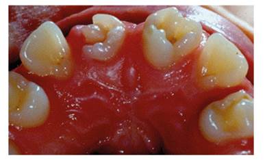
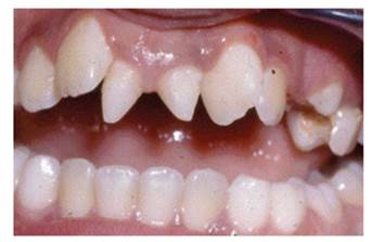
Figure 1 and Figure 2 show the front and occlusal images of an 11-year-old male patient who consulted the Universidad del Valle Clínica de Odontología Integral del Niño y del Adolescente. The patient does not have a history of relevant medical conditions or systemic conditions, nor background of dental anomalies of number in his family.
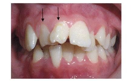
Figure 1 Front view. The arrows point to the supernumerary teeth; note that these are two supplemental dysmorphic teeth
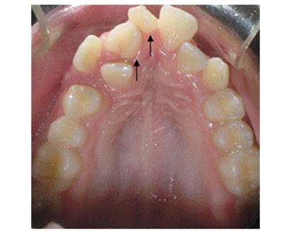
Figure 2 Occlusal view. The arrows point to the supernumerary teeth; note that these are two supplemental dysmorphic teeth
The patient had two mesiodens that have signifi cantly displaced the central incisors and lateral incisors. The mesiodens have the shape of incisors with multiple tubercles.
Case 2
Figure 3, Figure 4, Figure 5 and Figure 6 show the clinical images and x-ray image of a 10-year-old male patient who consulted the Emergency Clinic of Universidad del Valle School of Dentistry. They show two large molariform teeth in the upper anterior region in between the lateral incisors. The radiograph shows that the upper right mesiodens has moved the right central incisor to the region of the lateral right, and the left upper mesiodens is blocking the eruption of the left central incisor.
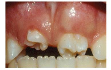
Note the presence of two molariform mesiodens blocking eruption of the upper central incisors.
Note the presence of two molariform mesiodens blocking eruption of the upper central incisors

CASE 3
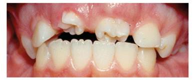
Figure 7 shows the front view of a 9-year-old male patient consulting the Clínica Integral del Niño y del Adolescente of Universidad del Valle School of Dentistry, with two conical-shaped mesiodens that have displaced the central and lateral incisors distally.
Note the presence of two conical-shaped mesiodens blocking eruption of the upper central incisors.
DISCUSSION
This article describes the clinical characteristics of three unusual cases of multiple supernumerary teeth located in the midline (mesiodens) of children 9 to 11 years old, with greater predominance of males.2)(5)(7)(15) Mesiodens are the most prevalent of supernumerary teeth in permanent dentition.22)(23)(24
The three cases described above are double mesiodens, which is a rare situation, since these cases have been recorded in 12.23% of cases according to Orhan et al.31 and Hernandez et al.21) Other studies on double mesiodens report an incidence ranging from 23.1% according to Gunduz et al, (22 13.2% to Salcido García et al, (23) and 5.1% to Ferres Padró et al. (24
It is not common to find erupted upper permanent incisors in relation to molariform, supplemental, or tuberculate mesiodens, contrary to what happens with conical-shaped mesiodens.10)(32)(34) As opposed to what was reported in the literature in case 1 reported here, the presence of two mesiodens did not block eruption of the lateral right central incisor, although it resulted in displacement of the upper right central incisor to the zone corresponding to the right upper canine, as well as marked palatalization of the upper right lateral incisor, affecting the patient′s occlusal harmony, appearance, and functionality.
In case 2, the molariform mesiodens were blocking the eruption of permanent incisors, agreeing with what has been reported in the literature,32)(33) affecting the room available for teeth in the upper anterior segment. In case 3, the conical mesiodens allowed the eruption of permanent central incisors with lateral displacement, as previously reported.2)(13
This type of dental anomaly occurs in some developmental disorders and syndromes such as cleft lip and palate, cleidocranial dysplasia, Gardner′s syndrome, condroectodermal dysplasia, Sturge- Weber syndrome, Down syndrome, Crouzon′s disease, oro-facial-digital syndrome, Hallerman- Streiffsyndrome, andFabry-Andersonsyndrome.1)(19)(36) In the three cases described here, patients do not have signs of cognitive impairment, development disorders, or any type of syndrome.
Management of the three cases involved immediate extraction of the mesiodens, since their presence was obstructing the correct location of teeth in the dental formula. Once the surgical procedures had been completed, the patients were referred to the Graduate Program in Pediatric Dentistry and Maxillary Orthopedics, where the interceptive orthodontic treatment was continued seeking to harmonize the occlusal conditions of each patient. Erupted mesiodens contribute to the displacement of teeth in the normal formula, cause alterations in the pattern of eruption of teeth moving towards the oral cavity and in some cases completely obstruct eruption,10)(11)(12) as seen in the three reported cases.
Early diagnosis of the presence of mesiodens and their extraction allows the spontaneous eruption of affected incisors, preventing associated complications.25)(34) The time it takes for a permanent tooth to erupt once a supernumerary tooth has been extracted varies from six months to three years.10)(34)(35) The factors influencing eruption are: type of supernumerary tooth, number of supernumerary teeth, displacement distance of the permanent tooth still to erupt, angle of impaction in relation to the midline, amount of root formation of permanent tooth, stage of root development of the supernumerary tooth, and space available in the dental arch for eruption of the permanent tooth yet to erupt.27)(30)(33
CONCLUSIONS
Mesiodens are the most prevalent form of supernumerary teeth in permanent dentition, being a clinical condition that must considered due to the complications of late intervention. This article reported three cases of double mesiodens: the first case involves two supplemental dysmorphic teeth, the second case involves two molariform teeth, and the third case two conical-shaped teeth. All three cases occurred in male patients, which is consistent with what has been reported in the literature.
The extraction of mesiodens in early mixed dentition helps the spontaneous alignment of adjacent teeth, avoiding other complications and further complex treatments. In the cases presented here, patients were extracted their mesiodens and were referred to interceptive orthodontic treatment.

