


 text in
  English (pdf)
 Article in xml format
 Article references
 Send this article by e-mail
 Cited by SciELO
  Cited by Google
  Similars in
SciELO
Similars in
SciELO  Similars in Google
 Permalink
INTRODUCTION
The growth of the human face is a continuous dynamic process involving different factors that are expressed in a three-dimensional manner.1 The changes observed throughout life have been widely documented by different diagnostic methods.2,3 To better understand craniofacial growth and development, it is necessary to assess the processes from the perspective of the three spatial planes: sagittal, vertical, and transverse. Each plane provides unique information on the extent and direction of the stage of growth, and ultimately helps planning and decision-making.1
Craniofacial morphogenesis begins with the transverse dimension, followed by depth or the sagittal dimension, and finally facial height.2 When the maxillaries fail to properly align in the transverse dimension, odontogenesis continues its process and teeth can erupt and align themselves in abnormal positions, causing complex situations, such as posterior unilateral or bilateral crossbites, severe malocclusions, scissor bite, open bite, dental malpositions, narrow arches, and deep palate.1,2,3,4
This alteration in the transversal growth of upper maxilla, diagnosed as a deficiency, forces posterior mandibular teeth into compensation, and mandibular molars show a strong “negative” inclination due to their compensatory lingual position, and therefore the maxillary molars take a vestibular inclined position5,6-a “positive” inclination that prevents the diagnosis of a possible deficiency in maxillary and mandibular growth.7 These compensatory positions of teeth can create occlusal alterations, seen as an accentuated curve of Wilson, interference in excursive movements, and a poor distribution of occlusal forces along the longitudinal axis of the tooth,5 that in the long run can become a risk factor and trigger periodontal alterations, such as gingival recessions and loss of vestibular bone height.8,9
In 2009, Tamburrino et al5 suggested that, in the presence of transverse discrepancies, besides the aforementioned dental and gingival characteristics, the literature has reported temporomandibular dysfunctions and occlusal alterations such as the curve of Wilson, shown as excessive inclinations of maxillary molars to compensate for insufficient maxillary width. This curve is very pronounced, so that the palatal cusps of maxillary molars are positioned on the buccal cusps of mandibular molars, suggesting that premature contacts between these cusps induce a habitual occlusion with displacements from the center.
In 2004, Podesser et al10 studied a new method to quantify a series of anatomical structures that could be related to the transverse dimension; some of the chosen structures were the nose, the maxillary bone, and the dental arches. To that end, they used a CT scan (Tomoscan 7000R, Philips, Eindhoven, The Netherlands), in order to evaluate the discrepancies and the possible conditions appearing with the orthodontic appliances in 10 patients aged 26 to 31 years. This allows establishing a line which is considered the lateral limit of the maxilla base, created from a vertical line of the alveolar process to a horizontal plane of the lower border of the zygomatic process, and the longitudinal axis of the first maxillary molars, thus establishing their average inclination, which is 101.34 ± 43.26 for the first upper right molar and 121.51 ± 18.25 for the first upper left molar.11
Many studies have attempted to assess the transverse dimension of maxillaries by different techniques with anteroposterior x-rays, which have many limitations due to their abundant distortion and their restrictions to locate anatomical points, as well as their little relation with the problem itself: maxillary discrepancy. Due to the limitations of anteroposterior x-rays and the analysis of study models, as well as the high doses of radiation in CT scans, diagnosis with cone bean computer tomography (CBCT) was introduced as it reduces the errors of two-dimensional cephalograms. This technique allows analyzing asymmetries, temporomandibular joint pathologies, the permeability of airways, and skeletal discrepancies with greater precision.12
This study seeks to relate the transversal maxillomandibular discrepancy with the buccolingual inclination of first maxillary and mandibular permanent molars in a population of patients aged 10 to 16 years, by means of CBCT. This study was formulated as a pilot test, so that future studies including a more representative sample can establish diagnostic protocols and guidelines for intervention in these patients.
METHODS
This was a descriptive observational crosssectional study in a sample of 48 bimaxillary CT scans belonging to patients of both sexes. The inclusion criteria were: bimaxillary CT scans of closed mouth in patients with no previous orthodontic or orthopedic treatment, who were seen at a specialized radiological orthodontic center in Pereira (Risaralda, Colombia). From the 48 initial CT scans, 23 were selected and 5 were excluded for not having the first four permanent molars erupted, for a total of 18 CT scans (14 women and 4 men) aged 10 to 16 years, with 13.28 years in average.
All CT scans were standardized and taken by a single operator on an I-CAT equipment (Imaging Sciences International Inc., Hatfield, USA), using a visual field of 160 mm by 100 mm high (16x10), 120 kV and 3-8 mA, with 26.9 s rotation time to get the images. The voxel size was 0.025 mm. A database was generated and stored on a hard drive of 1 terabyte, which was delivered to the radiological center. The scans were labeled with the ID number of and age of each patient, based on date of birth.
Then lines were drawn on the scans by a single operator previously calibrated with an expert and validated with the high coefficient of concordance (K > 0.8). The operator used the viewfinder OSIRIX (free 32-bit) version 5.8.2, with DICOM viewer for the Macintosh platform, with the Mac OSX operating system.
The calibration was carried out by Kappa concordance test between the pattern and the examiners. The following values of concordance were obtained: examiner 1 (k = 0,855), examiner 2 (k = 0.808), and examiner 3 (k = 0.509).) Examiner 1 was considered calibrated because he exceeded 80% concordance; this examiner recorded all the information.
The variables evaluated were: gender, age, intermaxillary distance (Mx-Mx), intermandibular distance (MGJ- MGJ), discrepancy rate (MX-MX) and (MGJ- MGJ), position of the first upper right molar (16) in relation to the palatal plane (PP-16), position of the first upper left molar in relation to the palatal plane (26) (PP-26), position of the first upper right molar (16) in relation to the occlusal plane (OP-16), position of the first upper left molar (26) in relation to the occlusal plane, position of the first lower right molar (46) in relation to the occlusal plane (OP-46), position of the first lower left molar (36) in relation to the occlusal plane (OP- 36), position of the first mandibular left molar (36) and mandibular plane (MP-36), position of the first mandibular right molar (46) and the mandibular plane (MP-46) and relationship between the maxillomandibular discrepancy and inclination Figures 1, 2, and 3.
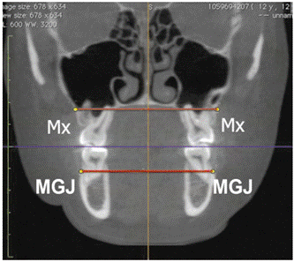
Figure 1 Maxillary transverse distance Mx (maxillary jugal point), mandibular transverse distance MGJ (mucogingival joint), and transverse discrepancy index ID (Mx to Mx) - (MGJ to MGJ)
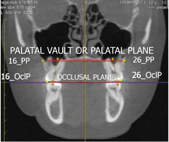
Figure 2 Bucco-lingual inclination of first maxillary molars in relation to the palatal plane and the occlusal plane.
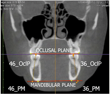
Figure 3 Bucco-lingual inclination of first mandibular molars in relation to the mandibular plane and the occlusal plane.
The information was entered in a database and processed with version 21.0 of the SPSS software (SPSS Inc., Chicago, IL). A descriptive statistical analysis was conducted by means of measures of central tendency (mean and median) and measures of dispersion (standard deviation), evaluating the distribution of quantitative variables with the Shapiro Wilk test. Student’s t test for parametric quantitative variables was used to determine relationships, and Pearson’s r test was used to establish correlations.
In terms of ethical considerations, this study complied with the standards established by the Ministry of Health of Colombia, Resolution No. 008430 of 1993 (October 4, 1993), which sets the scientific, technical and administrative standards for research in health. Authorization was requested to the Universidad Autónoma de Manizales Bioethics Committee, which by means of resolution No. 51 of 2015 approved this research project. This was considered a study with no risks, and the research team complied with the Committee’s recommendations regarding the procedures for using the CT scans by the radiographic center.
RESULTS
The sample consisted of 23 CT scans, but only 18 met the inclusion criteria. 77.8% of scans corresponded to women and 22.2% to men, aged 10 to 16 years, with 44.4% aged 10 to 13 years and 55.6% aged 14 to 16 years.
According to the obtained data, the average mandibular transverse distance was shorter (58.38 ± 2.92) than the maxillary transverse distance (61.53 ± 4.96). The Pearson’s test was used to compare the averages, yielding a correlation of 0,535 with 0.004 significance. On average, the lower molars had a larger inclination degree (102.57° and 103.77°) than the upper molars (80.01° and 81.80°) in relation to the occlusal planes. Regarding the palatal plane, the inclination of the upper first molars was larger than the lower ones Table 1. As for the discrepancy index, the average was 4.96 mm (±3.45).
Table 1 Variables descriptors: transverse maxillary distance measured in mm, inclination of the first molar measured in degrees, and result of the discrepancy index
| Variables | N | Media | Standard Dev. |
|---|---|---|---|
| Maxillary transverse distance (Mx-Mx) | 18 | 61.5294 | 4.95616 |
| Mandibular transverse distance (MGJ-MGJ) | 18 | 58.3828 | 2.92417 |
| Maxillomandibular discrepancy index | 18 | 4.96 | 3.45348 |
| Inclination of 1st right maxillary molar 16 with OP | 18 | 80.0089 | 7.11447 |
| Inclination of 1st left maxillary molar 26 with OP | 18 | 81.8083 | 6.20828 |
| Inclination of 1st left mandibular molar 36 with OP | 18 | 102.5722 | 5.33013 |
| Inclination of 1st right mandibular molar 46 with OP | 18 | 103.7744 | 6.01103 |
| Inclination of 1st right maxillary molar 16 with PP | 18 | 125.4833 | 5.00663 |
| Inclination of 1st left maxillary molar 26 with PP | 18 | 125.4928 | 3.52915 |
| Inclination of 1st left mandibular molar 36 with MP | 18 | 81.3972 | 7.13309 |
| Inclination of 1st right mandibular molar 46 with MP | 18 | 78.3983 | 6.17806 |
MGJ (Mucogingival Junction), OP (Occlusal Plane), PP (Palatal Plane or Palatal Vault) MP (Mandibular Plane), Mx (Jugal Point).
As for the maxillaries transverse distance, this was shorter among patients aged 10 to 12 years than among patients aged 14 to 16 years. The mandibular transverse distance showed a similar pattern. However, it was evident that the mandibular transverse distance was shorter than the maxillary transverse distance in all ages Table 2.
Table 2 Correlation between maxillary discrepancy and the inclination of molars segmented according to discrepancy type: positive (>5) and negative (< 5)
| Correlated variable | Positive discrepancy (n = 9) | Negative discrepancy (n = 9) | |||
|---|---|---|---|---|---|
| Pearson’s r correlation | P-value | Pearson’s r correlation | P-value | ||
| Inclination of 1st right maxillary molar 16 with OP | 0.679 | 0.044* | -0.589 | 0.095 | |
| Inclination of 1st left maxillary molar 26 with OP | 0.120 | 0.757 | -0.645 | 0.084 | |
| Inclination of 1st left mandibular molar 36 with OP | -0.462 | 0.297 | 0.351 | 0.355 | |
| Inclination of 1st right mandibular molar 46 with OP | -0.001 | 0.999 | -0.292 | 0.446 | |
| Inclination of 1st right maxillary molar 16 with PP | -0.813 | 0.0 * | 0.310 | 0.456 | |
| Inclination of 1st left maxillary molar 26 with PP | 0.335 | 0.378 | -0.105 | 0.788 | |
| Inclination of 1st left mandibular molar 36 with MP | 0.510 | 0.197 | -0.620 | 0.101 | |
| Inclination of 1st right mandibular molar 46 with MP | 0.614 | 0.079 | 0.797 | 0.018 * | |
Mx (Jugal Point), MGJ (Mucogingival Junction), OP (Occlusal Plane), PP (Palatal Plane or Palatal Vault)
The inclination of upper molars in relation to the occlusal plane shows that the greatest inclination occurred at the age of 14 and the smallest at the age of 11 years. The inclination of the lower molars with respect to the occlusal plane shows that the highest values are in the group of 10-year-olds and the lowest values in the 11-year-olds. As for inclination of the first maxillary and mandibular molars in relation to the palatal plane, the upper molars have smaller inclination degrees than the mandibular molars, but these degrees are very different among them, with the lowest degree at the age of 15 for the right upper molars and the age of 11 for the left ones, while the highest degree was for the age of 10 in the right side and the age of 15 in the left side.
The mandibular molars showed greater inclination. The lowest inclination was for patients aged 14 years for the right lower molars and for patients aged 11 for the left ones, while the highest values correspond to patients aged 16 years for the lower right molars and for patients aged 10 years for the lower left molars.
As to the inclination of maxillary molars in relation to the palatal plane and the mandibular plane, it remains with very similar changes between the ages of 10 and 16. In terms of discrepancy index of maxillary and mandibular width, a big difference can be seen among the ages, with the lowest rate for patients aged 14 to 16 years and the highest rate for patients aged 10 to 12.
The Shapiro Wilk tests showed normality among the distribution of variables. The variables met homoscedasticity conditions. Therefore, for the analysis of correlation between the transverse discrepancy index and the inclination of maxillary and mandibular first molars, Pearson’s correlation coefficient was applied. There was no statistically significant correlation, although the correlation between the discrepancy index and the inclination of the first lower left molar (36) and the first lower right molar (46) can be considered high (r = 0.95 and r = 0.76) Table 2.
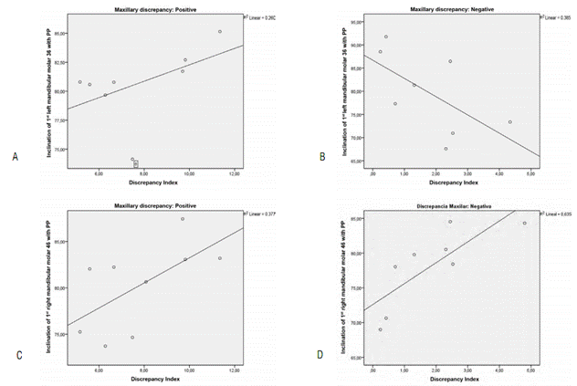
Dispersion charts between the transverse discrepancy index and the inclination of molars were drawn to identify clusters and the linear regression of the correlation in each variable Figure 4.
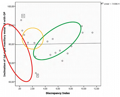
On the other hand, the correlations graph showed three clusters of points with different tendencies: the first point cloud is in the discrepancies between 0 and 5, the second includes the ones close to 5, and the third cloud is in discrepancies over 5 (ovals in Figure 4. Based on these results, an analysis was performed segmenting the groups into negative discrepancy (< 5°) and positive discrepancy (> 5°).
The analysis grouped by discrepancy type showed moderate and strong correlations between patients with positive maxillary discrepancy (> 5º) and the inclination of molars, except for 26 and 46 with OP. It was significant in both 16 with OP and 16 with PP. Regarding patients with maxillary negative discrepancy (< 5º), there was also moderate and strong correlation between discrepancy and molar inclination, except for 26 with PP.
On the other hand, the molar with the highest correlations was 16 in relation to OP and PP; these correlations were statistically significant (p = 0.044 and p = 0.01) and are shown in Figure 5. As to the planes of reference, the highest correlations were achieved with the palatal plane (PP), in contrast to the occlusal plane (OP), as shown in Figure 6.
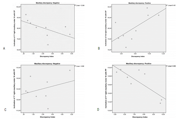
Figure 5 Dispersion charts between discrepancy and the inclination of 16 according to positive or negative discrepancy
On the other hand, in relation to the occlusal plane, the inclination of maxillary molars was in general vestibular, unlike the mandibular ones, where it was primarily vestibular, with some cases of lingual or neutral inclinations. In relation to the palatal plane, the inclination of maxillary molars was predominantly positive, and neutral in a lower proportion. The mandibular plane showed more vestibular inclinations, followed by neutral inclinations, and lingual inclinations in a lower proportion.
DISCUSSION
The diagnosis in orthodontics is mainly based on the morphological and quantitative description of craniofacial structures in the three planes of space, but greater attention has been given to the assessment of malocclusions in the sagittal and vertical dimensions, ignoring that the transverse plane is equally important to the final position of maxillary teeth and their coordinated function. 10 This study aimed at evaluating the transversal maxillomandibular discrepancy and correlating it with the bucco-lingual inclination of first maxillary and mandibular molars through bimaxillary cone beam computed tomography.
The diagnostic aid most commonly used in dental clinical research are lateral radiographs. In the case of the transverse dimension, the use of posteroanterior cephalometry has some limitations, such as the difficulty in standardizing the head position and the location of anatomical structures which overlap with each other. Currently, threedimensional imaging provides better information than conventional two-dimensional imaging, and studies using cone beam technology are becoming more popular since their costs have been reduced, equipment are more commonly available, and the control of radiographic exposure has been improved.13 On the other hand, tomography provides better reliability to assess craniometrics dimensions, including cross-cutting measurements, such as intermaxillary discrepancy.14 The present study evaluated 23 bimaxillary CTs, of which 18 met the selection criteria, measuring the transverse distances of maxilla and mandible and the inclinations of first molars in them.
The transverse maxillary distance had an average value of 61.53 ± 4.96 mm, being longer than the mandibular distance (58.38 ± 2.92 mm) in all cases. These results were expected from the point of view of growth and craniofacial development, as the dimensions of the maxilla, due to its position, are higher than the dimensions of the mandible. Once occlusion and full functionality begin, the mandible shows an occlusal adaptation to the position of the upper molars, regardless of the dimensions of the maxilla.
The average value of maxillary transverse distance obtained in this study (61.53 ± 4.96 mm) is slightly lower than the one reported by Ricketts,11 who states that the transverse distance of the maxilla is 62 to 66 mm in patients aged 9 to 16 years, using the same anatomical reference points. This slight difference can be explained because the studies were conducted in different populations, with high morphological variability. Hesby et al,3 on the other hand, analyzed maxillary width, finding out values ranging from 56.24 to 61.57 mm in 7-year-olds, with similar results to the present study.
Similarly, the mandibular transverse distance in this study was 58.38 ± 2.92 mm on average, measured from the most prominent point of the mandibular bone contour on each side over the area of the first permanent lower molar, which coincides with the most prominent part of the buccal alveolar bone seen from the occlusal surface, as well as with the mucogingival junction. In doing estimations, De Oliveira et al reported average values of 65.97 ± 3.42 mm in mandibular transverse distance.15
On the other hand, Andrews and Andrews 16) suggested that, to achieve an optimum position and inclination of molars, maxillary width should be 5 mm greater than mandibular width. This concept is validated by the results of the present study, showing an average of almost 5 mm. Also, this study established that, if the discrepancy rate was greater than 5 mm, surgically assisted expansion was needed, but if such discrepancy was equal or lower than 5 mm, orthodontic and orthopedic expansion was required.
Transverse discrepancy, described as the maxillary being 5 mm larger than the size of the mandible, helps preserve the root in the alveolar bone and to achieve vertical and bucco-lingual position inside its alveolus. When this discrepancy is within the limits of movement, it is essential to achieve a good straightening and intercuspidation of posterior teeth, in the presence of a lack of skeletal harmony. The risk of doing this is the possible effects to the periodontium. In case of trying to place the tooth in a more upright position and with good intercuspidation, in the presence of a discrepancy, theamount of soft tissueand bone overlying the roots becomes thinner, since teeth will not be centered in the alveolus. In severe transverse discrepancies, attempting to normalize the inclination of molars increases the risks of root fenestration and the loss in insertion becomes clinically evident.9
With respect to molars inclination, this study established angles based on cone beam tomography images, similar to those obtained by Tong et al (2012), who validated a method for determining buccolingual inclination based on tomographic images.17 The present study evaluated the inclination of molars taking the occlusal plane (OP) as a reference point, as reported in other studies that used tomography to evaluate the inclination of molars, taking the OP as a reference as well.17
Our results show that the average inclination of the first upper right molar was 80.00 ± 7.11°, in the upper left the inclination was 81.80 ± 6.20°, the lower left was 102.57 ± 5.33°, and the lower right was 103.77 ± 6.01°. This agrees with the findings by Gross et al, who identified the inclination in first molars as 88.49 ± 5.39°; 84.78 ± 5.99°; 104.247 ± 5.43°, and 103.63 ± 4.35°, respectively.18 A similarity may be established, especially in terms of mandibular molars, and some difference in the maxillary molars, which can be explained by variability in growth patterns.19
Finally, the correlation analyzed in this study showed that no significant relationships were found in evaluating overall discrepancies, but in grouping them according to whether the discrepancy (it is, the difference between transverse mandibular and maxillary length) is greater than 5 mm (positive) or shorter than 5 mm (negative), the study did find correlations with the inclination of both upper and lower molars, suggesting different behaviors in both groups. In the negative correlations, where the discrepancy is closer to 5 mm, the inclination of upper molars with respect to the OP tends to decrease, but when the discrepancy is positive, the tendency is that the more it increases and deviates from 5 mm, the bigger the angle tends to be. This situation is clinically relevant, since it guides clinicians in establishing final results to the treatment of molar inclination according to maxillary discrepancy.
This can also suggest, from a biomechanical perspective, that the inclination of both upper and lower molars seeks to establish the best possible inclination in discrepancies closer to 5 mm (where they also tend to be more stable), but the more they move away, either increasing or decreasing, the mechanical need is different in the two conditions and requires a compensatory response that is reflected in the change in molar inclination, which also creates other effects on the occlusion, such as cusp interference.20
Even though no other studies have correlated the variables evaluated in the present study, other researchers have demonstrated the clinical relevance of evaluating the transversal dimensions through different diagnostic aids. The study by Ricketts11 reported a moderate relationship between arch width and the inclination of canine and lower molars. Rongo et al21 found no association between transversal dimensions and vertical facial features. In 2016, Zhang et al identified an important relationship between transversal dimensions and the maxillary arch, the mouth, and the face.22 These data, together with the correlation reported in the present study between maxillary discrepancy and molar inclination, suggest that transversal lengths play an important role in craniofacial morphology, and that clinicians should analyze this role more carefully, not only for diagnosis and planning purposes, but also for follow ups.
Among the limitations of the present study is that, even though correlations and clusters could be established in terms of positive or negative discrepancy, the samples were not enough to create a cluster to analyze those patients whose discrepancy is close to 5 mm, so graphically they showed they might have different behaviors, and therefore the correlation with inclination could also be different.
Another limitation has to do with the fact that the sample was restricted since the only radiological center with cone beam CT scans available had only 18 bimaxillary CT scans of patients with first permanent molars completely erupted, with closed mouth and no apparatuses-inclusion criteria required for this study.
CONCLUSION
Transverse maxillomandibular discrepancy is related to the buccolingual inclination of first permanent maxillary and mandibular molars, in such a way that, the more maxillomandibular discrepancy, the more permanent upper molars lean towards vestibular and the less toward lingual in order to maintain occlusion.
The process of maxillary response requires further studies to understand the timing and extent of the adjustment.
RECOMMENDATIONS
The researchers recommend increasing the sample size to get stronger data and evaluate molar position based on inclination of the crown and the occlusal plane, and establish correlation with final tooth position along the longitudinal axis and its periodontal response.
It is also recommended to validate the transversal discrepancy of the maxillary in relation to facial type, by assessing and analyzing through cone beam tomography.

