


 text in
  English (pdf)
 Article in xml format
 Article references
 Send this article by e-mail
 Cited by SciELO
  Cited by Google
  Similars in
SciELO
Similars in
SciELO  Similars in Google
 Permalink
INTRODUCTION
The lipoma is a benign neoplasm of slow growth, composed of mature fatty cells, usually surrounded by a thin fibrous capsule.(1) Lipomas have been reported in all parts of the body, includingthe regions of the back, shoulders, neck, and extremities. They are the most common soft tissue neoplasm, but do not usually appear in the mouth, corresponding to 0.1 to 5% of benign tumors of the mouth.(2) About 15 to 20% of cases affect the region of the head and neck, while 1 to 4% affect the oral cavity.(3) Clinically, they show as nodular, asymptomatic, long-lasting bumps of soft consistency and covered by normal mucosa. They occur primarily in the areas of fat accumulation, especially in the cheek, followed by the tongue, the floor of the mouth, the buccal sulcus and buccal area, lip, palate, and less frequently in the gingiva. This pattern corresponds to the amount of fatty deposits in the oral cavity.(3,5,6) In general, lipomas occur in the fourth and fifth decades of life, without preference for one specific sex. Some studies, however, have shown predominance in males.(1,7,8,9)
The etiology of intraoral lipomas is not clear, but one possible pathogenetic mechanism is the “theory of hypertrophy”, which establishes that obesity and the unnoticed growth of adipose tissue can contribute to the formation of these oral lesions. This theory is less convincing to explain the lesions occurring in areas with no pre-existing adipose tissue. Another theory, known as “theory of metaplasia”, suggests that lipomas develop due to aberrant differentiation of mesenchymal cells in lipoblasts, since fatty tissue can originate from connective tissue cells that can mutate in almost any part of the body.(3) The clinical diagnosis of these tumors is not always easy, unless the yellow color of the tumor appears through the thin overlying mucosa. The size of the tumor depends on its location, but it rarely exceeds 25 mm in diameter; in general, it can vary from 0.2 to 1.5 cm in diameter, and tumors larger than 50 mm have been reported on the cheek. Large sublingual lipomas have also been found. They are usually asymptomatic, but in rare cases they can cause functional problems, such as dysphagia and difficulty in speech and mastication. It has been found that 5% of cases of lipomas are multiple and have been associated with certain syndromes, such as neurofibromatosis, Gardner’s syndrome, multiple painful subcutaneous lipomas, the obesity syndrome called Decrum disease, encephalocraniocutaneous lipomatosis, multiple familial lipomatosis, Proteus Syndrome, and Pai syndrome.(1,3)
The diagnosis of intraoral lipomas is usually clinical. Techniques such as xeroradiography and ultrasound are often used to define the anatomical extent of intraoral lesions; however, their accuracy tends to be limited. Computed tomography and magnetic resonance imaging allow the diagnosis of these tumors quite easily. Despite the availability of all these techniques, histopathology remains the gold standard in the diagnosis of lipoma.(3) Histologically, the tumor is composed of fatty cells which are subdivided into lobes by septa of fibrous connective tissue. According to microscopic characteristics, lipomas are classified as classic lipoma, fibrolipoma, angiolipoma, spindle cell lipoma, pleomorphic lipoma, myxoid lipoma, sialolipoma, and intramuscular lipoma.(10) Out of these variants, myxoid lipomas, intramuscular lipomas, and angiolipomas are rarely found in the oral cavity. There is diversity in histological pattern, including dense fibrous connective tissue, components of spindle-shaped cells, atypical mitotically active cells, presence of mature blood vessels, myxoid stroma, or even acinar salivary structures, which appear together with mature adipose tissue, depending on each variant.(3)
The histopathology of spindle cell lipoma is characterized by a mixture of mature adipose tissue, spindle cells, and fibrous collagen in the focal myxoid stroma.(11) Particularly, spindle cell lipoma (SCL) is a benign lipomatous neoplasia in the back of the neck and the back in older men, representing nearly 1.5% of cases. In adult men, the most common location of the classical oral and maxillofacial lipoma is the parotid region, followed by the oral mucosa. However, oral SCLs are rare, and only about 40 cases have been reported.(11) The differential diagnosis for intraoral lipoma includes oral dermoid cyst and epidermoid cyst, oral lympho-epithelial cyst, benign tumor of salivary gland, mucocele, benign mesenchymal neoplasia, ranula, ectopic thyroid tissue, and lymphoma.(3) Other differential diagnoses reported for SCL include myolipoma, schwannoma, myxoid neurofibroma, leiomyoma, myxoid solitary fibrous tumor, and atypical lipomatous tumor.(11) Lesions appear as a bump in the back of the tongue, and typically are similar to hemangioma, lymphangioma, rhabdomyoma, neuroma, or neurofibroma.(3)
SCLs have been very rarely reported in the oral cavity, and in those cases they usually show as a well circumscribed mass affecting different anatomical locations within the oral cavity, including the buccal mucosa, the tongue, the floor of the mouth, the hard palate, and the gingiva. They are characterized by loss of chromosomes 13q or 16q, or both.(12) The expression of androgen receptors has also been reported, suggesting a possible pathogenic role of steroid sex hormones; however, in cases of oral SCL, this correlation has not been sufficiently studied.(11) As for intramuscular or infiltrating lipomas, they are an unusual clinical variant of this neoplasm of adipose tissue, originating between the skeletal muscle bundles and infiltrating through intramuscular septum. They have a slight predilection for the tongue, because of the tight connection between the adipose tissue and the muscle layer. The infiltration of lipomas occurs consistently diffuse, with dissociation and entrapment of muscle fibers, some of which show degenerative changes. Muscle tissue is replaced by fat, which may extend beyond the muscle fascia in intermuscular connective tissue spaces. The fasciae, capsules joints, bones, and nerves can also be infiltrated. Infiltrating lipomas could suggest a false diagnosis of liposarcoma; however, the absence of cellular pleomorphism and nuclear hyperchromatism, and the low mitotic activity support the diagnosis of intramuscular lipoma.(3)
The main treatment for intraoral lipoma is complete surgical removal. Recurrence has been described after local extraction, but the infiltrative lipoma tends to recur after inadequate excision, as it is not encapsulated as a single lipoma. Even in recurrent cases, there has been no incidence of malignant transformation.(3) In spindle cell lipomas, lesions are rarely over 5 cm in diameter, and with adequate local excision, recurrence is very low.(11) The medical treatment of lipomas measuring less than 2.54 cm in diameter includes injections of steroids that cause local fat atrophy, resulting in a reduction in size. A local monthly injection of 1:1 mixture of lidocaine and triamcinolone acetonide in the core of the tumor can be useful in lesion regression.(3) What follows is the clinical case of a patient with a gingival lipoma in the buccal area of tooth 46.
METHODS
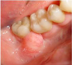
A 76-year-old female patient in good medical conditions, with no relevant medical or dental history, sought dental consultation in December 2011, showing a sessile exophytic lesion. It was a nodular pale pink lesion with yellow painless areas, that could move in mesiodistal direction and was located at the level of the interdental papilla and the buccal area between molars 46 and 47, measuring 1.3 cm in width and 1.0 cm in height. It was interfering with mastication and phonation (Figure 1).
With the informed consent by the patient, an excisional biopsy of the lesion was performed, prior application of infiltrating anesthesia of 1.8 ml of 2% lidocaine with epinephrine 1:80000 in the area. The lesion was gripped with Adson pliers, cutting with a #15 scalpel blade around the base of the lesion including healthy gingival tissue; the sample was sent to the Pathology Service for histopathological diagnosis. The Pathology microscopic description shows a non-keratinized stratified epithelium over a dermis, with a band of dense, mature connective tissue wrapped around a marked fat-cell hyperplasia. The changes are free of malignancy. The histopathological diagnosis is lipoma (Figure 2).
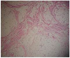
Figure 2 Histological evaluation showing “a lesion compromising gingiva and underlying mucosa with a band of mature dense connective tissue wrapped around a marked adipose cells hyperplasia, with one of its edges showing incomplete recession. The changes appear to be free of malignancy” (histopathology evaluation performed by Dr. Dolly Aristizábal).
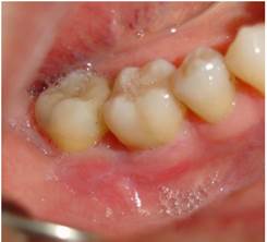
The clinical control was performed one week and one month post-surgery showing no evidence of recurrence (Figure 3).
DISCUSSION
This article reports a case in which a gingival lesion was removed by means of biopsy; the histopathological result shows a gingival lipoma on papilla and buccal area between molars 46 and 47, measuring 1.3 cm wide and 1.0 cm high. The diagnosis was therefore clinical and based on a consistent histopathological evaluation, as shown above.(13,14,15) Lipomas are the most common mesenchymal tumors, especially in the trunk and proximal portions of the extremities; they are also frequent in the region of the head and neck, being benign neoplasms of soft parts composed of mature adipose tissue, but they are rare tumors in the oral cavity.(1,3,8,16,17,18) The statistics show that this site is only affected in 1-4% of cases. In 2004, Furlong et al found only 125 cases of oral lipomas during a period of 20 years, which once again demonstrates the rarity of these oral tumors.(3,19)
The first description of an oral lesion was provided by Roux in 1848, in a review of alveolar masses he referred to as “yellow epulis”.(3,20) Lipomas are benign, congenital lesions resulting from multipotent cells of embryos that remain sub-clinically latent until they differentiate into fatty cells under hormonal influence during adolescence.(3,21) However, in some cases, trauma and chronic irritation can trigger the proliferation of soft tissue and play a role in the development of a lipoma.(3) Other lesions, such as cell granular tumor, neurofibroma, traumatic fibroma, and lesions of the salivary glands (mucoceles) can be included in the differential diagnosis.(1,7,8,9) In some cases, they may show as fluctuating nodules.(1,22) There are other lesions that should be considered in the differential diagnosis, including lymphoepithelial, dermoid and epidermoid cysts.(1,23) These may occur as either individual or multiple lesions.(1,16,17) Microscopically, the differential diagnoses are angiolipoma, liposarcoma and normal soft fatty tissue.(1,24) Palate lipomas are rare and most are developmental lesions-but true neoplasms of fatty cells are rare in this location-.(1)
Infiltrating lipomas are difficult to remove and show recurrence.(1,9,17)
A review of 26 cases analyzed in Brazilian population showed that the average age of onset is 54.6 years.(25) Fornage and Tassin reported that the peak incidence occurs in the fifth or sixth decade of life;(26) furthermore, this benign tumor occurs predominantly in women: the literature shows a similar distribution according to sex, with a 1:1,2 male-female ratio.(3,27) These findings are consistent with the reported case because it’s about a 76-year-old woman with a diagnosis of gingival lipoma. Rare cases of congenital lipomas have been reported in infants of 20 and 47 days of age. A study by Naruse et al, in which the authors analyze the clinical pathologic characteristics and proliferative activities of 24 cases of oral lipomas in a Japanese population, compared with the results of various studies reporting approximately 603 cases, revealed that the average age of onset of this lesion was 59 years, and out of the 24 cases in the study, 22 corresponded to gingival lipomas, while 2 cases corresponded to fibrolipomas,(28) which histologically differ from the classic variant in which mature adipose tissue is intercalated with bands of connective tissue. This benign tumor of soft tissue rarely occurs in the oral and maxillofacial region; however, if it happens, is most frequently present in the buccal mucosa and the buccal area, also showing a slight predominance in women.(28,29) In the study by Naruse et al, 23 cases were treated surgically, while one case was submitted to biopsy and follow-up. None of the cases showed recurrence.(28)

