


 text in
  English (pdf)
 Article in xml format
 Article references
 Send this article by e-mail
 Cited by SciELO
  Cited by Google
  Similars in
SciELO
Similars in
SciELO  Similars in Google
 Permalink
INTRODUCTION
In cosmetic dentistry, restorations in the anterior area are considered a very complex procedure, in which the clinician must choose the ideal treatment and select appropriate materials to achieve satisfactory and predictable cosmetic results. The treatment can be done by direct techniques, using composite resins to restore lost dental tissue in a fast, inexpensive, conservative way.(1) However, manufacturing these resins is complex, their color stability is not very predictable,(2) and the material’s abrasion is higher in comparison with ceramics and tooth enamel.(3) Indirect restoration procedures include the manufacturing of full ceramic or metal-ceramic crowns. In indirect procedures, metal-ceramic crowns have long been considered the gold standard in restorative dentistry, thanks to their high resistance to breakage, color stability, and a survival rate of 95.6% in 5 years,(4) providing also good aesthetic results.(5,6) However, in the anterior area, metal structures affect esthetics; light transmission decreases when it interacts with a metal-ceramic restoration compared to natural teeth, and the gingiva surrounding the restoration can become darker.(7) To avoid these situations, clinical studies have shown that full ceramic restoration systems have better optical properties and a better periodontal response than metal-ceramic restorations.(8,9)
As a result, the materials used in restorations with high esthetic demands, as well as the sophisticated adhesive techniques, have improved in recent decades to meet the needs of patients and clinicians who seek to preserve the largest amount of tooth structure and to obtain predictable results.(10) To perform adhesive restorations, it is necessary to use acid-sensitive ceramic systems, such as lithium disilicate-reinforced glass ceramic (IPS e.max) and leucite-reinforced glass ceramic (IPS Empress), which are indicated for veneers, inlays, onlays, and individual crowns. Clinical trials have shown high survival rates (onlays: 100% after 3 years(11) and crowns: 96.6% after 5 years)(12). Feldspathic ceramics with high concentrations of glass and silicate are generally used for coating ceramic and metal structures in order to achieve an adequate shape and to reach satisfactory aesthetic properties in the final restoration.(13)
On the other hand, there are the highly-resistant ceramic systems, such as yttria partially stabilized zirconia (Y-ZTP), which have little or no vitreous phase due to their polycrystalline nature, and therefore are not sensitive to acid etching. In addition, they have very low porosity (achieved during the manufacturing process in laboratory, based on CAD- CAM techniques), high opacity and high modulus of elasticity, over 900 MPa.(14) All these features make these materials suitable for manufacturing substructures for fixed dentures in the posterior area, and their high opacity allows masking pigmented or metal dental stumps. In addition, the high survival rate of 95.4% in 5 years makes them very reliable.(15)
Clearly, there is no one ceramic system that can be used in all clinical situations; therefore, it is necessary to understand some basic concepts of these systems in order to make an appropriate selection according to each clinical situation. While making an all-ceramic restoration, it is important to take into account the following criteria: location, type of restoration, dental remnant, desired final color, amount of dental remnant, design of the marginal end line, and cementing technique.(16) Therefore, when it is necessary to perform a restoration seeking excellent aesthetic results in an anterior tooth, it is recommended to use ceramics with a high content of glass, including feldspathic and monolithic ceramics such as lithium disilicate; if the restoration requires using fracture-resistant ceramics or masking pigmented or metal stumps, the clinician can choose opaque ceramics such as zirconia to manufacture the structure and cover it with a highly esthetic material such as feldspathic ceramic, due to its high content of crystals.(17)
Which ceramic system to choose?
It is important to take into account the following parameters when selecting a full ceramic system: the strength and longevity of the restoration, the ability to resemble adjacent teeth, the versatility of the material, and its clinical management.(13)
The most popular ceramic systems are those that have a zirconia structure, specifically yttria partially stabilized zirconia (Y-ZTP), with mechanical properties over 900 MPa, allowing its use in the anterior and posterior areas and in fixed prosthesis.(18) However, some studies suggest that the bi-layered (zirconia/veneer) ceramic systems must be used with some caution, since they have failures such as chipping and delamination of the veneering ceramic once they start to operate.(19) For monolithic single-tooth or full ceramic restorations there is lithium disilicate, with mechanical properties of 400 to 440 MPa and high survival rates for anterior and posterior single crowns and for inlay and onlay restorations.(20,21,22,23)
Ability to resemble adjacent teeth
The dental ceramist must have the ability to imitate the dimensions, textures, and contours of the teeth to be replaced. In addition, the material to be used must have a behavior similar to natural teeth when interacting with light, allowing translucence, opalescence, and metamerism.(24) The behavior of zirconium oxide and lithium disilicate varies in terms of translucency levels, since zirconium oxide is a ceramic with high content of crystals and therefore is less translucent than lithium disilicate.(25) Higher translucency levels allow for more light in the restoration, and if used with a light resinous cement, a more esthetic result can be achieved.(26) However, high translucency is not always desirable. There are situations that require ceramic materials with low translucency. Dark, pigmented or restored teeth with metal retainers need a ceramic that hides or masks these substrates.(27)
Clinical management and versatility
Restorations using zirconium oxide structures can be cemented with conventional cements or resin systems. While conventional systems are inexpensive and easy to handle during the cementation procedure, lithium disilicate restorations require the use of resin-based cements.(28,29) The ceramic-tooth adhesion increases the strength of both restoration and abutment.(30) However, resin-based cements are expensive and sensitive to the technique, their use involves many steps for the cementing process, and removal of excess material is difficult.(31) As for versatility of the material, yttria partially stabilized zirconia allows to manufacture restorations with full contour,(32) as well as crown structures and fixed prostheses. It is preferable in the latter because it reduces the risk of wear of antagonist teeth by direct contact with the material, as well as its thermal degradation when in contact with the oral environment.(33) Lithium disilicate is manufactured by means of injection and CAD-CAM, allowing the production of full crowns, structures for crowns, or restorations with partial coating. Its versatility makes this system very popular among clinicians and dental technicians.(13)
CASE REPORT
The following case describes the clinical application of three dental ceramics (lithium disilicate, zirconia, and feldspathic ceramic) in the anterior area according to clinical findings and the patient’s esthetic requirements.
A 53-year-old female patient sought consultation expressing dissatisfaction with the look of her upper anterior teeth. The clinical examination showed pigmented and maladjusted class III and class IV Black composite resin restorations on teeth 13, 11, 21 and 22, and abfractions on teeth 12, 22, 23 and 24, with a dental upper midline displaced 2 mm to the right with respect to facial midline, and discrepancies in teeth length. The periodontal evaluation showed no pockets or signs of active disease. Photographs were taken during this first appointment (Figures 1a, b, c), as well as intraoral x-rays, preliminary alginate impressions, bicondylar-maxillary registration with facial arch for semi-adjustable articulator and intermaxillary registration with Occlufast® (Zhermack).
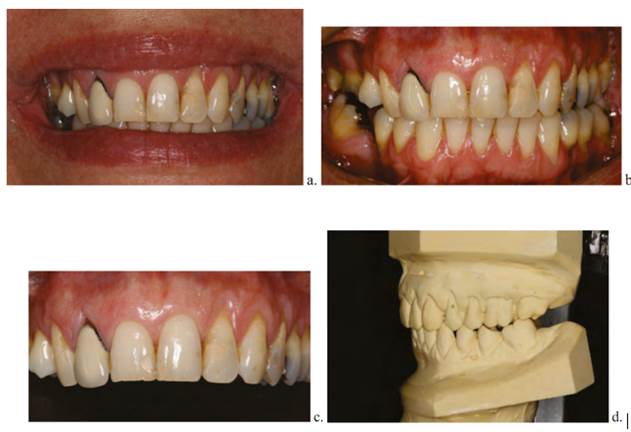
Figure 1 Initial records of patient (a, b, c). Intraoral photographs of anterior aspect and smile with multiple restorations in different materials. (d) Model mounted on semi-adjustable articulator.
The radiographic analysis showed a radiopaque zone in the cervical, middle and apical third of tooth 12’s root, compatible with an obturation material associated with a previous root canal treatment with proper length and condensation and an intraradicular metal cast retainer. The occlusal examination showed that the molar ratio was not correct, showing a canine relationship class III right and class II left. The desocclusions were carried out at the expense of canines and incisors without interfering with the posterior ones, as well as absence of signs or symptoms of a cranio-mandibular disorder.
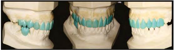
Smile analysis was performed with intraoral photographs. The models were mounted on a semi-adjustable articulator (Figure 1d), applying diagnostic wax up for proper planning of the prosthetic treatment (Figure 2). After diagnosing and planning, treatment alternatives were discussed with the patient, who emphasized on her desire of having very esthetic metal-free restorations. The proposed treatment involved removing the composite resin restoration and metal-ceramic crown of tooth 12, performing dental preparations, inserting provisional restorations, taking final impressions, manufacturing full ceramic restorations for teeth 13, 12, 11, 21, 22, 23, performing final cementation, adjusting occlusion, and performing periodical follow-ups.
Description of the procedure
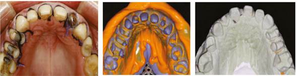
Using waxed diagnostic, silicone matrices were made, conducting dental preparations trying to preserve as much structure as possible (Figure 3). The provisional restorations were made using a matrix with silicone and bis-acrylic resin (Luxatemp® DMG), and were adapted, polished, shined, and temporarily cemented with translucent triclosan dual-cure resin (TempBond Clear®; Kerr) (Figure 4). The patient kept the provisional restorations for four weeks, evaluating comfort, phonation, and aesthetics during this period. For the final impression, the double-cord technique was used in the gingival sulcus (Ultrapack®; Ultradent); the cords were soaked with astringent solution (25% aluminum sulfate Viscostat® - Ultradent) (Figure 5). The impression material used was silicone adhesive (Elite HD + Putty Soft Normal Set® - Zhermack) with the single-step double-mix technique.
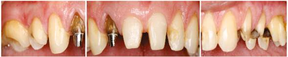
Figure 3 Dental preparations, in which teeth 13, 11, 21, 22 and 23 are prepared for a veneer ceramic in lithium disilicate, and tooth 12 has a metal core and is prepared for a zirconia crown.
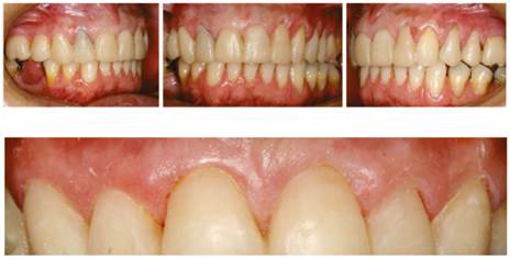
Figure 4 Placement of provisional material, showing adequate adaptation of the provisional material with respect to the gingiva, proper management of emergence profiles, and removal of excess temporary cement to keep the stability and health of periodontal tissues.
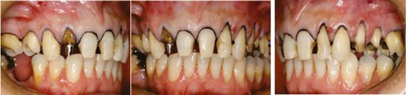
Figure 5 Placement of spacing cords. The double-cord technique was used to achieve adequate retraction of gingival tissues at the time of final impression.
To take the final impression (Figure 6), the first cord was removed, placing light impression material in the gingival sulcus of each tooth with a dispenser provided with a mixer tip and an intraoral tip. A metal tray previously polished with adhesive for tray (VPS Tray Adhesive® 3M ESPE) was loaded with heavy impression material and placed in the patient’s mouth for 5 minutes; it was then removed. Finally, the impressions of facial and intermaxillary arch were taken and mounted to the semi-adjustable articulator in the dental laboratory.
Manufacturing of final restorations in laboratory
To perform full ceramic restorations, individual models and dies of the teeth were prepared in type IV plaster (WhipMix) and mounted on the semi-adjustable articulator (WhipMix 2240). All restorations to be manufactured were fully waxed, verifying anterior fit, posterior fit, and functional movements with no interferences (Figure 7). The restorations of teeth 13, 11, 21, 22 and 23 were manufactured using an injection technique with the Programat EP 5000® system (Ivoclar-Vivadent). A zirconia structure was prepared for tooth 12 using the Ceramill® system (Amann Girrbach), seeking to mask the pigmented stump (Figure 8 and tables 1 and 2). Due to its high concentration of glass, a fluorapatite-based veneering ceramic (IPS e.max Veneer) was used for restorations of teeth 12, 11, 21 and 22, in order to achieve more esthetic results. The veneers for teeth 11, 21 and 22 were made using a cut back technique on the lithium disilicate structure to achieve the necessary space for an esthetic veneering ceramic (1.5 mm), which was heated in a Programat P-300® furnace (Ivoclar - Vivadent). Zirliner® (Ivoclar-Vivadent) was applied to the zirconia structure of tooth 12, which was sintered at the temperature recommended by the manufacturer; the powder/liquid layering technique was used for the veneering ceramic (Figure 9), which was inserted in the ceramic Programat P-300® furnace (Ivoclar-Vivadent). During the last heating cycle (“glaze”), a slow cooling protocol was used to reduce residual stress and to avoid chipping of the veneering ceramic.(34,35)
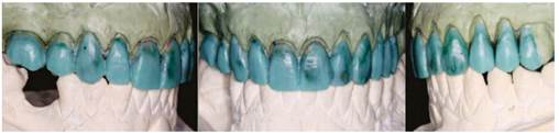
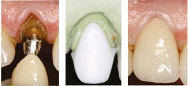
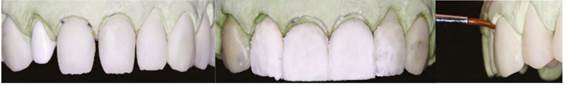
Figure 9 Powder-liquid technique for the veneering ceramic of teeth 12, 11, 21 and 22; and staining technique for the monolithic restorations of 13 and 23
Table 1 . Selection of restoration type and material according to the clinical characteristics of the case
| Tooth | Material | Restoration type |
| 13, 23 | Lithium Disilicate | Monolithic veneers |
| 11, 21, 22 | Lithium disilicate/Ceramic with fluorapatite | Laminated veneers |
| 12 | High opacity zirconia (Y-ZTP)/Ceramic with fluorapatite | Crown with double-layer technique |
Table 2 Composition of the different types of dental ceramics
| Brand | Material | Manufacturer | Chemical composition |
| IPS e-max Press | Lithium disilicate | Ivoclar Vivadent, Schaan, Liechtenstein | SiO2, Li2O, K2O, MgO, ZnO, Al2O3, P2O5 and other oxides |
| IPS e-max Ceram | Fluorapatite-based Veneering ceramic | Ivoclar Vivadent, Schaan, Liechtenstein | SiO2, Al2O3, ZnO2, Na2O, K2O, ZrO, CaO, P2O5, fluoride and pigments |
| Ceramil Multi X | Zirconia oxide | Amann Girrbach, AG, Koblach, Austria | ZrO2 + HfO2 + Y2O3: > 99.0 Y2O3: 4.5-5.6 HfO2: < 5 Al2O3: < 0.5 other oxides: < 0.5 |
The restorations of teeth 13 and 23 were made using the monolithic technique in lithium disilicate, benefiting from the mechanical properties of the material. The staining technique with pigments was used for color characterization (Ivoclar.Vivadent) (Figure 10).
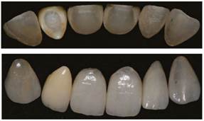
Figure 10 Image showing the different degrees of translucency and opacity in final restorations. Veneers in lithium disilicate (13 and 23: monolithic technique with pigments; 11, 21 and 22: trimming with veneering ceramic technique). Full crown of teeth 12 with a zirconia cap and veneering ceramic.
For the selection of ceramics
Final cementation
The patient returned for final cementation, which includes the following sequence of procedures: removal of provisional restorations, prophylaxis of dental preparations with pumice, verification of the adaptation of restorations using a probe, and aesthetic and functional evaluation using the proof paste RelyX Veneer® by 3M ESPE. Once agreement and acceptance of both patient and clinician was achieved, the actual final cementation was conducted, as shown in tables 3 and 4.
Table 3 Preparation of restoration for adhesive cementation
| 1 | Conditioning of the inner side of the restoration with 9% hydrofluoric acid for 20 seconds |
| 2 | Profuse washing with water for 1 minute |
| 3 | Ultrasonic washing with isopropyl alcohol for 5 minutes, aerating for thorough drying |
| 4 | Application of silane (thin layer), leaving to act for 1 minute |
| 5 | Application of bonding agent (not curing) |
| 6 | Final cementation with resinous cement or dual-cure cement |
Table 4 Preparation of dental substrate for adhesive cementation
| 1 | Cleaning tooth surface with baking soda |
| 2 | Conditioning of enamel with 37% phosphoric acid for 15 s. |
| 3 | Profuse washing with water for 1 minute, aerating without desiccating dentin |
| 4 | Application of bonding agent (not curing) |
In order to comply with the appropriate adhesive cementation protocol, the lithium disilicate restorations of teeth 13, 11, 21, 22 and 23 were internally etched with 9% hydrofluoric acid (Ultradent) for 20 s.; they were thoroughly washed with water, placed in ultrasonic washer with 90% isopropyl alcohol for 5 minutes, thoroughly dried with a triple syringe, silanized applying Silane Primer® (Kerr) with a micro-brush for 60 s., and applied a layer of Single Bond® adhesive (3M ESPE). They were finally applied light curing resinous cement RelyX Veneer® (3M ESPE) (Figure 11).(36)
The dental preparations were subjected to enamel and dentin etching for 20 and 10 s. respectively with Ultraetch® (Ultradent) 37.5% phosphoric acid; they were washed with plenty of water and dried with paper towel. The adhesive system used was Single Bond (3M ESPE), which was rubbed against the surfaces of preparations for 15 s. They were aerated, the adhesive material was thinned with a triple syringe, and were not light-cured (Figure 12).(37) The restorations were slowly put in place taking into account their insertion paths and applying gentle digital pressure. After removing excess cement with a spatula, they were light cured for 2 s., keeping a slight pressure on the restoration to avoid any movement. Dental floss was used and final light curing for 40 s. was performed on the palatal, incisal, and buccal surfaces respectively. Excess cement remainders were removed with a #12 surgical blade. Final polishing was performed during the next appointment (Table 5).(38)
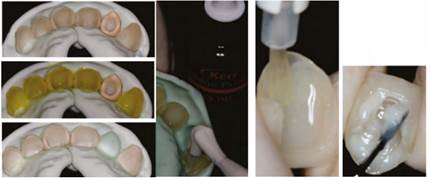
Figure 11 Conditioning of the internal surface of lithium disilicate veneers using the adhesive cementation protocol
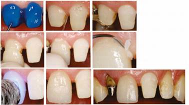
Tooth 12 was subjected to prophylaxis with baking soda and dried with paper towel. The zirconia crown was loaded with dual-cure resin cement (RelyX U200®, 3M ESPE), and secured on the stump by applying a slight pressure; excesses were removed with a metal spatula and dental floss after a period of 40 s. (Figure 13).(39)
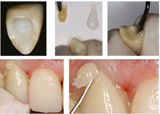
Table 5 . Materials for the adhesive cementation technique
| Brand | Material | Manufacturer | Chemical composition |
| Ultra-etch | 35% phosphoric acid | Ultradent Products Inc., South Jordan, USA | 38% phosphoric acid, Cobalt aluminate blue spinel, Cobalt zinc aluminate blue spinel |
| Single Universal Bond | Adhesive “all in one” bottle | M ESPE AG, Seefeld, Germany | MDP Phosphate Monomer, Dimethacrylate resins, HEMA, Vitrebond™ copolymer, filler, ethanol, water, initiators, silane |
| Porcelain etch | 9% hydrofluoric acid | Ultradent Products Inc., South Jordan, USA | Hydrofluoric acid |
| Monobond S | Silane | Ivoclar Vivadent, Schaan, Liechtenstein | Ethanol, 3-trimethoxysilylpropyl methacrylate, Methacrylated phosphoric acid ester |
| RelyX Veneer | Resinous cement | 3M ESPE AG, Seefeld, Germany | |
| RelyX U200 | Resinous cement | 3M ESPE AG, Seefeld, Germany | Alkaline (basic) fillers, silanated fillers, phosphoric acid modified methacrylate monomers, methacrylate monomers, initiators |
| Luxatemp | Bis-acrylic temporary material | DMG America Englewood, NJ. USA | Bisacril |
Occlusion control (Figure 14) was done with AccuFilm® articulating paper, verifying functional movements. A protective maxillary splint in acrylic was provided for night use. Periodical clinical and radiographic follow-up was recommended (Figure 15).
CONCLUSION
The perfect material does not exist; it cannot be stated that there is a universal dental ceramic applicable in all clinical situations. Sometimes, different materials can yield similar results; however, in other circumstances one of these materials can stand out as the best choice. Therefore, the clinician’s responsibility and criteria play an important role in choosing the appropriate material for each case.

