


 English (pdf)
 Article in xml format
 Article references
 Send this article by e-mail
 Cited by SciELO
  Cited by Google
  Similars in
SciELO
Similars in
SciELO  Similars in Google
 Permalink
INTRODUCTION
Elastics and elastomers-along with wires-are force-storing devices that produce orthodontic movement by releasing force (F) on teeth and transmitting it to the periodontium. Latex elastics are isopropyl polymers made of natural rubber with favorable characteristics for orthodontics, such as a high elastic limit, easy handling, and low cost compared to other active elements.1,2 However, latex elastics can have an allergen potential in 0.1% to 6% of the population, mainly because of the addition of chemicals used in the vulcanization process, such as ammonia and various antioxidants.3,4 Therefore, synthetic elastics-also called non-latex or latex-free elastics-have been developed. These are equally useful in orthodontic biomechanics and are commercially presented as materials that maintain mechanical conditions similarly to latex elastics.5
Despite the advantages of elastics in orthodontic treatment, both types of devices have shown different behavioral changes in terms of mechanical properties in different clinical and experimental situations.6 Clinically, changes occur because of exposure of the materials to the environment of the oral cavity. Several studies suggest that saliva and bacteria can infiltrate the molecular structures, weakening the rubber surfaces of latex and resulting in discoloration and elongation.7-10 It has also been proved that these elastics lose more force with temperatures above 45 °C due to a loss in rigidity of the material.11 In addition, more stretching means greater force loss,12,13 which occurs in the first 24 hours.10,12 Some in vitro studies on the behavior of latex and non-latex elastics show a great variability in the material used for the latter, which depends on the manufacturing and production process used.13-15 A force loss of up to 50% of the initial force of non-latex elastics has been reported after 24 hours of activation.15,16 Elastics generally show a greater force loss in moist environments compared to dry environments. In the same environment, non-latex elastics show greater force loss than latex elastics.17-19 However, no significant differences have been found in the force loss of latex and non-latex elastics during static testing.20
In in vivo studies, latex elastics retain higher force levels between 0 and 12 hours, but no significant differences are observed with non-latex elastics after 24 hours.21 While some distributors present both types of elastics as similar,5 they do not offer tables for correct application according to these parameters. This information is critical and could be used in the construction of scientific, evidence-based clinical practice guidelines to improve patient safety through reasonable use of medical devices.22
The aim of this study was to evaluate the mechanical behavior in vitro (initial release and force degradation up to 24 hours) of two types of elastics differently manufactured (latex and non-latex) from a single manufacturer.
MATERIALS AND METHODS
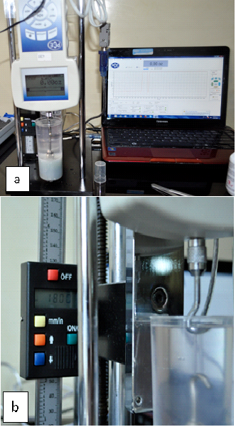
This in vitro study used 20 latex and 20 non-latex intermaxillary elastics of ⅟4" 6 oz in force (170.10 g; 1.67 N) (Forestadent® FOR Elastics-Fruit-Line intraoral [Ref. 650-1022, 650-4022], Forestadent Bernhard Förster GmbH, Pforzheim, Germany). Elastics with recent manufacturing date and of the same lot which had not been subjected to fatigue during manipulation were included. Elastics showing manufacturing alterations, with deformities, or adhered to each other were excluded. To measure the force released by the elastics, a PCE-FG test dynamometer for force-displacement measurement was used, placed in a PCE-FTS50 base (PCE Instruments DE, Germany). The measuring device was added a fixed container to keep the elastics immersed for 24 hours in artificial saliva (Salivar® solution, Farpag S.A.S., Colombia. Composition: magnesium chloride: 0.015 g, calcium chloride: 0.015 g, potassium chloride: 0.120 g. Excipients: potassium monophosphate, 70% sorbitol, hydroxyethyl-cellulose, methyl and propyl paraben, red color #2, purified water q.s. 100.0 mL). The study was approved by the Research Committee of Universidad Antonio Nariño (Act # 0113 of 2013).
All the measurements used in this study were taken by a single researcher, using cotton pliers to bring the elastic on the active tip of the test stand hook to the active extension tip of the dynamometer, stretching the elastic by means of the manual handwheel up to three times its internal diameter (18 mm) (Figure 1). Force was recorded at 0, 6, 12, 18, and 24 hours. The elastic was then removed from the appliance and discarded. These steps were repeated for the entire sample. Once the data were obtained, the percentage of force relaxation (%R) was calculated for both latex and non-latex elastics, as shown in Equation (1).
 Equation (1)
Calibration between the observer and a reference method was conducted, calculating intraclass correlation coefficient (CCI), which yielded a concordance index of 0.912 with 95% confidence interval and limits of 0.955 and 0.956, indicating a very good concordance. Descriptive statistics and a non-parametric test (Kruskal-Wallis) were conducted to compare the force applied by latex and non-latex elastics, as well as force degradation over time (p < 0.05), using version 17 of the SPSS® software for Windows (SPSS®, USA).
RESULTS
The initial force provided by both types of elastics was higher than that reported by the manufacturer (Table 1). The latex and non-latex elastics showed an initial force of 2.11 ± 0.11 N and 1.83 ± 0.11 N respectively, while the manufacturer reports an initial force of 1.67 N. At 6 hours of activation, force reduced to 2.00 ± 0.11 N in the latex elastics and 1.72 ± 0.11 N in the non-latex elastics. After 12 hours, the reduction in force decreased by half the initial reduction (latex: 1.94 ± 0.13 N; non-latex: 1.69 ± 0.11 N). At 18 and 24 hours of activation, both types of elastics kept the same reduction values as those reported since 12 hours of activation (18 hours: latex: 1.92 ± 0.12 N; non-latex: 1.67 ± 0.09 N; 24 hours: latex: 1.91 ± 0.13 N; non-latex: 1.66 ± 0.10 N) (Table 1). A statistically significant difference was found, being favorable to latex elastics in all post-activation evaluations (p < 0.05) (Table 1). The non-latex elastics showed values slightly below those approved by the distributor at 24 hours (Table 1 and Figure 2).
Table 1 Initial force and force loss over time in latex and non-latex elastics (Forestadent®)
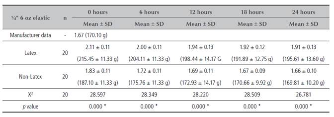
* Kruskal-Wallis test (p < 0.001)

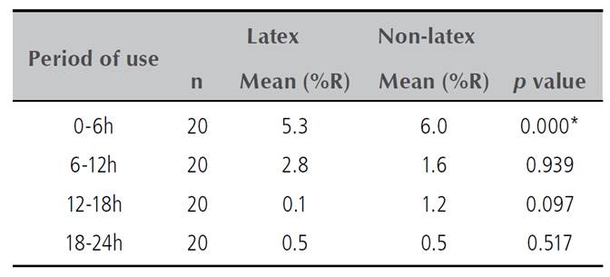
Regarding force reduction in latex and non-latex elastics, statistically significant differences were found between 0 and 6 hours only (p < 0.05) (Table 2). The percentage of force relaxation in latex (8.7%) and non-latex (9. 3%) elastics at 24 hours of activation indicated that force loss was greater in the latter.
DISCUSSION
Finding out if there are differences between latex and non-latex elastics helps identify whether the forces produced by the elastics fit the biomechanical needs of each patient, thus achieving greater accuracy and efficacy in the orthodontic treatment. Since it was not clear whether the force produced by the elastics was reported by a given manufacturer to a given amount of stretch, nor how much force loss the elastics suffered during prolonged elongation, this study evaluated the force behavior of latex and non-latex elastics of the same manufacturer when stretched three times their diameter. The elastics’ force was measured at 0, 6, 12, 18, and 24 hours, allowing to evaluate their behavior during the entire time a patient keeps them in the mouth as recommended by the orthodontist.
In the aforementioned experimental conditions, the initial forces of latex and non-latex elastics are above the values given by the manufacturer. This increased initial force provided by both elastic types can only be a compensation for the force loss given by environmental factors in the oral cavity. In the study by Hershey and Reynolds,22 the initial force supplied by three commercial houses was substantially different, if each commercial house has its own formula for the elastic’s manufacturing material. This concept was corroborated by Alavi et al.23 This would explain findings such as that of Kersey et al,17 who found that the initial forces of latex and non-latex elastics were below the force reported by the manufacturer, when stretched three times their inner diameter.
The elastics’ force is closely related to their stretching,14 but the decrease in force is not proportional to this stretching.24 The non-latex elastics showed a higher percentage of force relaxation compared to the latex elastics. In this study, the samples experienced a higher, statistically significant percentage of force loss in non-latex elastics (6.1%), compared to 5.3% in latex elastics during the first 6 hours of elongation. These percentages are very similar to those reported by Alavi et al23 (4-7.5%) but lower than those reported by Russell et al13 (15-20%) and Aljhani et al20 (10-12%). However, force relaxation after 6 hours was lower, and no statistically significant differences were found between the two types of elastics. Russell et al13 found that GAC® non-latex elastics showed a higher percentage of force relaxation. However, Aljhani et al20 found no significant differences in the force provided by the two groups of elastics in static periods, but in periods with movement.
One of the variables taken into account in this study was the elastics exposure to artificial saliva, a factor that can affect the force provided by them. López et al18 found a greater force loss in latex and non-latex elastics in a humid environment compared to a dry environment, and a significant difference between the two types of elastics in a dry environment. This can be explained by the structure and composition of non-latex elastics, which contain synthetic polymers with molecular joints that maintain the integrity of the structure, compared to the covalent cross-links of latex elastics, leading to a deficient behavior of non-latex elastics.20 Liu et al24 established that the extension of all elastics because of anatomical needs would be in the range of 20 to 50 mm. Accordingly, the normal extension range would be given by an elongation of three times the diameter of the elastics, according to Bell’s rule of the triple stretch (1951).25 This rule was used in this study to achieve a standard elongation.
This study was carried out with in vitro tests, controlling variables such as immersion in saliva and the elastic’s extension; however, in the oral cavity elastics are exposed to other factors, such as the pH of food,26 the use of oral antiseptics27 and the activity of the jaws, among others7 that were not taken into account in the study. This is why it is recommended to take the results with caution, perhaps performing in vivo studies to observe the actual behavior of elastics with these variables.27
The orthodontist should use measuring instruments to verify that the elastics are producing the expected level of force,18 and replace elastics several times a day, if necessary, to maintain higher constant forces during treatment, as recommended by Alavi et al.23
CONCLUSIONS
The force provided by latex elastics stretched three times their internal diameter and immersed in artificial saliva was greater than that provided by non-latex elastics at all evaluated times. The initial force given by latex and non-latex elastics was higher than that announced by the manufacturer. The force of non-latex elastics decreases to values similar to that reported by the manufacturer at 18 hours.
The non-latex elastics used in this study can be used in clinical situations. However, the use of latex and non-latex elastics should be kept under strict control to make orthodontic mechanics efficient, since the period of greatest elastic instability occurs between 0 and 6 hours.

