


 English (pdf)
 Article in xml format
 Article references
 Send this article by e-mail
 Cited by SciELO
  Cited by Google
  Similars in
SciELO
Similars in
SciELO  Similars in Google
 Permalink
INTRODUCTION
Assessing an individual’s dental development helps compare it with a reference group and thus determine whether the person is advanced or delayed compared with other subjects of the same age group,1-3 which is useful in dentistry, orthodontics, and maxillary orthopedics.4-6 Also, in bioarchaeology it allows investigating regional or ethnic differences among populations,3,7 and in forensic sciences it helps estimate dental age (DA) when age is unknown in deceased people or in living individuals with no valid identification documents.1,8-10 This is increasingly relevant nowadays due to the expansion of illegal immigration2 related to natural disasters or war conflicts.
The development of permanent teeth occurs in a series of sequential and predictable events in both sexes, with the radiographic study of this process being the basis for DA estimation methods, which determine age according to the morphological changes observed.1,11,12 The method of Demirjian et al,1,11 which was standardized in a French-Canadian sample, is one of the most commonly used to assess dental maturation and to estimate DA.2,13,14 In this method, the seven mandibular teeth on the left side are assessed, with the exception of the third molar, and classified into development stages identified with letters A to H.; each stage is then given a score according to a table available for each sex, adding up the scores to obtain a dental maturity index (DMI), which is transformed into a DA expressed in years and tenths of year, using reference tables. In 1976, the authors published a revised version11 in which they incorporated stage 0 (when tooth calcification cannot be observed), expanded the sample (enabling to calculate new scores for the seven-tooth method), and presented an updated dental maturation curve that can be used for age calculation. They also proposed two new systems that consider four teeth in the left lower quadrant. The first method includes the evaluation of premolars and permanent molars, while the second uses the lateral incisor, the two premolars and the second molar. Scoring tables and their respective maturation curves are presented for both methods.
On the other hand, in 2005, Chaillet et al12 carried out a multicentric study with samples originating in eight countries in order to calculate scoring tables for the maturation stages of the method by Demirjian et al,1 creating dental maturation curves used in the assessment of dental development and the estimation of DA in individuals of unknown ethnic origin. Both methods have been used in different populations in order to determine whether they can be accurately applied in DA estimation. In this sense, a method can be considered accurate when the difference between chronological age (CA) and estimated DA is closed to zero.15
Regarding the method of Demirjian et al,1,11 a consistent overestimation of age (CA-DA difference) has been found, with average values of -0.60 years in males (0.23 to -3.04) and -0.65 years in females (0.10 to -2.82 years), when used in individuals with ethnic and socioeconomic characteristics that differ to those of the sample used to standardize the method.5,7,13-20 Studies in Latin American subjects, particularly Argentinians,6 Brazilians,4 Chileans,21 Paraguayans,10 and Peruvians9 have reported age overestimation, while a tendency to underestimation was observed in Colombian individuals.22 In Venezuelans, age overestimation has been found in samples originating in the center and the west of the country3,23,24 and underestimation in subjects from the Andean region.2 With respect to Chaillet et al,12 age overestimation has been seen in Bosnian25 and Spanish2 individuals, as well as underestimation in Koreans13 and French subjects.7
Consequently, the aim of this study was to assess the applicability of the methods of Demirjian et al1 in its revised version11 (DEM) and Chaillet et al (CHA)12 in a sample of Venezuelan children and adolescents of both sexes from Maracaibo, Zulia, in the estimation of the DA.
MATERIALS AND METHODS
Sample
This is a descriptive, retrospective, field study using a convenience sample of 516 panoramic radiographs from the clinical records archive of the Center for Comprehensive Child Care (Centro Integral de Atención al Niño, CIAN) and the archive of the Forensic Dentistry Unit of the Research Institute at Universidad del Zulia School of Dentistry, Maracaibo, Zulia, belonging to individuals of both sexes (243 males and 273 females) with chronological ages of 6 to 18 years. Since the panoramic radiographs come from different radiological centers of the region, they were selected according to the following inclusion criteria, in order to guarantee quality and suitability in representing the object of study: images with adequate contrast and density and minimal distortion, from subjects with no systemic diseases and with size and weight according to the CA as stated in the clinical records, presence of the seven permanent mandibular teeth on the left side, and without extensive pathologies or anomalies in number, shape, size, or position that could alter the odontogenesis. In the case of tooth agenesis, the counterpart on the opposite side was considered.
Age groups were formed by at least 10 individuals with differences of 11 months among them. Actual age was calculated by subtracting the date of birth from the date the X-ray was taken, and expressed in years and tenths of years.
Procedures and techniques
Due to distance and availability reasons, the selected analogue panoramic X-rays from the CIAN were digitized using a camera (Sony Cyber-shot DSC-W650, Sony Corporation, Tokyo, Japan) with a resolution of 300 dpi. The images were saved in a computer and transformed to grayscale. Adobe Photoshop CS6 (Adobe System Incorporated, San Jose, CA, USA) was used, with the operator using tools like brightness, contrast, and magnification.
Selected analogue panoramic X-rays from the Forensic Dentistry Unit were physically available. For their evaluation, they were placed on a desk viewbox with a black mask in a light-reduced environment. All radiographs were simultaneously examined by two calibrated observers, who concurred in identifying the stages and only knew the sex of each subject.
To apply the method of Demirjian et al,1 the stages were selected following the procedure described by the authors. DA was calculated using the scoring tables of the revised version for the seven-tooth method and the 50 percentile of the corresponding maturation curve for each sex.11 Concerning the CHA method,12 the stages previously selected by DEM and the proposed scoring tables were used to obtain the IMD, which was converted to DA using the values of the 50 percentile of the available conversion table by sex. The data were recorded on a sheet designed for that purpose.
Statistical analysis
Version 15.0 of the SPSS® Statistical Package (Statistical Package for the Social Sciences, SPSS Inc. Chicago, Il, USA) was used for statistical analysis, calculating mean dental ages as suggested by each method for each age group in both sexes. Mean differences between CA and estimated dental ages were determined by means of a Student’s related-samples t test. This is why in this study a negative symbol represents age overestimation and a positive symbol represents underestimation. The level of significance was p < 0.05.
RESULTS
The analyses yielded high and statistically significant correlations between CA and the dental ages estimated in both methods. In the DEM method,11 such correlations were 0.91 for boys and 0.90 for girls, while CHA,12 showed values of 0.92 for boys and 0.91 for girls.
Demirjian et al method11
Age underestimation was observed in the entire sample, since the difference between CA and the estimated DA was statistically significant (Table 1). Age was underestimated in both sexes, with lower underestimation in males, while the CA-DA difference was significant in females. When considering age groups, males showed age overestimation up to the age of 13 years (Table 2). The differences were significant for the groups of 6 to 8 years and 16 to 18 years. In females, overestimation was seen up to the group of 11 years, with significant CA-DA difference in the groups of 6 to 9 years (Table 3). Significant age underestimation was observed from the age of 12 years.
Table 1 Means and mean difference between chronological age and dental age estimated by the two methods under study
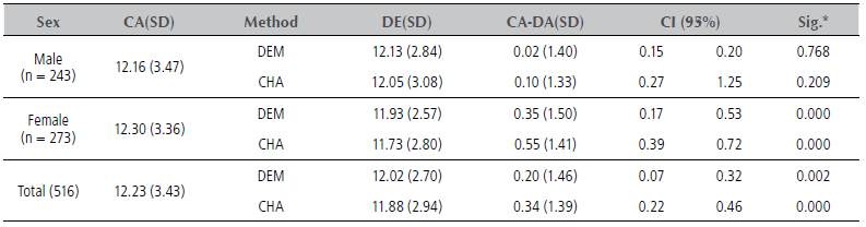
* Related-samples t-test. DEM: Demirjian et al method, CHA: Chaille et al method, CA: chronological age, DA: dental age, SD: standard deviation, CI: confidence interval, Sig: significance (p < 0.05).
Table 2 Means and mean difference between chronological age and dental age estimated by the methods of Demirjian et al and Chaillet et al by age group, males
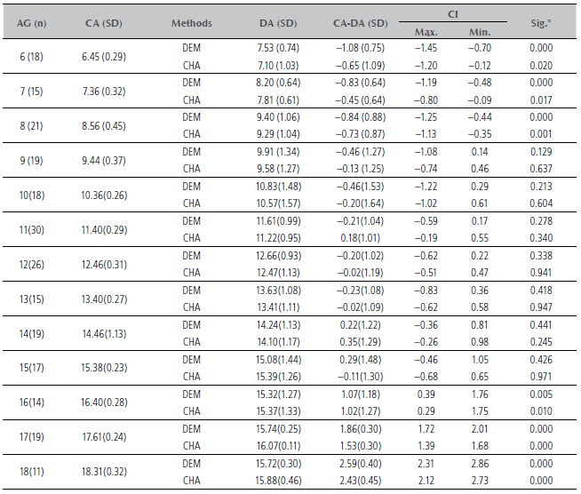
* Related-samples t-test. DEM: Demirjian et al method, CHA: Chaille et al method, AG: age group, CA: chronological age, DA: dental age, SD: standard deviation, CI: confidence interval, Max.: maximum, Min.: minimum, Sig: significance (p < 0.05).
Table 3 Means and mean difference between chronological age and dental age estimated by the methods of Demirjian et al and Chaillet et al by age group, females
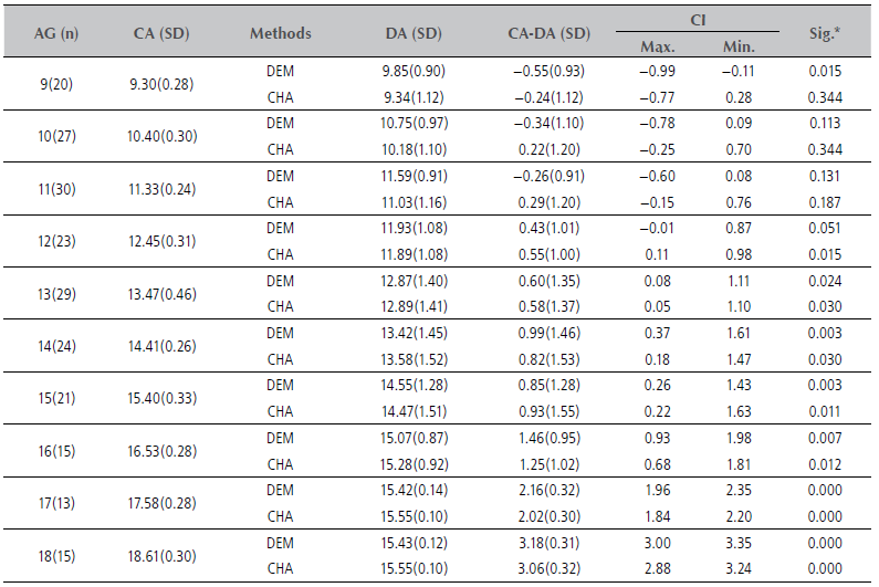
* Related-samples t-test. DEM: Demirjian et al method, CHA: Chaille et al method, AG: age group, CA: chronological age, DA: dental age, SD: standard deviation, CI: Confidence interval, Max.: maximum, Min.: minimum, Sig: significance (p < 0.05).
Chaillet et al method12
Age underestimation was seen in the entire sample, with a significant difference between CA and DA (Table 1). Age was underestimated in both sexes, with lower underestimation in males. The CA-DA difference was significant in females. Age was overestimated in males up to the age of 10 years and in the groups of 12, 13, and 18 years (Table 2). Differences were significant between 6 and 8 years of age and since the age of 16 years (Table 3). In females, age overestimation occurred in the groups of 6 to 9 years, with a significant difference between CA and DA in the group of 8 years. Age underestimation was also found since the age of 10 years, with a significant difference in the group of 11 years.
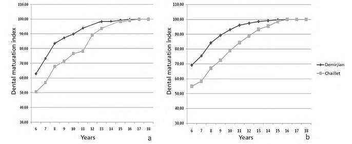
These results are displayed in Figure 1, showing age overestimation in the younger age groups in both sexes, while the estimated dental ages are similar to CA around puberty, but consistent underestimation is seen later in older age groups (16-18 years).
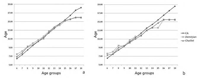
Figure 1 Comparison between mean chronological age (CA) and mean dental ages estimated by the methods of Demirjian et al and Chaillet et al. (a) male (b) female
In view of the results found in both sexes, a Student related-samples t-test was performed using a subsample up to the age of 15 years, in order to confirm whether the underestimation seen in the older groups had an influence on the differences found in the entire sample. The subsample yielded age overestimation by DEM,11 regardless of sex (-0.23 p = 0.000), while DA calculated by CHA12 was underestimated (0.08 p = 0.894). Both methods overestimated age in males (DEM -0.42 p = 0.00; CHA -0.24 p = 0.00). In females, DEM overestimated age (-0.05 p = 0.500), while CHA underestimated it (0.22 p = 0.01).
Figure 2 shows the relationship between the dental maturation index (DMI) of each method and CA. In males, 100% of dental maturation was reached around the age of 15 years in both methods, while in females it was evidenced from the age of 15 years by DEM11 and 16 years by CHA.12
DISCUSSION
Morphological methods to assess dental development in X-rays in order to estimate DA have been used for clinical purposes and are also useful in forensic science as part of age-diagnosis protocols. It is therefore relevant to know if the method being used is accurate and useful for the matter. The purpose of this study was to determine whether the revised method of Demirjian et al11 and that of Chaillet et al12 could be applicable in a Venezuelan sample.
In considering the total number of individuals, age calculated by both methods was underestimated; however, when assessing a subsample of the group up to 15 years of age using the 1.25 to 3.18 years ratio CA-DA difference found in older groups, there was an overestimation of age calculated by DEM11 in both sexes, as reported in previous studies2,5,7,13,15-17,19,20 using the revised version of the method. The values of the CA-DA difference in this study are higher than those observed in Korean,13 Iranian,5 and Tunisian15 males, lower than those reported in Australian,14 Spanish,2 Chinese,20 French,7 Turk,16 and Yugoslav19 males, as well as in Iranian5 and Tunisian females,15 and similar to those found by Abesi et al18 in another sample of Iranian origin. Table 4 shows the difference in values between CA and estimated DA found in the aforementioned studies, enabling a comparison with those obtained in the present study.
Table 4 Summary of the chronological age-dental age difference in the various samples in which the methods of Demirjian et al (1976) (11) and Chaillet et al12were used
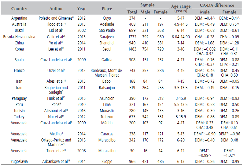
CA: chronological age, DA: dental age, DEM: Demirjian et al method, CHA: Chaille et al method, ^ calculation obtained from table in original article, * original version of the Demirjian method (1973)
As for research findings in Latin American individuals, it could be noted that the obtained differences are lower than those observed in Brazilians,4 Paraguayans,10 and Peruvians,9 but similar to those seen in Argentine males.6 In samples of Venezuelan origin, age overestimation in the present study was lower than that observed in children of the metropolitan area of Caracas3 and other samples of Zulians (western area of the country),23 in contrast with the report by Cruz-Landeira et al,2 who showed a delay in dental development in the sample studied (Table 4).
The age overestimation calculated by CHA12 in males in the present study was lower than that verified in Spanish2 and Bosnian males,25 in contrast to the age underestimation reported in Korean13 and French7 males. In females, age underestimation was lower than that observed in Korean13 and French subjects,7 unlike the results observed in a sample of Spanish origin,2 as well as in Bosnian25 and French7 girls, in which the method overestimated age. Concerning other Venezuelans, the findings in a sample from the Andean region2 agree with the age underestimation in the females of the present study, and differ with that of males, in whom age was overestimated by the method (Table 4).
The CA-DA value difference reported in the various samples and those obtained in this study may be an expression of the influence of genetic, ethnic, socioeconomic, and climatic factors on human development, as well as sample size, the studied age range, the number of subjects in each group, and the statistical approach.14,18,19,26 In this sense, our findings agree with the results of a previous study in which DEM11 was used in a sample of Zulians, where the CA-DA difference was higher than that reported in the present study; therefore, adding around 200 X-rays and modifying the arrangement of age groups seemed to have an impact on the obtained values.
As for age groups, both methods yielded age overestimation up to the age of 12 years in boys and 11 years in girls, with significant CA-DA differences in younger age groups. During puberty, males showed an advanced age with respect to the samples used for standardization of the methods, since the CA-DA differences were not significant, whereas in females the delay was consistent and statistically significant, which may reflect the variability in maturation process between the samples.
With DEM,11 100% dental maturation was observed around the age of 15 years in both sexes, that is, one year later than the sample of Andean origin of Cruz-Landeira et al.2 With CHA,12 this was observed at the age of 15 years in males and 16 years in females, allowing for a higher range of estimation of DA in the latter. In comparing these results with those observed in the Andean children population, Zulian boys reached 100% maturation one year and eight months later and girls eight months earlier, which may express regional differences in terms of the environmental, nutritional, and cultural conditions in which individuals develop.14
The results obtained with the subsample regardless of sex indicate that the CHA12 method showed a lower CA-DA difference; however, DEM11 showed more precision in boys and CHA12 in girls. It is important to note that the differences verified in both methods are within the range of ±0.5 to ±1 year, which is acceptable in forensic sciences for age estimation in growing individuals, just like values of ±0.6 years of delay or advance is acceptable in clinical disciplines.1,13,26
CONCLUSIONS
This study assessed the applicability of the methods of Demirjian et al (DEM)11 and Chaillet et al (CHA)12 in a sample of Venezuelan children and adolescents to determine dental age, finding out that both methods are similar in precision; however, the Demirjian et al11 method was more accurate in boys and that of Chaillet et al12 in girls. Because no maturation indicator by itself can reflect an individual’s developmental status, it is recommended to assess the estimated dental age in conjunction with other indicators such as bone age, height, weight, and the presence of secondary sexual characters.

