


 English (pdf)
 Article in xml format
 Article references
 Send this article by e-mail
 Cited by SciELO
  Cited by Google
  Similars in
SciELO
Similars in
SciELO  Similars in Google
 Permalink
INTRODUCTION
A cyst is defined as a pathological cavity covered with epithelium, which may contain liquid and semi-solid or gaseous material. It is not strictly necessary for it to have an epithelial coating to be diagnosed as a cyst; in fact, there are cysts without this feature.1 Residual cysts are usually asymptomatic and are accidentally discovered during routine examinations. According to the recent classification of maxillary cysts proposed by the World Health Organization (WHO) in the fourth version of the consensus for the classification of head and neck tumors: maxillofacial and odontogenic bone tumors (2017), they are divided into developmental cysts or those of uncertain origin on one hand, including dentigerous cyst, odontogenic keratocyst, lateral periodontal cyst, gingival cyst, glandular odontogenic cyst, and odontogenic cyst, and inflammatory cysts on the other, which are the radicular/residual cyst and the inflammatory collateral cysts (paradental and buccal bifurcation mandibular cyst).1-3 Residual cysts are inflammatory, originate in a radicular cyst that remains in its entirety or in rests of it, and develops after an extraction of an affected tooth.1 The average age at which it appears is 52 years; they are most often located in the maxilla, and males are the most affected, with a 3:2 ratio.
Residual cysts represent approximately 10% of the cases of odontogenic cysts.4 They originate from chronic inflammation of the periradicular tissues, producing a periapical granuloma and the subsequent stimulation of proliferation of the epithelial rests of Malassez. This is followed by central degeneration and necrosis, producing a cavity. The histological features include a stratified squamous epithelium lining and a connective tissue wall in contact with bone. They usually show intense inflammation; in addition, there can be cholesterol crystals, macrophages, hemosiderin, keratin, and cellular remains inside the cavity. Even if the source of inflammation has been removed, the wall of a residual cyst may mature and show less inflammation or no inflammation at all, and the epithelial lining becomes thin and regular.1 Treatment alternatives for cystic lesions include enucleation, curettage, marginal resection, and endoscopic surgery.5 The aim of this article is to describe the treatment of a residual cyst located in the upper maxilla.
CLINICAL CASE
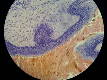
In 2009, a 64-year-old male patient requested an evaluation of the area of dental units 11 and 12. The patient did not go on with this evaluation, and he returned to the school of dentistry two years later, already with units 11 and 12 extracted, reporting the presence of a tumor in the edentulous area. The stomatological examination showed an increased bulk in the edentulous area of 11 and 12, covered by a healthy mucosa with a translucent area in the adhered gingiva, giving it a bluish appearance. The increased area was asymptomatic, depressible, measuring 1.0 x 1.5 cm, with no signs of secretion and defined margins, causing expansion of the vestibular cortical, with 2 years of evolution (Figure 1A). The panoramic X-ray showed a well-circumscribed radiolucent image located between 12 and 13, with no defined borders, producing divergence of adjacent teeth (Figure 1B). Exploratory puncture was performed, finding some yellowish, serous liquid with traces of hematic components and an oily consistency. The cystic lesion was intervened using anesthesia for the upper front alveolar rami. A full-thickness complete Newman flap was performed from 13 to 23, as well as osteotomy of the vestibular wall. The cystic cavity was accessed, performing enucleation and curettage, bone plasty, irrigation, and suture (Figure 2A). 100 mg nimesulide tablets were prescribed orally every 12 h for 4 days, as well as 500 mg amoxicillin capsules orally every 8 h for 5 days. Post-surgical control was performed 7 days afterwards, as well as follow-ups 1, 2 and 6 months after the procedure. The results of the histopathological study confirmed the residual cyst diagnosis (Figures 3A, 3B, and 4). Radiographically, after 2 months of surgery there was trabeculated bone in the edentulous area of 11 and 12 (Figure 2B).
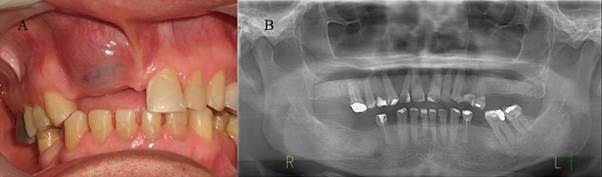
Figure 1 A) Clinically, there is an increased bulk in the edentulous area at the level of 11 and 12, with a healthy, translucent, bluish mucous lining. B) Initial panoramic radiograph showing a well-circumscribed radiolucent image with no defined margins located between dental organs 12 and 13, causing radicular divergence; in addition, there is a coronal radiopacity compatible with poorly adapted restorations.
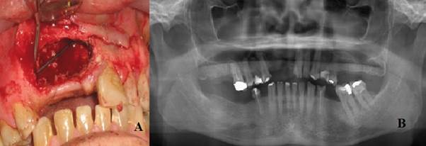
Figure 2 A) A Newman flap was followed by osteotomy to reach the cystic cavity and later by enucleation. Note the cavity after enucleation of the cystic lesion. B) Panoramic radiograph 2 months after surgery, showing trabeculated bone in the edentulous area of dental organs 11 and 12, compatible with healthy bone in the process of regeneration.
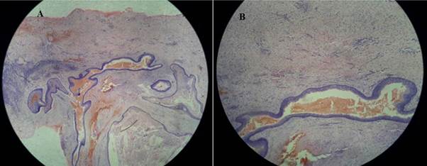
Figure 3 A) histological image showing a cavity covered by thin, non-keratinized epithelium, with a capsule of mature fibrous tissue (HE, 10x). B) A more detailed image of the cystic capsule containing moderate inflammatory infiltrate. The cavity contains intralesional hemorrhage.
DISCUSSION
Residual cysts are odontogenic cysts of inflammatory nature resulting from the incomplete removal of cystic tissue after the extraction of a non-vital tooth with a radicular cyst in its periapical area.1 In 2014, García et al stated that the presence of epithelial cell rests of Malassez in apical and lateral cysts stimulated by an inflammatory process triggers their proliferation mechanism until producing an epithelial cystic cavity.6 Other authors, like Sridevi et al, in 2014 published a case of residual cyst associated with calcifications in an edentulous patient. The authors claim that dystrophic calcifications usually appear in long-lasting chronic cysts, are generally rare in the presence of a residual cyst, and may occur in cases of chronic lesions due to the precipitation of calcium salts in the inflammatory areas.4 In 2012, Jamdade et al suggested that residual cysts occur in the upper and lower jaws, but are more commonly seen in the mandible, just above the lower alveolar nerve duct; in addition, the presence of these lesions at growth may cause displacement or resorption of the adjacent tooth. Radiographs generally show a circumscribed, unilocular, well-defined area of radiolucency overlapping the edentulous region.7,8 In 2015, Sukegawa et al reported that odontogenic cysts can produce carcinomatous changes, with residual cyst being one of the few that can have a malignant transformation. This is because odontogenic cysts result from epithelial lining, which has the potential to transform various types of odontogenic cysts into malignant tumors, but this happens on rare occasions.9
For the therapeutic approach of residual cysts, it is advisable to take a complete clinical history, conduct a good clinical stomatologic examination, and carry out radiographic analysis to achieve a correct diagnosis; however, differential diagnoses should be considered, including keratocyst and adenomatoid odontogenic tumor. All these measures are required to perform the treatment of choice, consisting on enucleation of the lesion.10,11
CONCLUSION
A residual cyst is a benign inflammatory condition usually identified in routine evaluations as in the case presented, in which the patient apparently did not receive a complete initial treatment and consequently developed a cystic pathology; it is therefore recommended to thoroughly analyze each case before performing a dental extraction to prevent the emergence of underlying cystic lesions.

