SciELO - Scientific Electronic Library Online

 
vol.30 número2Estimación del sexo a partir de medidas coronales de caninos permanentes en una población mestiza contemporánea de Manizales, ColombiaGenes involucrados en la amelogénesis imperfecta. Parte II índice de autoresíndice de materiabúsqueda de artículos
Home Pagelista alfabética de revistas  

Servicios Personalizados

Revista

Articulo

Indicadores

Links relacionados

  • En proceso de indezaciónCitado por Google
  • No hay articulos similaresSimilares en SciELO
  • En proceso de indezaciónSimilares en Google

Compartir


Revista Facultad de Odontología Universidad de Antioquia

versión impresa ISSN 0121-246X

Rev Fac Odontol Univ Antioq vol.30 no.2 Medellín ene./jun. 2019

https://doi.org/10.17533/udea.rfo.v30n2a8 

Literature review

Clinical practice guidelines for the surgical endodontic management of post-treatment periapical disease** ***

Guía de práctica clínica para el manejo quirúrgico en endodoncia de la enfermedad periapical postratamiento

Lina María García-Serrano1 

Diana Carolina Lara-Rodríguez1 

Claudia C. García-Guerrero2  * 

1 DDS, Endodontics Specialist, Grupo de investigación INVENDO, Facultad de Odontología, Departamento de Ciencias Básicas y Medicina Oral, Universidad Nacional de Colombia, Bogotá DC. Colombia. Email: dianalara86@hotmail.com, linmari@hotmail.com

2 DDS, Endodontics Specialist, MSc in Dentistry. Grupo de investigación INVENDO, Facultad de Odontología, Departamento de Ciencias Básicas y Medicina Oral, Universidad Nacional de Colombia, Bogotá DC. Colombia. Email: ccgarciag@unal.edu.co


ABSTRACT

Introduction:

in Colombia, persisting post-endodontic disease has been reported by up to 45%, validating the use of secondary alternative therapies, like endodontic microsurgery (EM). The aim of this study was to systematically-and with reliable scientific evidence-develop de Novo Clinical Practice Guidelines for the surgical endodontic management of post-treatment periapical disease (PPD), with more accurate recommendations for therapeutic decisions and preferences consulted with both practitioners and patients.

Method:

the guidelines developers team identified EM as a topic in the literature and established the scope, objective, questions, and outcomes, which were analyzed using the scientific evidence reported in secondary or primary clinical studies. A first screening identified titles and abstracts for each question asked. The validity of the selected studies was quantified with tools like AMSTAR or SIGN. Finally, the strength of recommendations and the quality of evidence were confirmed with GRADE.

Results:

concepts like PPD, EM indication, use of local anesthetics, antibiotics and presurgical anti-inflammatory drugs, effect of magnification, implementation of cone beam computed tomography, hemostasis, retrograde filling, and control time were integrated, supporting each topic with relevant evidence, experts’ recommendations, and even good practice points.

Conclusions:

this document is considered a tool with sufficient evidence for clinical decision-making in EM.

Keywords: apicectomy; endodontics; clinical practice guidelines as subject; evidence-based dentistry; periapical periodontitis; periapical tissue.

RESUMEN

Introducción:

en Colombia, la persistencia de enfermedad posendodoncia ha sido reportada hasta en un 45%, lo cual justifica propuestas terapéuticas secundarias, como la microcirugía endodóntica (ME). El objetivo del presente estudio consistió en desarrollar sistemáticamente, y con evidencia científica confiable, una Guía de Práctica Clínica de Novo para el manejo quirúrgico en endodoncia de la enfermedad periapical postratamiento (EPP), con las recomendaciones más acertadas frente a decisiones y preferencias terapéuticas consultadas a profesionales y pacientes.

Método:

el grupo desarrollador de la guía identificó el tópico ME y estableció el alcance, el objetivo, las preguntas y los desenlaces, analizados mediante la evidencia científica registrada a partir de estudios clínicos secundarios o primarios. Un primer tamizaje identificó títulos y resúmenes para cada pregunta formulada. La validez de los estudios seleccionados se cuantificó con las herramientas AMSTAR o SIGN. Finalmente, la fuerza de las recomendaciones y la calidad de la evidencia se constataron con la herramienta GRADE.

Resultados:

se integraron los conceptos de EPP, indicación de la ME, uso de anestésicos locales, antibióticos y antinflamatorios prequirúrgicos, efecto de la magnificación, implementación de la tomografía computarizada de haz cónico, hemostasia, materiales de retroobturación y el tiempo de control, sustentando cada tópico con la evidencia, la recomendación de los expertos y en ocasiones con las premisas de las buenas prácticas.

Conclusiones:

el presente documento se considera una herramienta con suficiente evidencia para la toma de decisiones clínicas en ME.

Palabras Clave: apicectomía; endodoncia; guías de práctica clínica como asunto; odontología basada en la evidencia; periodontitis periapical; tejido periapical

INTRODUCTION

Post-treatment periapical disease (PPD) is defined as the onset or persistence of periapical pathology once an endodontic treatment has been completed.1 According to the literature, endodontic failure occurs in 38% of all cases on average,(2) requiring a secondary endodontic intervention. In this situation, there are two therapeutic alternatives: orthograde retreatment or endodontic microsurgery (EM)-indicated as a surgical alternative when retreatment fails or cannot be performed-.3 With a success rate ranging from 78 to 92%, EM can access periapical tissue with magnification, illumination, ultrasonic instruments, and regenerative retrograde filling materials.4,5

In Colombia, the mandatory health insurance plan (plan obligatorio de salud, POS), under Resolution 5857 of 2018,6 states that specialized dentistry includes “the excision procedure on an odontogenic lesion […] and other corrective endodontic procedures”, as a practice subjected to agreements by Health Maintenance Organizations (HMOs) (entidades prestadoras de salud, EPS).6 In order to promote a controlled practice, the Ministry of Health and Social Protection states that “All induced and mandatory activities, procedures, and interventions must design or adopt technical standards and healthcare guidelines for relevant diseases”,7a statement that validates the development of Clinical Practice Guidelines (CPGs), defined as evidence-based documents aimed at establishing the most appropriate health care procedures when dealing with a clinical condition.7,8

Evans et al (2012)9 published some EM standards, but methodologically speaking the document cannot be considered a CPG to be adapted nationwide, and therefore a de novo10,11 CPG needs to be developed for the surgical endodontic management of PPD (CPGEM).

That being said, in recognizing the prevalence of PPD2 and understanding the preservation of natural teeth as the main objective of dental specialties, the purpose of this study was to make evidence-based recommendations to support the practice and quality of EM as an effective treatment of PPD.

The final CPGEM document will be a product with an impact on dental schools, endodontics programs, practitioners, endodontic service providers, and especially on patients consulting health care reference centers for EM therapy because of PPD.

METHODS

The development of this document was approved by the Universidad Nacional de Colombia Ethics Committee (Minutes 20-15). The Guidelines Developing Group (GDG) was formed with an endodontist and two graduate students leading the projects within the Graduate Endodontics Program at the School of Dentistry (Posgrado de Endodoncia de la Facultad de Odontología Universidad Nacional de Colombia, PE- FOUN). The stages for the development of the document are shown in Figure 1, including the selection of “Microsurgical treatment of PPD” as subject, and the formulation of objective, scope, and 11 questions in either PICO (Patients-Inter-vention-Comparison-Outcome) or PECOT (Population-Exposure-Comparison-Out- come-Time).11

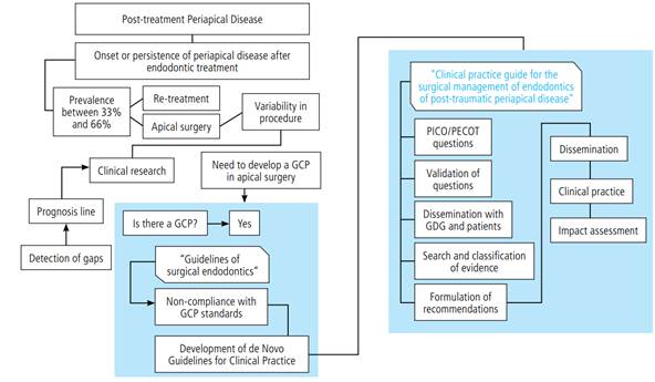
Figure 1 Algorithm representing the phases for the development of the CPGEM. Adaptation of the original in: Methodological directions for the creation of clinical practice guidelines with economic assessment in Colombia’s General Social Security System in Health. Final full version - Methodological Guidelines Update Group 2014-11 

The methodology complied with the principles of clinical validity and reliability through a multidisciplinary review of each question and dissemination for correction and re-formulation by endodontic experts who served as consultants in CPG methodology10,11 and two PPD patients in need of EM seen at PEFOUN.

OUTCOME CATEGORIES

In accordance with the Grading of Recommendations Assessment, Development and Evaluation (GRADE),12 each question’s expected impact was quantified by identifying the outcomes, like this: 1. “critical”, for anesthetic effect, pain control, infection control, hemostatic effect, and treatment success or failure. 2. “significant but not critical”, for reduction of healing time.

The GDG conducted a systematic search of literature in the period October 2015-October 2017 in the following databases: Science Direct, Medline via PubMed, Embase via OVID, Lilacs, SciELO via Bireme, Trip Database, as well as in the tables of content of endodontic journals. The search sought highly sensible answers to the questions asked, selecting secondary studies first, like meta-analysis (MA) and systematic literature reviews (SLR) and then primary clinical studies, like randomized controlled clinical trials (RCT), cohorts, and case controls, all with EM as subject.6

A search equation was explored for each question, and a first screening by title identified the abstracts to be evaluated. Differences between evaluators were resolved by a third evaluator, finally selecting the articles that were read in full text. For each selected publication, validity was determined according to methodological standards and relevance for answering the questions. Tools like Measurement Tool to Assess Systematic Reviews (AMSTAR)(13) for SLR and MA, and The Scottish Intercollegiate Guidelines Network (SIGN)(14) for clinical studies helped qualify the validity of each study. Finally, GRADE was used to rate the quality of the evidence and the strength of the recommendations, as high, moderate, low, or very low.12

On October 26, 2016, a consensus meeting with endodontic experts, professors, and members of scientific organizations disseminated the document and submited it for external evaluation, generating recommendations for the established outcomes. In the absence of scientific evidence for certain topics, good clinical practice points were considered when the desirable effects outweighed the undesirable effects, as “clinical common sense”.11

RESULTS

RECOMMENDATION

DISCUSSION

The construction of these CPGEM aimed to establish agreed, evidence-based behaviors in controversial topics that have a direct influence on the clinical outcome of EM as an alternative treatment of PPD.

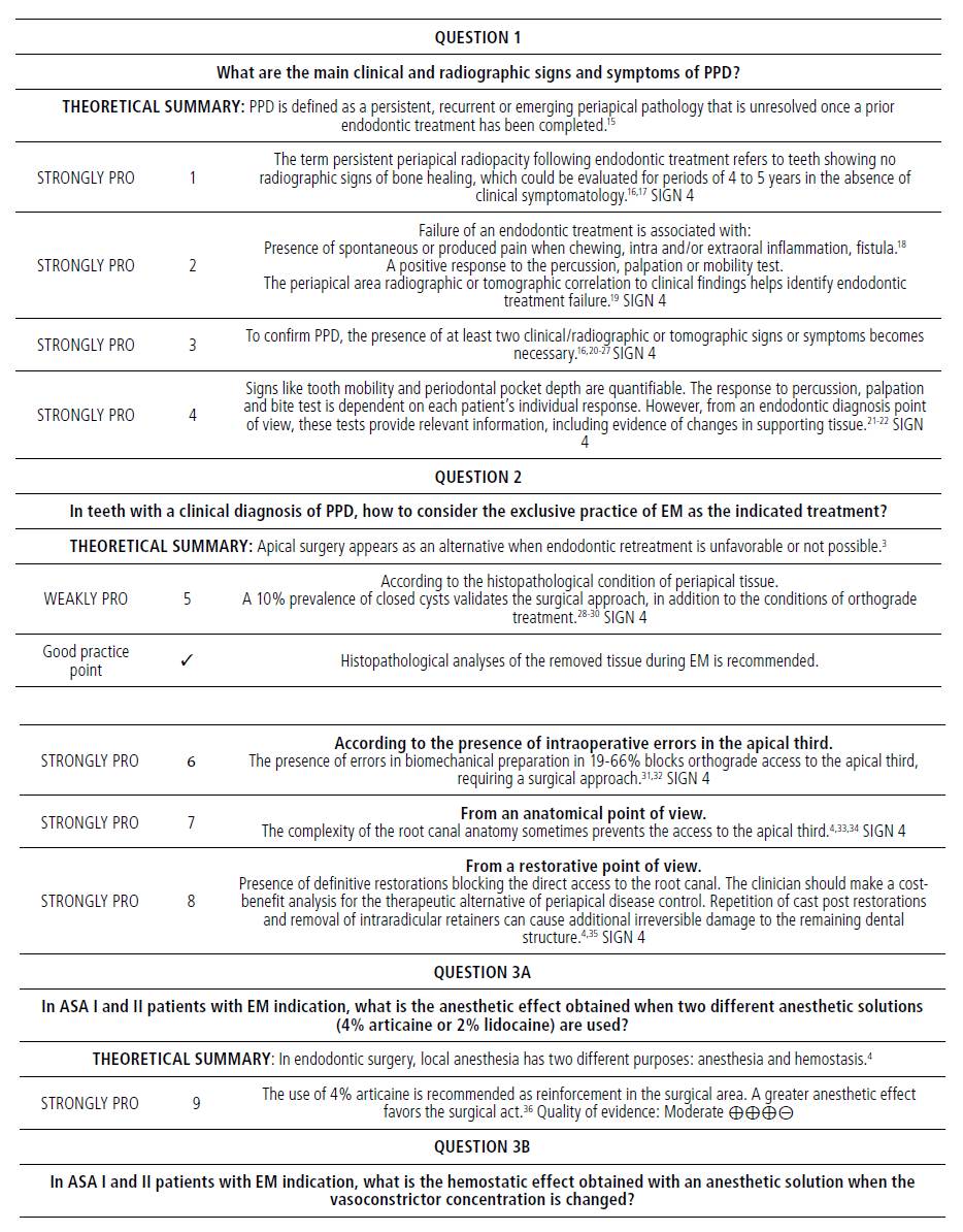
There was unanimous consensus in the identification of PPD,15-16 with categories like pathological process,28-30 occurrence of intraoperative accidents,31,32 anatomical conditions,33,34 and existing restorations35 indicating the practice of EM generally accepted and clarified by experts and interns, all of whom understood the recommendations (questions 1 and 2).

As for the implementation of different anesthetic solutions (question 3), the evidence65,66 exclusively focuses on either inferior dental nerve block in symptomatic pulp pain or extraction of third molars. This shows the lack of evidence for anesthetic options in EM.

The use of magnification systems, ultrasound technology, and regenerative retrograde filling materials is common in EM; 4 however, access to such technologies is limited in the country (questions 4 and 6). The legalization of bioceramics in Colombia67 overcomes opportunity barriers in terms of use, dissemination and future production of knowledge to support the evidence.

As an intraoperative factor, bleeding control (question 5) is a fundamental step for EM success; however, the evidence is not conclusive or innovative. The exploration of new alternatives like laser beam or natural polysaccharides is not yet demonstrated by the available experience due to the limited access to these techniques or because of the little existing evidence.

The consensus, based on existing literature and experience, does not recommend using regenerative techniques in EM,54,55 as it increases costs and unnecessarily triggers periapical bone repair (question 7). As an additional contribution, the discussion also included the influence of the pre-surgical state of supporting tissue as a risk factor to EM failure.64

On the other hand, the pre-surgical use of analgesics and antibiotics does not establish an effect on the outcome. The American Endodontic Association highlights the indiscriminate use and lack of clarity in criteria.68 The consensus unanimously decided against presurgical medication (questions 8 and 9) in systemically stable patients.

Finally, the recommendation of pre-surgical CBCT imaging, as a predictor of success in EM, was considered a “good practice point”. Even though the evidence in this regard is still low, the sensitivity and information offered by a prior three-dimensional image was considered a step in the good direction before the surgical approach. In addition, the existence of time-dependent factors indicates the need to evaluate EM at periods longer than one year.

CONCLUSIONS

Carried out by a multidisciplinary team, the CPGEM can be considered a supporting reference tool for general dentists, endodontists, and patients in need of therapeutic decisions for PPD. It includes recommendations, good practice points, and healthcare barriers as a support to university units and reference centers promoting the practice of EM. The CPGEM contributes sufficient evidence on issues affecting decision-making, case selection and quality of procedure.

RECOMMENDATIONS

The authors suggest implementing and adhering to the recommendations of these 11 indicators that support the practice of EM. Future projects should focus on validating the impact of the implementation of the CPGEM on qualified healthcare facilities.

ACKNOWLEDGEMENTS

The construction of the clinical questions in this document was possible thanks to the participation of PEFOUN professors José Manuel González Carreño, Javier Niño Barrera, Julio César Avendaño Rueda, Sara Quijano Guauque, and Freddy Jordán Mariño; methodological advisors Dairo Javier Marín Zuluaga, María Claudia Naranjo, and Mauricio Rodríguez, from the group of PEFOUN interns in the period 2016-2017, and patients Daniel Carrillo and Sandra Acuña, who benefitted from the surgical management of post-treatment periapical disease.

Finally, for the construction of the recommendations, the CPGEM was disseminated among coordinators, professors and interns in the Endodontic Graduate Programs of the following universities: del Valle, de Antioquia, del Bosque, Javeriana de Bogotá, Nacional and Fundación Universitaria CIEO.

REFERENCES

1. Siqueira JF Jr, Rôcas IN, Debelian G, Carmo FL, Paiva SS, Alves FR et al. Profiling of root canal bacterial communities associated with chronic apical periodontitis from Brazilian and Norwegian subjects. J Endod. 2008; 34(12): 1457-61. DOI: https://doi.org/10.1016/j.joen.2008.08.037Links ]

2. Hülsmann M. Epidemiology of post-treatment disease. Endod topics. 2016; 34: 42-63. DOI: https://doi.org/10.1111/etp.12096Links ]

3. Tsesis I, Rosen E, Taschieri S, Telishevsky Strauss Y, Ceresoli V, Del Fabbro M. Outcomes of surgical endodontic treatment performed by a modern technique: an updated meta-analysis of the literature. J Endod. 2013; 39(3): 332-9. DOI: https://doi.org/10.1016/j.joen.2012.11.044Links ]

4. Kim S, Kratchman S. Modern endodontic surgery concepts and practice: a review. J Endod. 2006; 32(7): 602-23. DOI: https://doi.org/10.1016/j.joen.2005.12.010Links ]

5. von Arx T, Jensen SS, Hänni S, Friedman S. Five-year longitudinal assessment of the prognosis of apical microsurgery. J Endod. 2012; 38(5): 570-9. DOI: https://doi.org/10.1016/j.joen.2012.02.002Links ]

6. Colombia. Plan obligatorio de salud POS. Bogotá: Ministerio de Salud y la Protección Social, 2019 [ Links ]

7. Sistema Nacional de Salud. Biblioteca de Guías de práctica clínica del sistema nacional de salud [Internet]; 2009. Available in: http://portal.guiasalud.es/emanuales/elaboracion/documentos/Manual%20metodologico%20-%20Elaboracion%20GPC%20en%20el%2Links ]

8. National Institute for Health and Clinical Excellence. The guidelines manual. London [Internet]; 2009. Available from: http//www.nice.org.ukLinks ]

9. Evans G, Bishop K, Renton T. Guidelines for surgical endodontics. Faculty of dental surgery. London: Royal College of Surgeons of England; 2012 [ Links ]

10. España, Ministerio de Sanidad y Consumo Elaboración de guías de práctica clínica en el sistema nacional de salud: manual metodológico [Internet]. Madrid: Ministerio de Sanidad y Consumo; 2007. Available in: http://www.madrid.org/bvirtual/BVCM017418.pdfLinks ]

11. Colombia. Ministerio de Salud, COLCIENCIAS. Guía metodológica para la elaboración de guías de práctica clínica con evaluación económica en el sistema general de seguridad social en salud colombiano: versión completa final. Bogotá: Ministerio de Salud y Protección Social; 2013 [ Links ]

12. Schünemann H, Brozek J, Oxman AE. GRADE handbook for grading the quality of evidence and strength of recommendations. Version 3.2.. [Internet]. The GRADE Working Group; 2009. Available in: http://www.cc-ims.net/gradeproLinks ]

13. Shea BJ, Grimshaw JM, Wells G a, Boers M, Andersson N, Hamel C, et al. Development of AMSTAR: a measurement tool to assess the methodological quality of systematic reviews. BMC Med Res Methodol. 2007; 15: 7-10. DOI: https://doi.org/10.1186/1471-2288-7-10Links ]

14. SIGN. Sign 50: a guideline developer’s handbook [Internet]. Edinburgh: Scottish Intercollegiate Guidelines Network; 2011. p. 111. Available in: http://www.sign.ac.ukLinks ]

15. Friedman SJ. Considerations and concepts of case selection in the management of post-treatment endodontic disease (treatment failure). Endod Topics. 2002; 1(1): 54-78. DOI: https://doi.org/10.1034/j.1601-1546.2002.10105.xLinks ]

16. Abbott, P. Diagnosis and management planning for root-filled teeth with persisting or new apical pathosis. Endod Topics. 2011; 19: 1-21. DOI: https://doi.org/10.1111/j.1601-1546.2010.00252.xLinks ]

17. Yu VS, Khin LW, Hsu CS, Yee R, Messer HH. Risk score algorithm for treatment of persistent apical periodontitis. J Dent Res. 2014; 93(11): 1076-82. DOI: https://doi.org/10.1177/0022034514549559Links ]

18. Martínez P, Marín DJ, Suárez LC, García CC. Signos y síntomas clínicos predictores de cicatrización apical 12 meses después de microcirugía endodóntica. Univ Odontol. 2015; 34(73): 87-96. DOI: http://dx.doi.org/10.11144/Javeriana.uo34-73Links ]

19. Kirkevang L-L, Wenzel A. Risk indicators for apical periodontitis. Community Dent Oral Epidemiol. 2003; 3(1): 59-67 [ Links ]

20. Weissman J, Johnson J, Anderson M, Hollender L, Huson T, Paranjpe A et al. Association between the presence of apical periodontitis and clinical symptoms in endodontic patients using cone-beam computed tomography and periapical radiographs. J Endod. 2015; 41(11): 1824-9. DOI: https://doi.org/10.1016/j.joen.2015.06.004Links ]

21. Levin LG, Law AS, Holland GR, Abbott PV, Roda RS. Identify and define all diagnostic terms for pulpal health and disease states. J Endod. 2009; 35(12): 1645-57. DOI: https://doi.org/10.1016/j.joen.2009.09.032Links ]

22. Klausen B, Helbo M, Dabelsteen E. A differential diagnostic approach to the symptomatology of acute dental pain. Oral Surg Oral Med Oral Pathol. 1985; 59(3): 297-301. DOI: https://doi.org/10.1016/0030-4220(85)90170-7Links ]

23. Venskutonis T, Daugela P, Strazdas M, Juodzbalys G. Accuracy of digital radiography and cone beam computed tomography on periapical radiolucency detection in endodontically treated teeth. J Oral Maxillofac Res. 2014; 5(2): e1. DOI: https://doi.org/10.5037/jomr.2014.5201Links ]

24. Fernandez R, Cadavid D, Zapata S, Alvarez L, Restrepo F. Impact of three radiographic methods in the outcome of nonsurgical endodontic treatment: a five-year follow-up. J Endod. 2013; 39(9): 1097-103. DOI: https://doi.org/10.1016/j.joen.2013.04.002Links ]

25. Low K, Dula K, Bürgin W, von Arx T. Comparison of periapical radiography and limited cone-beam tomography in posterior maxillary teeth referred for apical surgery. J Endod. 2008; 34(5): 557-62. DOI: https://doi.org/10.1016/j.joen.2008.02.022Links ]

26. Abella F, Patel S, Duran-Sindreu F, Mercadé M, Bueno R, Roig M. Evaluating the periapical status of teeth with irreversible pulpitis by using cone-beam computed tomography scanning and periapical radiographs. J Endod. 2012; 38(12): 1588-91. DOI: https://doi.org/10.1016/j.joen.2012.09.003Links ]

27. Metska M, Parsa A, Aartman I, Wesselink P, Rifat Ozok A. Volumetric changes in apical radiolucencies of endodontically treated teeth assessed by cone-beam computed tomography 1 year after orthograde retreatment. J Endod. 2013; 39(12): 1504-9. DOI: http://dx.doi.org/10.1016/j.joen.2013.08.034Links ]

28. Nair PN, Pajarola G, Schroeder HE. Types and incidence of human periapical lesions obtained with extracted teeth. Oral Surg Oral Med Oral Pathol Oral Radiol Endod. 1996; 81(1): 93-102 [ Links ]

29. Nair PN. New perspectives on radicular cysts: do they heal? Int Endod J. 1998; 31(3): 155-60 [ Links ]

30. Nair PN. Pathogenesis of apical periodontitis and the causes of endodontic failures. Crit Rev Oral Biol Med. 2004; 15(6): 348-81 [ Links ]

31. Mozayeni MA, Asnaashari M, Modaresi SJ. Clinical and radiographic evaluation of procedural accidents and errors during root canal therapy. Iran Endod J. 2006; 1(3): 97-100 [ Links ]

32. Haji-Hassani N, Bakhshi M, Shahabi S. Frequency of iatrogenic errors through root canal treatment procedure in 1335 charts of dental patients. J Int Oral Health. 2015; 7(Suppl 1): 14-7 [ Links ]

33. Jafarzadeh H, Abbott PV. Dilaceration: review of an endodontic challenge. J Endod. 2007; 33(9): 1025-30. DOI: https://doi.org/10.1016/j.joen.2007.04.013. Epub 2007 May 23Links ]

34. Kim S, Pecora G, Rubinstein R. Comparison of traditional and microsurgery in endodontics. In: Color atlas of microsurgery in endodontics. Philadelphia: W.B. Saunders; 2001. 5-11 [ Links ]

35. Gil-García CD, Quijano-Guauque SB, Marín-Zuluaga D, Marín-Zuluaga DJ, García Guerrero CC. Remanente de la obturación endodóntica en dientes restaurados con retenedor intra-radicular y su relación con la condición periapical post-tratamiento. Acta Odontol Col. 2016; 6(2): 31-44 [ Links ]

36. Kung, J, McDonagh M, Sedgley CM. Does articaine provide an advantage over lidocaine in patients with symptomatic irreversible pulpitis? a systematic review and meta-analysis. J Endod. 2015; 41(11): 1784-94. DOI: https://doi.org/10.1016/j.joen.2015.07.001Links ]

37. Kim S. Principles of endodontic microsurgery. Dent Clin North Am. 1997; 41(3): 481-97 [ Links ]

38. Rubinstein RA, Kim S. Short-term observation of the results of endodontic surgery with the use of a surgical operation microscope and super-EBA as root-end filling material. J Endod. 1999; 25(1): 43-8. DOI: https://doi.org/10.1016/S0099-2399(99)80398-7Links ]

39. Carr GB. Microscope in endodontics. J Calif Dent Assoc. 1992; 20(11): 55-61 [ Links ]

40. Carr GB. Surgical endodontics. In: Cohen S, Burns R editor. Pathways of the pulp, 6th ed. St Louis: Mosby, 1994. 531 [ Links ]

41. Pecora G, Andreana S. Use of dental operating microscope in endodontic surgery. Oral Surg Oral Med Oral Pathol. 1993; 75(6): 751-8. DOI: https://doi.org/10.1016/0030-4220(93)90435-7Links ]

42. Setzer FC, Kohli MR, Shah SB, Karabucak B, Kim S. Outcome of endodontic surgery: a meta-analysis of the Literature - Part 2: comparison of endodontic microsurgical techniques with and without the use of higher magnification. J Endod. 2012; 38(1): 1-10. DOI: https://doi.org/10.1016/j.joen.2011.09.021Links ]

43. Del Fabbro M, Taschieri S. Endodontic therapy using magnification devices: a systematic review. J Dent. 2010; 38(4): 269-75. DOI: https://doi.org/10.1016/j.jdent.2010.01.008Links ]

44. Von Arx T, Peñarrocha M, Jensen S. Prognostic factors in apical surgery with root-end filling: a meta- analysis. J Endod. 2010; 36(6): 957-73. DOI: https://doi.org/10.1016/j.joen.2010.02.026Links ]

45. Menendez-Nieto I, Cervera-Ballester J, Maestre-Ferrín L, Blaya.Tárraga J, Peñarrocha-Oltra D, Peñarrocha- Diago M. Hemostatic agents in periapical surgery: a randomized study of gauze impregnated in epinephrine versus aluminum chloride. J Endod. 2016; 42(11): 1583-87. DOI: https://doi.org/10.1016/j.joen.2016.08.005Links ]

46. Jang Y, Kim H, Roh BD, Kim E. Biologic response of local hemostatic agents used in endodontic microsurgery. Restor Dent Endod. 2014; 39(2): 79-88. DOI: https://doi.org/10.5395/rde.2014.39.2.79Links ]

47. Clé-Ovejero A, Valmaseda-Castellón E. Haemostatic agents in apical surgery. A systematic review. Med Oral Patol Oral Cir Bucal. 2016; 21(5): e652-7. DOI: https://dx.doi.org/10.4317%2Fmedoral.21109Links ]

48. Kim S, Song M, Shin SJ, Kim E. A randomized controlled study of mineral trioxide aggregate and super ethoxybenzoic acid as root-end filling materials in endodontic microsurgery: long-term outcomes. J Endod. 2016; 42(7): 997-1002. DOI: https://doi.org/10.1016/j.joen.2016.04.008Links ]

49. Zhou W, Zheng Q, Tan X, Song D, Zhang L, Huang D. Comparison of mineral trioxide aggregate and iRoot BP plus root repair material as root-end filling materials in endodontic microsurgery: a prospective randomized controlled study. J Endod. 2017; 43(1): 1-6. DOI: https://doi.org/10.1016/j.joen.2016.10.010Links ]

50. Shinbori, N, Grama AM, Patel Y, Woodmansey K, He J. Clinical outcome of endodontic microsurgery that uses endoSequence BC root repair material as the root-end filling material. J Endod. 2015; 41(5): 607-12. DOI: https://doi.org/10.1016/j.joen.2014.12.028Links ]

51. Karring T. Regenerative periodontal therapy. J Int Acad Periodontol. 2000; 2(4): 101- 9 [ Links ]

52. Von Arx T, Alsaeed M: The use of regenerative techniques in apical surgery: a literature review. Saudi Dent J. 2011; 23(3): 113-27. DOI: https://dx.doi.org/10.1016%2Fj.sdentj.2011.02.004Links ]

53. Bottino MC, Thomas V, Schmidt G, Vohra YK, Chu TM, Kowolik MJ et al. Recent advances in the development of GTR/GBR membranes for periodontal regeneration-a materials perspective. Dent Mater. 2012; 28(7): 703-21. DOI: https://doi.org/10.1016/j.dental.2012.04.022Links ]

54. Tsesis I, Rosen E, Tamse A, Taschieri S, Del Fabbro M. Effect of guided tissue regeneration on the outcome of surgical endodontic treatment: a systematic review and meta-analysis. J Endod. 2011; 37(8): 1039-45. DOI: https://doi.org/10.1016/j.joen.2011.05.016Links ]

55. Deng Y, Zhu X, Yang J, Jiang H, Yan P, The effect of regeneration techniques on periapical surgery with different protocols for different lesion types: a meta-analysis. J Oral Maxillofac Surg. 2016; 74(2): 239-46. DOI: https://doi.org/10.1016/j.joms.2015.10.007Links ]

56. Lindeboom JAH, Frenken JWH, Valkenburg P, van den Akker HP. The role of preoperative prophylactic antibiotic administration in periapical endodontic surgery: a randomized, prospective double-blind placebo-controlled study. Int Endod J. 2005; 38(12): 877-81. DOI: https://doi.org/10.1111/j.1365-2591.2005.01030.xLinks ]

57. Kvist T, Reit C. Postoperative discomfort associated with surgical and nonsurgical endodontic retreatment. Endod Dent Traumatol. 2000; 16(2): 71-4 [ Links ]

58. Vanegas H, Schaible HG. Prostaglandins and cyclooxygenases in the spinal cord. Prog Neurobiol. 2001; 64(4): 327-63 [ Links ]

59. Aznar-Arasa L, Harutunian K, Figueiredo R, Valmaseda-Castellón E, GayEscoda C. Effect of preoperative ibuprofen on pain and swelling after lower third molar removal: a randomized controlled trial. Int. J. Oral Maxillofac Surg. 2012; 41(8): 1005-9. DOI: https://doi.org/10.1016/j.ijom.2011.12.028Links ]

60. Suebnukarn S, Rhienmora P, Haddawy P. The use of cone-beam computed tomography and virtual reality simulation for pre-surgical practice in endodontic microsurgery. Int. Endod J. 2012; 45(7): 627-32. DOI: https://doi.org/10.1111/j.1365-2591.2012.02018.xLinks ]

61. Matherne RP, Angelopoulos C, Kulild JC, Tira D. Use of cone-beam computed tomography to identify root canal systems in vitro. J Endod. 2008; 34(1): 87-9. DOI: https://doi.org/10.1016/j.joen.2007.10.016Links ]

62. Leonardi Dutra K, Haas L, Porporatti AL, Flores-Mir C, Nascimento Santos J, Mezzomo LA et al. Diagnostic accuracy of cone-beam computed tomography and conventional radiography on apical periodontitis: a systematic review and meta-analysis. J Endod. 2016; 42(3): 356-64. DOI: https://doi.org/10.1016/j.joen.2015.12.015Links ]

63. Grondahl HG, Huumonen S. Radiographic manifestations of periapical inflammatory lesions. Endod Topics. 2004; 8: 55-67. DOI: https://doi.org/10.1111/j.1601-1546.2004.00082.xLinks ]

64. García GCC, Quijano GS, Molano N, Pineda GA, Nino B JL, Marín ZDJ. Predictors of clinical outcomes in endodontic microsurgery: a systematic review and meta-analysis. G ital endod. 2017; 31(1): 2-13. DOI: https://doi.org/10.1016/j.gien.2017.03.001Links ]

65. Nagendrababu V, Pulikkotil SJ, Suresh A, Veettil SK, Bhatia S, Setzer FC. Efficacy of local anaesthetic solutions on the success of inferior alveolar nerve block in patients with irreversible pulpitis: a systematic review and network meta-analysis of randomized clinical trials. Int Endod J. 2019; 52(6): 779-89. DOI: https://doi.org/10.1111/iej.13072Links ]

66. Shahi S, Rahimi S, Yavari HR, Ghasemi N, Ahmadi F. Success rate of 3 injection methods with articaine for mandibular first molars with symptomatic irreversible pulpitis: a CONSORT randomized double-blind clinical trial. J Endod. 2018; 44(10): 1462-66. DOI: https://doi.org/10.1016/j.joen.2018.07.010Links ]

67. Colombia. Ministerio de Salud y Protección Social, Instituto Nacional de Vigilancia de Medicamentos y Alimentos - INVIMA. Resolución No. 2017044599 de 23 de octubre de 2017: por la cual se concede un registro sanitario. Bogotá, Ministerio de Salud; 2017 [ Links ]

68. Germack M, Sedgley CM, Sabbah W, Whitten B. Antibiotic use in 2016 by members of the American Association of Endodontists: report of a national survey. J Endod. 2017; 43(10): 1615-22. DOI: https://doi.org/10.1016/j.joen.2017.05.009Links ]

** The development of these guidelines was sponsored by a couple of local fellowships: “Convocatoria estímulo a la investigación Facultad de Odontología 2016 - 1er Corte” Date: First semester of 2016, and “Convocatoria nacional de proyectos para el fortalecimiento de la investigación, creación e innovación de la Universidad Nacional de Colombia 2016-2018”.

***García-Serrano LM, Lara-Rodríguez DC, García-Guerrero CC. Clinical practice guidelines for the surgical endodontic management of post-treatment periapical disease. Rev Fac Odontol Univ Antioq. 2019; 30(2): 211-223. DOI: http://dx.doi.org/10.17533/udea.rfo.v30n2a8

Received: December 03, 2017; Accepted: March 19, 2019

*CORRESPONDING AUTHOR Claudia Carmiña García Guerrero Universidad Nacional de Colombia (+57) 1 316 5000 Ext 16018 ccgarciag@unal.edu.co Carrera 30 #45-03 Edificio 210 Bogotá. Colombia

CONFLICTS OF INTEREST

The authors state that they have no conflict of interest.

Creative Commons License This is an open-access article distributed under the terms of the Creative Commons Attribution License