


 English (pdf)
 Article in xml format
 Article references
 Send this article by e-mail
 Cited by SciELO
  Cited by Google
  Similars in
SciELO
Similars in
SciELO  Similars in Google
 Permalink
INTRODUCTION
Rehabilitation with removable partial prosthesis facilitates treatment and the subsequent resolution of the patients’ complex clinical problems, such as large toothless spaces and absence of distal parts, or even the recovery of occlusal vertical dimension due to tooth loss (collapse) in the posterior occlusal table. This requires performing the entire treatment in a relatively short time and at a substantially lower operational cost compared to other treatment modalities.1 However, dissatisfaction has been observed in a considerable number of patients with this type of dental prosthesis.
The design and development of removable partial prostheses often involves the use of retainers for direct retention. These can have various disadvantages, favoring the appearance of bacterial plaque accumulation sites, the risk of abrasion of tooth surfaces that are in close contact with each other, and the deformation of the prosthetic structure over time. In addition, prosthetic appearance has often been reported as a key element in patient’s acceptance of removable partial prosthetic rehabilitation.4,5,6 The appearance of removable partial prostheses is highly important to patients, and the structures that are visible when opening the mouth are a common cause of rejection.2
A critical analysis by Santana et al reveals that dissatisfaction can be avoided in most cases if prostheses are made in such a way that the basic functional requirements are addressed with minimal exposure during mastication and phonation, incorporating patient’s aesthetic demands, which are usually disregarded.2) It is unrealistic to assume that by achieving an acceptable chewing capacity and therefore adequate retention, stability and prosthetic support, patients will accept poor esthetic appearance.1,3
This study aims to find a possible solution to one of the most used dental treatments in the Chilean population.16 Certainly, the biggest challenge is how to better deal with the esthetic aspect of a removable partial prosthesis without affecting its functionality, especially the retentive capacity. In short, this in vitro study suggests that by achieving adequate parallelism of the guiding planes in biomechanical preparation, there will be no loss of functional retention when removing anterior retainers in Kennedy Clasification type III.
METHODS
Operational definitions: this is an in vitro study
Presence/absence of anterior retainers
This categorizes the presence or absence of anterior retaining clasps of the retaining systems, dividing them into two classes: denture bases with anterior retainers and bases with no anterior retainers. It is therefore a categorical variable.
Type of anterior attachments
An evaluation was conducted, classifying attachments into two groups: attachments of the same type if the same type of part is found on both sides of the arcade, and attachments of different type if they differ from each other on both sides of the arcade.
Retention
The retention value on each denture base was assessed as the maximum tensile strength obtained in each evaluation, which is a force value recorded in Newton as an international unit.
Type of edentulism
Seven edentulism types were determined, with the most being Kennedy Class III subdivision 1, with anterior abutments corresponding to first premolars and/or canines, and a toothless area delimited at the most by a second molar as a posterior abutment.
Sample collection
To carry out the proposed study, 7 ideal maxillary models with different levels of partial tooth loss were obtained in dental plaster type IV, based on a silicone pattern. The abutment pieces were replaced on them with similes of aladdinite (Nissin®), thus obtaining 7 partially edentulous jaw models, all of whom corresponded to Class III Kennedy subdivision 1 of partially toothed arches. Each model included anterior attachments with esthetics affected by retainers, canines and first premolars. All tooth loss possibilities that were part of the classification and that considered aesthetic compromise were previously determined.
For each obtained model, the metal base most suited to its edentulism was designed and drawn, considering all the parts of the removable partial prosthesis, determining the position of the retaining complex after further analysis with a paralleling surveyor (a high-precision auxiliary equipment for the design and processing of prostheses, used to determine, create and confirm the parallelism of two or more surfaces) in order to identify the guiding planes as well as prosthetic and dental equators. This design was also modified in the prosthetic site area, where two metal washers were symmetrically located, one per side, in the center of each void. This was done to make sure that the traction tests were correctly performed.
Each model was individually analyzed on the surveyor to determine the most suitable insertion and removal axis, establish the location of the model’s elements, and determine the necessary modifications for the abutments, in order to carry out the carving of guiding planes according to each case.
Later, a handpiece (rotating instrument) was positioned on the vertical arm of the paralleling surveyor, using a diamond cylindrical drill. The corresponding carvings were made to establish the guiding planes on the position determined for each model according to the conclusions from the analysis.13
The biomechanical preparation for each model was carried out. This involved the preparation and carving of each bed to support the abutment teeth, following the indications of the proposed model for each case (Figure 1).
Once the above procedure was completed, the depth of each retaining area of the anterior abutments was measured, as well as the length of the guiding planes carved on the abutment in each model under study (Figure 2).
Source: by the authors
Figure 2 Measuring the convergence angle that determines the retention degree of the abutment tooth
Next, models were sent to the prosthetic laboratory to manufacture the metal bases as designed, using Dentaurum® preformed waxes to wax up the structures, and casting in cobalt-chromium alloy. Each base corresponds to a conventional removable partial prosthesis model, including cast posts of the anterior region.
Once the prosthetic bases were obtained, it was verified that they were in proper condition, evaluating the presence of a correct adjustment, meaning that both the beds and the guiding planes were in close contact with their respective prosthetic elements (occlusal support and minor connector).
Source: by the authors
Figure 3 Evaluation of the prosthetic base, including proper adaptation and adjustment to the plaster model
To perform the tensile tests, it was necessary to adjust the metal bases with a metal circular washer designed for this purpose and made of the same alloy of the base (Cr-Co), measuring 0.8 mm in diameter (Figure 4). These were laterally welded to the base and placed equidistant from the abutments of each model under study.
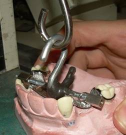
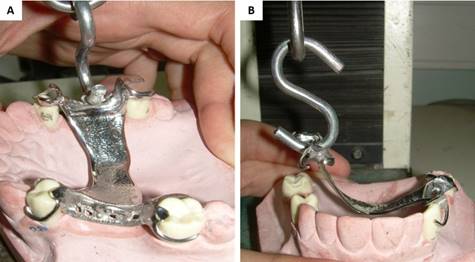
The bases were subjected to tensile analysis on a Tinius Olsen H5K-S14 universal traction machine (Figure 5). This machine works by vertically loading the metal bases located on the models from the point established in the prosthetic saddles (washers). First, the bases bilateral tractions were carried out with a device specially designed to create equidistant and simultaneous bilateral traction. Then the unilateral tractions were performed, arbitrarily choosing the left side for all models (Figures 6 and 7). The obtained results indicate the maximum necessary force (in Newtons) to traction the metal base vertically until surpassing the prosthetic equator from its final location.

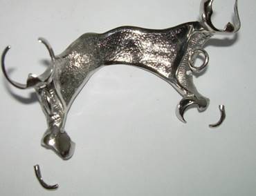
For each metal base, 10 bilateral and unilateral measurements were made, obtaining 20 force values per base. Once the measurements in the 7 models were taken, the retaining clasp of each retaining complex of anterior abutments of the metal bases was cut, thus eliminating the unattractive element.
The bases were then set, carrying out the same previous procedure, i.e., 10 bilateral and 10 unilateral measurements for each base, thus completing 40 different force values for each metal base. In total, 280 measurements were obtained, with n = 70 for each analysis (Figure 9).
Statistical Analysis
The obtained data were subjected to descriptive statistical analysis with the use of histograms, as well as the Shapiro-Wilkins and Kolmogorov-Smirnov descriptive tests, yielding a normal distribution of data, which allowed to choose the most appropriate statistical test.
The results were analyzed with Student’s T statistical test of independent variables and different variances with a 0.05 significance, comparing the mean and standard deviations between two groups of data, and determining whether the differences between groups are statistically significant or just random differences, and whether they are within the ideal ranges of retention force values required in RPD.
RESULTS
Figure 10 shows the differences in the measured force averages for the entire sample (n = 70) in each.
Retention (Newton)
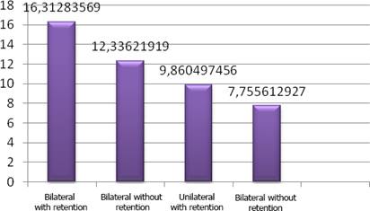
Source: by the authors
Figure 10 Retention of metal prosthetic bases with and without retainers in the anterior region when subjected to uni- and bilateral traction
The average tensile force value required to displace the metal base bilaterally in bases with anterior retainers was 16.93 N, with a standard deviation of 3.43 N and a range of 17.67 N, determined by the difference between the minimum and maximum values, 10 and 27.67 N respectively, as shown in Table 2.
Table 2 Bilateral traction with anterior retainers
| n | Average | Standard Deviation | Variance | Minimum | Maximum | Range |
|---|---|---|---|---|---|---|
| 70 | 16.93 | 3.43 | 11.79 | 10.00 | 27.67 | 17.67 |
Source: by the authors
The average tensile force value required to displace the metal base bilaterally in bases without anterior retainers was 12.84 N, with a standard deviation of 2.379 N, a minimum value of 8.666 N and a maximum of 19 N, determining a force range of 10.334 N, as shown in Table 3.
Table 3 Bilateral traction without anterior retainers
| n | Average | Standard Deviation | Variance | Minimum | Maximum | Range |
|---|---|---|---|---|---|---|
| 70 | 12.84 | 2.38 | 5.663 | 8.67 | 19.00 | 10.33 |
Source: by the authors
The average tensile force value required to displace the metal base unilaterally in bases with anterior retainers was 10.42 N, with a standard deviation of 2.08 N, a range of 9 N, determined by a minimum force value of 6.5 N and a maximum of 15.5 N. These results can be seen in Table 4.
Table 4 Unilateral traction with anterior retainers
| n | Average | Standard Deviation | Variance | Minimum | Maximum | Range |
|---|---|---|---|---|---|---|
| 70 | 10.42 | 2.08 | 4.33 | 6.50 | 15.50 | 9.00 |
Source: by the authors
Table 5 shows that the result of the average tensile force value required to displace the metal base unilaterally in bases without anterior retainers was 8.07 N, with a standard deviation of 1.92 and minimum and maximum force values of 4.67 N and 12.83 N respectively, determining a range of 8.17 N.
DISCUSSION
This in vitro study aimed to evaluate a new alternative RPD treatment in which retention is not affected by removing retainers from anterior abutments in Kennedy’s Class III dentures, subdivision 1.
The results obtained in this study showed that the tensile force values in the presence of anterior retainers were higher in the tensile tests with retainers compared to the tests with no retainers, as expected. These differences are statistically significant (p = 0.0000) in both unilateral and bilateral traction. However, the retention values yielded by the bases with no retainers appear to be sufficient and acceptable to prevent the displacement of a prosthetic apparatus in which-according to the literature-a conventional RPD apparatus needs a force of 500 Gf, or 4.90 N, to perform adequately.8
This value is exceeded, based on the results obtained in anterior retainer-free bases, which were on average 12.84 N and 8.07 N in bilateral and unilateral tractions, respectively. These values were compared to the standard value (4.90 N), yielding statistically significant differences (p = 0.0000) for both uni- and bilateral tractions, in the absence of anterior retainers. The retention action offered by carved guiding planes with controlled parallelism and their close contact with the minor RPD connectors is likely to provide key assistance in retention when the biomechanical preparation prior to RPD installation is properly done.6-9
The unilateral tensile tests were carried out to verify how the prosthetic apparatus would work during the unilateral masticatory process. The force that opposes the unilateral displacement of a prosthesis is basically determined by the retention complexes of the working side (the one that produces traction), provoking on the opposite side a rotation around the retaining clasp contact with the abutment, which undoubtedly generates deleterious forces on it and its periodontium, which cannot be contained because rotation disables any function of the elements that cause reciprocation.6,13,15
In addition, the force values obtained on dental bases bilaterally loaded in the absence of anterior retainers were higher than those in bases that had all the retaining clasps but unilaterally loaded. This perhaps happens because, by generating traction from both sides, the anti-displacement force is provided by both the retaining systems and the guiding planes, which provide additional friction retention. However, this is nullified in unilateral traction.3,6,7,12,15
CONCLUSIONS
This in vitro study aimed to compare the dental loading retention in partial removable prosthesis, both in the presence and absence of anterior retainers with parallel guiding planes, analyzing the possibility of eliminating them and thus solve a critical aesthetic problem of RPDs with anterior abutments. The obtained results lead to conclude that:
There are statistically significant retention values differences in denture bases with no anterior retaining clasps compared to bases with clasps in both bilateral and unilateral tractions.
There are statistically significant retention values differences in denture bases with and without bilateral traction retainers with respect to the values reported in the literature as sufficient to retain a removable prosthetic device, with significantly higher values as the results of this study show.
Statistically significant differences were also detected when comparing denture bases with and without retainers in unilateral tractions, compared to the standard value reported in the literature, also in favor of the results of this study.
The obtained strength values do not decrease as the literature states as a sufficient and acceptable value to retain a prosthetic device, and it has been shown that the retaining strength is sufficient to retain a prosthesis and to allow a smooth functioning as shown in this in vitro study.

