INTRODUCTION
Amelogenesis Imperfecta (AI) refers to a group of genetic alterations of the normal structure of the dental enamel that disturbs its clinical appearance. AI is classified as hypoplastic, hypocalcified, and hypomaturation. These abnormalities may exist in isolation or associated with other systemic conditions in the context of a syndrome1. AI’s presentation can vary depending on the type and severity2, teeth affected may be discolored, sensitive or prone to disintegration3. This condition is characterized by abnormal enamel formation caused by gene mutations that alter the quality and/or quantity of enamel4. Impaired esthetics, tooth hypersensitivity, wide pulp chamber, higher risk of dental caries, and decreased occlusal vertical dimension are among the challenges faced in the management of AI. Dental anomalies associated with AI include enamel disorders, anterior open bite, pulpal calcifications, delayed eruption of teeth, pathological root and crown resorption, and taurodontism5,6.
In an effort to classify the disease, particular phenotypes have been defined but this approach can be confounded by mixed phenotypes. Hypoplastic AI describes thin but mineralized enamel, or in extreme cases, the complete absence of enamel, that results from failure during the secretory stage. Hypomineralized AI is caused by maturation stage failure; giving rise to enamel that is of full thickness but is weak and fails prematurely. The hypomineralized phenotype can be further subdivided into hypomaturation and hypocalcified AI. The former is caused by incomplete removal of protein from the enamel matrix and produces brittle enamel, while the latter is characterized by insufficient transport of calcium ions into the developing enamel and produces soft enamel7,8,9.
AI passes down through families as a dominant trait. This condition is characterized by abnormal enamel formation caused by gene mutations that alter the quality and/or quantity of enamel. The literature review showed that mutations in 16 genes (AMELX, AMBN, ENAM, LAMB3, LAMA3, ACPT, FAM83H, C4ORF26, SLC24A4, ITGB6, AMTN, MMP20, KLK4, WDR72, STIM1, GPR68) are responsible for non-syndromic hypoplastic, hypocalcified, or hypomaturation AI1. FAM20A is essential for amelogenesis, but is dispensable for dentinogenesis10. Amelogenin is the most abundant enamel matrix protein, most of which is encoded by the amelogenin gene in the X-chromosome (AMELX). More than 16 alternative splicing transcripts have been identified in the murine Amelx gene2.
Clinically the presentation of the AI can have variety of presentation. This condition can impact both primary and permanent dentition7. The clinical implications of AI are significant for patients and clinicians. They include esthetic involvement, hypersensitive teeth, mastication impairment and psychosocial affections. The varying etiology of AI conjures a wide array of clinicals features whose restorative management poses a challenge for dentist11.
The treatment for AI depends on the severity of the problem and age of the patient. It is crucial to plan a proper intervention, which requires collaboration among dental specialties to execute comprehensive dental treatment in order to provide a long-term solution with adequate esthetics4. The diagnosis and planification of plans for coverage can be significative in the life of a patient with AI, while some of them have received proper canalization others can spend many times without satisfying results.
Currently, there are no defined therapy recommendations available for patients suffering from AI. The mentioned therapies are more or less equal but no comprehensive therapy recommendation is evident. When treating patients suffering from AI, a comprehensive therapy of almost every dental discipline has to be considered. The earlier the diagnosis of AI is confirmed, the better the outcome is12. Management of a patient with AI involves a complex interplay of various factors5,13. To design an appropriate treatment plan, factors such as the quality and quantity of the enamel, age and socioeconomic status of the patient, the type and severity of the disorder, and the intraoral status should be taken into consideration14.
In order to achieve these goals a stepwise approach to treatment planning is required starting with the most conservative but aesthetically acceptable treatment15.
Having AI has a marked impact on the psychosocial health of affected people comparable with the impact of systemic health conditions, especially at younger ages16. Negative emotions that occur in adolescents when disagreement is perceived in the dentolabial aesthetics, will affect the social and intellectual life of the teenager. Multidisciplinary team is useful to collect, manage knowledge of self-esteem, self-concept and psychological formation of these patients17.
Case presentation
This is a case report of a male patient who is 12 years old, born of non-consanguineous parents. He is the product of first birth of a mother of 33 years old, whose prenatal and medical history was non contributory. The mother reported a normal pregnancy and delivery without complications. As an important antecedent she informed that the father had discolored teeth, but not so marked as the son. No other sibling or family had the enamel defect. He did not report any renal disease, a renal ultrasound showed no images of renal affection and laboratory exams, including calcium, phosphate, and creatinine levels were below the average.
The current case report presents a patient affected by AI whose diagnosis was unclear and confused with a trauma since the first visit to dentist while the patient had primary teeth. Then after each time he went to dental visit, the diagnosis was dental caries in all his dentition.
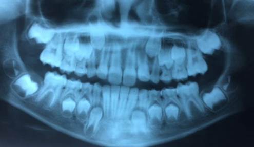
The orthopantomogram at 8 years old showed the presence of unerupted lower second and third molars and upper second molar, normal pattern of eruption of all teeth, without signs of periodontal injuries. A generalized thinning of enamel on all tooth surfaces was detected (Figure 1).
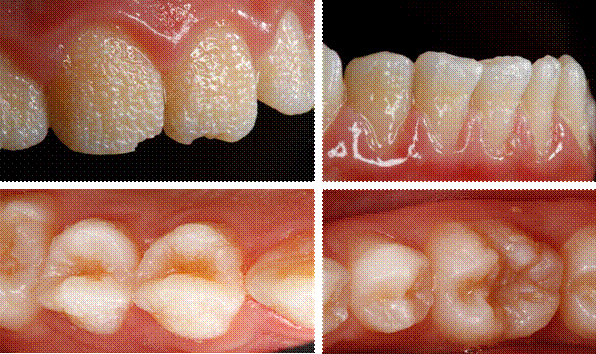
The mother provided consent for treatment and publication of the case. Diagnosis was done through history, clinical examination and imaging. The AI type in this case report was classified according to the clinical features (phenotypes) and radiographic images. The patient showed: Hypoplasic Amelogenesis imperfect. Thin and smooth enamel was noted affecting both primary and permanent dentition. The color was discolored light cream with particular shapes with absence of enamel similar as pits in incisive and deep fissures in molars, phenotype described as a quantitative defect hypoplastic type of Amelogenesis imperfecta (Figure 2 y 3). The diagnosis of hypoplasia (type I AI) was made.

By: the authors
Figure 3 View of upper incisors, lower incisors, premolar and molar with Amelogenesis imperfecta
Initially the treatment procedures included the diagnosis of Amelogenesis imperfecta done by a pediatric dentist, who refer the teenager FOR an evaluation with a pediatric nephrologist. Following this, the patient was evaluated by a psychologist, who applied a projective Drawing of the Human Figure test, Koppitz method. The next intervention was the rehabilitation plan by a prosthodontic joined with a dental technician. The treatment plan was carried out in step by step fashion until positive results were reached.
Intervention
Histopathological reports confirms that when enamel hypoplasia is the predominant clinical finding, the enamel is reduced in thickness. The enamel-dentin junction may show exaggerated scalloping. Areas of homogeneous aprismatic enamel or fused indistinct prisms are seen18. The histology phenotype described by Witkop showed that the most marked defects in the enamel were seen in the outer half19. For these reasons it was proper to restauration with direct resin veneers.
Minimal preparation was performed on lateral incisors and canines. In the central incisors no preparation was done. Red ring fine diamond bur was used in proximal faces and gingival area, a preparation with a marginal chamber finish for was done. Chamfer preparation design was complete in remaining enamel, nothing in dentin.
After teeth preparation Ultradent® Ultrapack retractor cord was used with Ultradent® Fischer’s Ultrapak packers. The adhesive technique includes deproteinization of the enamel with 5.25% sodium hypochlorite in solution for one minute and rinse. The enamel surface was treated with 35% phosphoric acid gel (Ultradent®) for 10 seconds. Then it was rinsed with water for 30 seconds and the excess water was removed. The Ultradent® peak adhesive was applied, and then air dried during 20 seconds, and then it was light cured in accordance with the manufacturer’s instructions. Freehand direct resin veneers, Ultradent brand, Vit-l-escence®. Color A2, A1, TM were done and polished with Jiffy System by Ultradent®, only mechanical. Polishing system was made with Jiffy System by Ultradent®, only mechanical. No final liquid was used. Finally, the occlusion was evaluated (Figure 4). In premolars and molars no intervention was done, because of the hygiene of the patient although is not common to leave the posteriors teeth without restorations.
A 12-month follow-up with clinical and radiographic examinations revealed no evidence of any undesirable effects of the treatment on the restored teeth or their supporting tissues.
DISCUSSION
This case report showed the challenge represented in the management of a patient with AI for the clinician. Although it has limitations, the dentist can see many variables that must be taken into consideration to provide a comprehensive intervention for the patient with this condition. The patient with AI not only has an enamel defect with problems of color or shape, but there are also lots of complications, that they may have during all their life and the scope giving after a dental treatment are numerous. The treatment options vary considerably depending on several factors such as the age of the patient, socio-economic status, periodontal condition, loss of tooth structure, severity of the disorder, and, most importantly, the patient’s cooperation. The clinician has to consider the long-term prognosis of the outcome20. Treatment of a patient with AI presents a real problem from both functional and esthetic points of view. An esthetic intervention will also result in an improvement in the patient’s quality of life21.
Treatment not only restored function and esthetic, but also showed a positive psychological impact and thereby improved perceived quality of life22. Particularly in this case because the recovery of the patient’s self-confidence was allowed by a multidisciplinary team. Studies in children with enamel defects have showed prior to treatment, that children reported high levels of worry, embarrassment and a perception that their teeth looked yellow and discolored. Following treatment, children thought their teeth looked much better, felt happier and more confident. They also felt very positive about their clinical experiences, rating the staff as extremely friendly and kind and reporting that procedures were clearly explained. Simple non-invasive dental treatment can have a positive effect on appearance-related satisfaction23.
The preventive, psychological, and curative measures of a young child with AI are notable25. The successful management of AI during childhood requires the cooperation and motivation of the patient and parents24. The esthetic and psychosocial aspects of a child growing are extremely important. It is known that young people may make negative psychosocial judgements on the basis of enamel appearance25.
On the other hand, when rehabilitation area is involved, intervention includes a wide variety of options like removal of surface stains, reducing sensitivity, maintaining the vertical dimension of occlusion, and the esthetics22. Indirect composite restorations can be a valuable tool for improving occlusion, esthetics and function in the treatment of children and adolescents. In most cases, direct composite fillings can be placed. However, in severe cases, these restorations may be more challenging and error-prone, especially when occlusal adjustments are necessary. Since composite materials do not require specific lamination strength and are easy to repair, they can be applied using the indirect technique, enablingthe conservation of more sound hard tissue than is possible when conventional restorations are used26. Previous reports indicate indirect restorations should be preferred over direct restorations in patients with AI27.
Composite resin restorations have been advocated to mask discoloration and improve dental esthetics with minimal or no tooth preparation to preserve tooth structure and provide satisfactory esthetics28. In this case report direct restorations were an excellent option for the patient because the greatest challenge was in the anterior sector. In the posterior zone, there was no intervention due to the poor oral hygiene of the patient.
Attention should first be given to the patient’s level of oral hygiene and dietary habits, which can compromise the rehabilitation procedures. Poor oral hygiene is a recognized problem in patients with AI, due to the rough enamel surface which causes biofilm retention and the sensitivity experienced when brushing29. Therefore, oral hygiene instructions are essential for the successful treatment30. Although a study by Kammoun et al. showed that hypoplasic AI patients have a low DMFT index, an alkaline pH of saliva enriched with Bacillus spp, Enterococcus faecalis and Enterococcus faecium and dental tissues more easily invaded by Lactobacilli than Streptococci. The combination of these bacteria seems to give AI patients protection against dental caries31. However, dental treatment in patients with AI should focus on restorations as soon as possible27.
It is important to highlight that the longevity of dental restorations in patients with AI is limited. Pousette et al. compared oral health and longevity of dental restorations in a group of young patients with AI to a control group. They included 82 patients with AI, 40 boys and 42 girls, 6 to 25 years old and a control group matched in age, gender and residential area. The longevity of dental restorations was significantly lower in the patients with AI. Patients with hypomineralized/hypomaturized AI have restorations of shorter longevity than those with hypoplasic AI32. In this particular case with hypoplasic AI, restorations can be durable due to the specific type of defect. Although further laboratory and clinical studies should be achieved to investigate the performance of minimally invasive indirect restorations bonded to enamel in patients with AI27.
Management is relevant at various stages of the developing dentition but it also should be considered the compromises which may have to be made depending on the child’s ability to cope. The importance of early diagnosis, prevention and the benefits of a multi-disciplinary approach are crucial33.
Early recognition followed by appropriate preventive and restorative care is essential in the successful management of AI. A multidisciplinary approach with careful planning from early childhood will maximize the treatment options available for the permanent dentition and optimize the final outcome6.
There is an urgent need of knowledge of causes, diagnosis and treatment of AI and also to detect the differential diagnosis with fluorosis, molar incisive hypomineralization or isolate hypoplasia. Even among the pediatric dentist who are the first to receive these patients, there is a need to improve the comprehension of the enamel defects34.
This case has a strength in the fact that the caregivers are aware to find a solution and are advocated to keep to all the appointments. Although the full rehabilitation of the patient was very expensive, we found grants with Ultradent®. The limitations of this case report are the challenges that represents covering all the patient’s needs and the late diagnosis done. It is important to underline that the interdisciplinary management needs to contemplate a genetic shift to identify potential mutations in these patients. Although the cost is expensive, it is necessary to involve this kind of test to have better follow-up and diagnosis of the family.
Another important consideration of patients with AI is the association between renal disease and AI which has been reported decades ago35,36. The contributions provided by this case are represented in the fact that this information will help dentists to identify this condition and understand the importance to refer patients with amelogenesis imperfecta to nephrologists as soon as possible. Pediatric dentists should obtain a detailed family history and past medical history with particular reference to urinary tract and should be refer for medical examination including renal function test and ultrasonography. The same recommendation is done to pediatric nephrologists that need to know the association that exists between amelogenesis imperfecta and renal disease37,38.
This treatment showed to improve occlusion, esthetics and self-image of the teenager. The satisfactory clinical result has made it possible to avoid more invasive and expensive treatments.


