


 Article in xml format
 Article references
 Send this article by e-mail
 Cited by SciELO
  Cited by Google
  Similars in
SciELO
Similars in
SciELO  Similars in Google
 Permalink
Introduction
The changes that take place during menopause, such as the loss of estrogen, have measurable effects. These include a decreased response to oxidative stress and cognitive deterioration (1,2). The loss of 17β-estradiol (E2) during menopause is partly responsible for these changes, since its intercellular receptors ERα and Erβ, and possibly G protein-coupled estrogen receptor 1 (GPER1), possess a regulatory function to consolidate memory at the level of the hippocampus in animal models (3). In peri-menopausal women, hypothalamic-pituitary insensitivity to estrogens (4) as well as blood dyscrasia have been observed. These induce aberrant cell cycles, which cause the re-entry of post-mitotic neurons in the cell cycle. These changes then may facilitate the development of diseases affecting cognition (5). Additionally, estrogens such as 17β-estradiol (E2) participates in the transcription of genes for cerebral antioxidant enzymes such as catalase, glutathione peroxidase as well as manganese superoxide dismutase, in which the “A” phenolic ring participates in electron donation and free radical uptake, thus preventing lipid peroxidation induced by cellular damage (6). Therefore, it is probable that the loss of estrogens makes postmenopausal women highly susceptible to oxidative damage and memory loss. Additionally, the generalized use of mobile telephones, which generates exposure to radiofrequency radiation (RFR) and other radiations, may accelerate these symptoms (7-10).
The International Agency for Research on Cancer (IARC) categorized radiation from mobile telephones as a possible human carcinogen (Group 2B). Large-scale carcinogenicity studies in rodents exposed to RFR mimicking human lifetime exposure have demonstrated significant increases in schwannoma, malignant glioma, and chromosomal damage rates. Previous studies have reported that exposure to electromagnetic radiation causes an increase in reactive oxygen species (ROS), a reduction in antioxidant systems, brain and liver damage, and abnormal fetal development in pregnant rats exposed for 1 hour a day for 5 days per week (8). Given the damage that RFR may cause, especially in postmenopausal women, it has become necessary to investigate new alternatives for drugs or food products that may offer memory protection and reduce the oxidative damage to proteins, carbohydrates, lipids, and nucleic acids that build cells (9). One potential target is maca (Lepidium meyenii Walp.), a well-studied native Andean plant.
Maca, a food plant in the Brassicaceae family, contains compounds of interest such as alkaloids, amino acids, glucosinolates, fatty acids, and macamides that have immunomodulating effects on the female reproductive system, as well as energizing, antioxidant, and nutritional properties (10-12). Therefore, it is likely that daily consumption of maca may reduce memory and acquired learning loss and reduce oxidative stress caused by mobile phone radiation.
This study aimed to investigate whether maca consumption affects memory and/or cerebral oxidative damage in female rats (Rattus rattus var. albinus) with simulated menopause. This effect will be compared with standard estradiol treatment.
Materials and methods
Materials
A lyophilizate maca extract from red ecotype was obtained from CAYENATUR E.I.R.L, (Individual Company of Responsibility Limited). Lima, Perú. Mobile phones used in the experiments were second generation (900 MHz band, specific absorption rate of 1.58) (13). Estrogen valerate (PubChem CID: 13791) was obtained from Progynova, Bayer as 2 mg-tablets.
Study animals
Thirty-six female rats (Rattus rattus var. albinus) between 200 and 250 g were obtained from the animal facility at the National Institute of Health in Lima, Peru. The rats were housed individually with free access to water and balanced food (NUTRIMAX®) in a ventilated environment with a 12-hour light (7:00-19:00) and a 12-hour dark (19:00-7:00 cycle. Experiments were performed between the hours of 9:00 and 18:00. All animals were treated according to the recommendations of Directive 2010/63/EU of the European Parliament and of the Council of 22 September 2010 on the protection of animals used for scientific purposes (14) as well as the ethical norms of the National University of Trujillo.
Experimental design
Rats were distributed randomly in 6 groups with 6 individuals per group. The treatments were: ovariectomized (O+), ovariectomized + estrogen (O+E+), ovariectomized + estrogen + irradiated (O+E+I+), ovariectomized + maca (O+M+), and ovariectomized + maca + irradiated (O+M+I), and the control group was not ovariectomized (O-). Rats in the O+ groups were ovariectomized following the Kumar’s technique (15); rats were permitted to recuperate for 80 days post-surgery. Rats in the E+ and M+ groups were treated with estradiol valerate (200 µg/kg/day) or lyophilized maca (2 g/kg/day) dissolved in 1 mL water 81 to 140 days post-surgery, respectively. Treatments were administered using a tracheal cannula (#18, Fisher Scientific, Pittsburgh, Pennsylvania). The rats in the I+ groups were exposed to RFR from second-generation mobile phones (13) for 7 days (days 134 to 140 post-surgery). In order to allow contact with the cellphone, a backpack with a pocket to accommodate the mobile phones was made to simulate the way people carry these devices. A mobile phone was placed in the pocket, and the backpack was installed on the animal’s back. The animal was immobilized manually (13) during the 30-minute daily exposure, where the phone was used to receive a telephone call.
Morris water navigation task (Morris water maze)
Spatial memory of all groups was evaluated using the Morris water navigation task (also called the Morris water maze). All groups were subjected to the tasks one week before ovariectomy and after exposure to mobile phone radiation (16,17,18). A black circular pool (120 cm diameter, 56 cm height) was filled with water (18 to 27 ˚C) and divided into four imaginary quadrants. A black platform (12 cm diameter, 19 cm height) was submerged 2 cm below the water’s surface in the center of the northeast quadrant. Each test took 5 days to complete and consisted of two phases: the acquisition (time latency) phase took the first 4 days, and retention of memory was measured on the fifth day (time spent).
Acquisition Phase
In this phase, the platform was present in the northeast quadrant of the pool. The subject was deposited in the pool with the nose pointed toward the pool wall in a different quadrant each day, except the northeast quadrant. Subjects were allowed to swim freely for 120 seconds in order to learn the platform’s location using visual and non-visual signals. After this time, if the animal did not find the platform, it was placed on the platform for 15 seconds, removed, dried with a paper towel, and allowed to rest for 120 seconds. The test was repeated up to two times, each one lasting 120 seconds. The time required to find the platform was recorded. If the subject found the platform in less than 120 seconds, the test was terminated.
Retention phase
In this test, the platform was absent. The subjects were placed in the pool to swim for 60 seconds for one time. The time it took each subject to reach the northeast quadrant (where the platform was in the previous tests) was measured in seconds.
Determination of malondialdehyde (MDA)
The rats were sacrificed on day 141 at the end of the navigation task experiments. The spectrophotometric method described by Pretel-Sevillano (18) was used to determine malondialdehyde concentration in cerebral tissues. The samples were prepared by extracting 1 g right hemisphere brain which was subsequently homogenized with 5 ml of Krebs solution, at low temperatures and corresponding reagents. In tubes (t) numbered from t1 to t4, containing 1.6 ml, 1.9 ml, 1.7 ml, and 1.5 ml Krebs solution was added. Then, 0.1 ml of the sample (except in t3), 0.1 ml of Fe (0.05 mM) and 0.2 ml of ascorbate (0.4 mM) (except in t2) were added. The tubes were allowed to incubate at 37 ° C for 30 minutes. Then, 1 ml of 20% trichloroacetic acid was added, bringing the water bath to 100 ° C for 30 min. The supernatant was decanted, transferred to a centrifuge, and centrifuged at 1,700 rpm. for 20 min. Then, 1 ml of 1% thiobarbituric acid (TBA) was added, bringing the water bath to 100 ° C for 30 minutes. They were centrifuged at 1,700 rpm. for 20 min. The samples were read with the spectrophotometer at an absorbance of 532 nm.
Statistical Analysis
The SPSS statistical package was used for calculations (SPSS Inc. Released 2007. SPSS for Windows, Version 16.0., Chicago). The differences between groups were tabulated by analysis of variance (ANOVA), and Tukey’s post-hoc HSD test, with P < 0.05 considered statistically significant. Furthermore, a comparison of two slopes for independent lines was evaluated.
Results
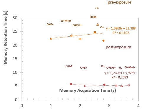
Results from the acquisition and memory retention time duration for both pre-and-post-RFR exposures are presented in Figure 1. The treatment groups that were not irradiated were tested at the same time as the irradiated groups. The time reported as the memory acquisition phase indicates the average time required by the subjects to learn the quadrant’s location of the platform. The time reported as the memory retention phase indicates the time required by the subjects to arrive at the northeast quadrant. Thus, the data represents the total sum of times a subject passed through the northeast quadrant during the 60-second retention phase test. If the animal remembered the platform’s location, then it would spend more time swimming in that particular quadrant.
There was not statistically significant difference between groups for retention times, as revealed by the ANOVA test. However, Table 1 shows a significant difference between pre-and-post-exposure groups, which indicates that at least one group differs from the others. Additionally, the comparison between pre-and-post RFR exposure acquisition and retention phases indicated a statistically significant difference between groups O+E+I+ and O+M+I+.
Source: self made
Figure 1 The time duration of Memory Acquisition and Memory Retention in rats subject to pre- and-post radiation treatments. Rats were distributed randomly in 6 groups with 6 individuals per group: Control group or not ovariectomized (O-), ovariectomized (O+), ovariectomized + estrogen (O+E+), ovariectomized + estrogen + irradiated (O+E+I+), ovariectomized + maca (O+M+), and ovariectomized + maca + irradiated (O+M+I). Experimental units in the I+ groups were rats exposed to RFR from second-generation mobile phones for 7 days. The animal was manually immobilized during the 30-minute daily exposure, where the phone was used to receive a telephone call.
Table 1 Comparison of the study group. (One-way ANOVA)
| Acquisition time (s) | Retention time (s) | |||||||||
|---|---|---|---|---|---|---|---|---|---|---|
| Total test | Σ2 | df | RMS | F | Sig. | Σ squared | df | RMS | F | Sig. |
| Between groups | 0.633 | 6 | 0.106 | 75.2 | 0 | 54.838 | 6 | 9.14 | 14,031 | 0 |
| Within groups | 0.048 | 34 | 0.001 | 22.147 | 34 | 0.65 | ||||
| Total | 0.681 | 40 | 76.985 | 40 |
RMS: Root Mean Square), df: degrees of freedom, Sig: significance at α=0.05.
*There was no statistically significant difference between groups before exposure to RFR (p = 0.5703) and (p = 0.3640); both (p >= 0.05); ANOVA. The difference of the means is significant at the 0.05 level in retention time.
* *There was no statistically significant difference between groups after exposure to radiation (p=0.6977) and (p=0.4212), both (p >= 0.05).
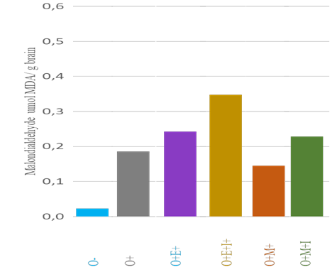
Figure 2 Average levels of Malondialdehyde (µmol MDA/g brain, λ=532 nm) by study group. The vertical bars indicate standard deviation for each group (n=5 for O-, n=6 for the rest). The group with the lowest concentration is O-, followed by O+M+, while the highest concentration was observed for group O+E+I+. Tukey's HSD (honestly significant difference) test indicated that the following pairs of groups were statistically different from one another (p <0.05, CI=95%): O- and O+E+, O- and O+M+I+, O- and O+E+I+, as well as O+M+ and O+E+I+. The other pairs of groups gave p>0.05.
Discussion
The current study has evaluated the effect of Lepidium meyenii (maca) ecotype red on spatial memory parameters and MDA levels in brain tissue. The results suggest that additions of Lepidium meyenii (maca) in the food at doses of 2 g/kg provides a protective effect on memory and an antioxidant effect in rats. It was found that the latency or learning times acquired in the first four days were significantly different. Animals that were not exposed to the mobile phones’ radiation, such as ovariectomized + maca (O+M+), responded better to memory tests than ovariectomized + estrogen (O+E+). In contrast, ovariectomized + estrogen + irradiated (O+E+I+), responded more favorably than did ovariectomized + maca + irradiated (O+M+I). However, with the test comparing groups, this is not attributed to the variable but considered random.
The maca provided a protective effect on the exposure to electromagnetic waves, responsible for oxidative stress generated under the study conditions (19). Furthermore, showing better properties than other reported Peruvian plants studied with the MWM test, including the Physalis peruviana (Solanaceae; aguaymanto), where the capacity of uptake of the radicals, exerted by the crude extract in doses of 3 and 4.5 g ES/kg, did not improve the latency time (20).
Previous studies have shown that maca did not exert hormonal or immune biological action on a study involving a small cohort of patients. However, it appeared to reduce symptoms of depression and improve diastolic blood pressure in Chinese postmenopausal women (21).
The learning process to find the platform submerged underwater aimed to measure the alteration of the medial temporal lobe function, which is typical in old age. Those mentioned above demonstrated that the subjects did not present motor or perceptual alterations to find the platform but a deterioration of the spatial learning per se. This alteration in ancient rats (22) occurred in the reference (or long term) memory, shown by the difficulty in evoking the spatial position of the platform (which was constant throughout all sessions), and in the working memory when the animal was required to learn the new location of the platform each day (23).
It is suggested that the hippocampus participates in the consolidation of the contextual information because this deterioration was observed only in ancient rats; this also occurred with longer intervals between learning and injury, suggesting the existence of an additive effect of aging and injury on the hippocampus (23).
The existence of place or spatial cells in the hippocampus, which are activated when the animal is in a specific location within an environment, shows the importance of this structure in spatial learning. The hippocampus contains a cognitive map formed by place or spatial cells, which the animal could use to move through its surroundings. These cells have been described in rats and mice and appear to encode a polymodal representation of the site. In another study, the findings indicated the difficulty of inferring the occurrence or absence of behavioral learning and the difficulty of causally linking the action of particular receptor populations with the formation of specific memories. Aging and stress could modify the activation pattern of place cells, although the data are not entirely conclusive (24-26).
In previous studies, the brain-selective 17β-oestradiol prodrug demonstrated early-stage efficacy in a mouse model without showing a systemic impact, which is relevant to Alzheimer's disease. Therefore, it deserves additional evaluation as a potential therapeutic candidate (27). These concepts are corroborated with the latency or learning time results of the present study, where it was observed that ovariectomized + estrogen (O+E+) rats arrived at the platform in a shorter time than the ovariectomized + estrogen + irradiated (O+E+I+) rats and the control (O-) rats. However, maca administered in ovariectomized + maca (O+M+) rats and ovariectomized + maca + irradiated (O+M+I) rats exerted improvement of spatial-memory parameters concerning permanence or memory retention. This is a spatial preference test in which, if the animal has learned, it will swim longer in the target quadrant, where the platform was previously located (19). Time spent in the target quadrant in seconds for ovariectomized + maca (O+M+), and ovariectomized + maca + irradiated (O+M+I), was higher, with a level of significance of P <0.05 compared to the ovariectomized + estrogen (O+E+) and ovariectomized + estrogen + irradiated (O+E+I+).
Comparative group tests show that these results are highly significant and may be attributed to maca’s use in the food. The data suggests that maca may have exerted a protective effect on these animals. Our results are in agreement with other previous studies (19,27,28).
Estrogens had demonstrated to modulate oxidative and antioxidative processes, resulting in a decrease of free radicals production, an increase in the expression of antioxidative enzymes, and as an antioxidant molecule in itself. The administration of estrogen decreases oxidative damage to both DNA and lipids in postmenopausal women. The oxidation status of lipids may be inversely related to estrogen levels in postmenopausal women (29). Age-associated memory impairment is higher in females and is related to an estrogen deficiency according to the antioxidant mechanisms. Treatment with Hormone Replacement Therapy (HRT) could be employed; however, when it is contraindicated, the use of phytoestrogens, which are natural chemical compounds derived from certain plants, has been favored (30).
The cerebral tissues of ovariectomized + estrogen + irradiated (O+E+I+) rats subjected to radiation by mobile phones had higher levels of malondialdehyde than ovariectomized + maca + irradiated (O+M+I) rats. In the group comparison test, the results are highly significant. Our results agree with other previous studies using plants with antioxidant and neuroprotective effects where the concentrations of MDA decrease, and there are positive effects on learning and memory; thus, the effects can be attributed to antioxidant properties (31).
Previous studies have related irradiation by mobile phones to the production of more free radicals. Animals such as guinea pigs that received irradiation from mobile phones presented physiological changes such as neuronal demyelination. The reason is that the second-generation cell phone waves can reach a value of 900 MHz, which is the range of microwave radiations and causes oxidative stress with the formation of reactive oxygen species on the galactocerebroside, which is the main lipid of the myelin sheath of guinea pigs. Thus, free radicals cause damage, as they affect unsaturated fatty acids causing lipid peroxidation, which represents a form of tissue damage (32). In this investigation, these levels have been generated by ovariectomy and mobile phone radiation, demonstrating that maca significantly decreases MDA levels (P <0.005). This suggests that irradiation promotes the MAD’s formation and that the protection provided by maca is latent and is only active during stressful periods, like irradiation. Additionally, the maca treatment groups presented lower MAD concentrations than similar groups treated with estrogen.
The phytochemical components of maca found in a methanol extract contain (1R, 3S) -1-methyltetrahydro-beta-carboline-3-carboxylic acid. This is a molecule that promotes activities in the central nervous system. In previous studies, 18 compounds of maca were isolated, including seven alkaloids, four fatty acids, and seven other compounds. An investigation on the tuber constituents of maca (33,34) found that it possesses macamides (a distinct class of secondary metabolites), which until now have only been found only in this plant (12). In addition, maca has more glucosylate content than other ecotypes (black and yellow). It contains the secondary metabolite benzylglucosinolates, which is most abundant in the hypocotyls, seeds, and leaves. However, it is difficult to determine which particular secondary metabolite of maca would act specifically at the neuronal level (10,35).
Conclusions
The present study results suggest that rats fed with red maca at 2 g/kg have improved spatial memory retention and decreased malondialdehyde concentration in cerebral tissue compared to estrogen replacement. Additional research is necessary to ascertain whether some maca components have an active causal effect on the observed improvements.

