


 English (pdf)
 Article in xml format
 Article references
 Send this article by e-mail
 Cited by SciELO
  Cited by Google
  Similars in
SciELO
Similars in
SciELO  Similars in Google
 Permalink
Introduction
When a radiopharmaceutical is administered to a pregnant woman, the estimation of absorbed dose to the uterine wall/fetus is essential due to the high sensitivity of developing fetal tissues [1].
Radiation exposure to the uterine wall/fetus occurs when the administered radiopharmaceuticals cross the placental barrier spreading in the tissues of the uterine wall/fetus; if the administered radiopharmaceuticals do not cross the placental barrier, the exposure is due to external irradiation from radiopharmaceuticals present in the mother's organs and tissues. The chemical and biological properties of radiopharmaceuticals are the determining factors for crossing the placental barrier [2].
99m Tc is the most suitable radioisotope for the labeling of different pharmaceutical products that allow obtaining a wide range of studies. 99m Tc minimizes the absorbed dose in patients [3].
Radiopharmaceuticals 99m Tc-(MAG3), 99m Tc-(DTPA) or 99m Tc-(DMSA) were used to perform renal function studies. These labeled compounds are distributed in the patient's organs according to their biokinetics (organs of biokinetics). In pregnant patients, the uterine wall and the fetus are exposed to radiation emitted by 99m Tc, radiation from the female biokinetics organs, or the labeled compound from its placental transfer.
The dosimetric behavior of radiopharmaceuticals is related to their residence times in the organs/tissues, as well as their biokinetics, which in turn is related to the differences in the biological and physical excretion mechanisms.
The biokinetic models used in the estimation of the absorbed dose for organs/tissues, as well as the radiopharmaceutical data are available in the literature [4,5]. To estimate the absorbed dose to the uterine wall and the fetus was used the MIRD method and the Stabin's anthropomorphic representation of organ biokinetics [6,7]. Fetal uptake and placental transfer of the administered radiopharmaceutical are included in the biokinetic data in terms of residence time, organs/tissues from Oak Ridge National Laboratory [8].
The purpose of the present study was to estimate the lowest absorbed dose of radiation in the uterine wall and fetus of a patient with three months of gestation who performs renal studies with 99m Tc-DTPA, 99m Tc-DMSA, and 99m Tc-MAG3. To determine the procedure that administers the lowest absorbed dose, the MIRD methodology and the anthropomorphic representation of maternal organs and tissues described by Stabin were used.
Materials and Methods
The 99m Tc, which is the most suitable radioisotope for the labeling of different drugs, decays through isometric transition by gamma emission, with an energy of 140 keV and a half-life of 6 hours. Gamma radiation can transfer energy directly to one of the most strongly bound electrons, expelling it from the atom, a process that is called internal conversion [9].
The photons and particles produced ionize and excite matter, but their interaction mechanisms are different, as well as their scope within tissues. In the MIRD procedure, the uterine wall and fetus were assumed as target organs and the absorbed dose per unit activity of the administered radiopharmaceutical was calculated. The MIRD equation can be expressed as:
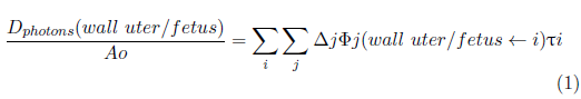
On the right side of the equation, the absorbed dose represents the dose to the uterine wall and fetus due to source organ i, Δ j is the average energy of photon j emitted by Tc 99m by decay, Фj (wall/fetus←i) is the fraction of energy emitted by organ i that is absorbed by the wall/fetus; it is also known as the specific absorbed fraction (SAF), and тi is the residence time of the radiopharmaceutical in the source organ i.
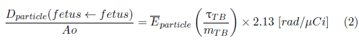
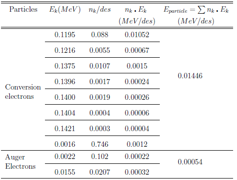
The absorbed dose, in rad/µCi, in the fetus due to the electron conversion and Auger electrons were calculated using the equation:
where
 particle
is the average energy of the particle, тTB is the residence time of the Radiopharmaceuticals in the whole body and m
TB
is the mass of the whole body of an adult. In the previous equation we consider 1 [rad/µCi] = 270 [mGy/MBq].
particle
is the average energy of the particle, тTB is the residence time of the Radiopharmaceuticals in the whole body and m
TB
is the mass of the whole body of an adult. In the previous equation we consider 1 [rad/µCi] = 270 [mGy/MBq].
The SAF of the uterine wall and fetus and of the biokinetics source organs, such as the kidneys, bladder, placenta, and the "rest" of the tissues were obtained from Stabin [7].
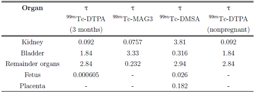
The residence times for 99mTc-DTPA, 99mTc-DMSA, and 99mTc-MAG3 in kidney, bladder, placenta, fetus, and the "rest" of organs used in the calculations are shown in Table 1 [8].
TABLE 1 Residence times, in hours, for the organs of biokinetics of the 99m Tc-DTPA, 99m Tc-DMSA, and 99m Tc-MAG3 radiopharmaceuticals.
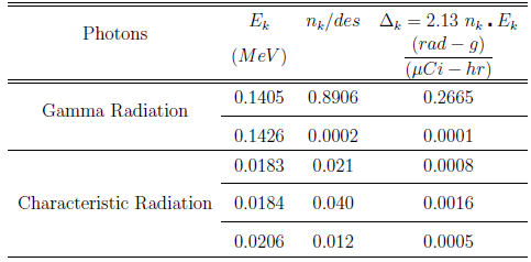
Tables 2 and 3 show the characteristics of the photons and particles emitted during the 99mTc decay that were used in the calculation of the dose [10].
The mass of the organs included in the biokinetics of the pregnant woman used in the calculations indicates that the mass of the fetus (m fetus ) is 458 g and the mass of the whole body (m TB ) is 58000 g (ORNL/TM - 12907).
Results
Using the MIRD methodology and the biokinetic characteristics of 99m Tc-DTPA, 99m Tc-DMSA, and 99m Tc-MAG3 in the target organs (uterine wall and fetus) and source organs (bladder, kidneys, the rest, and the placenta), the absorbed dose was calculated in the uterine wall and fetus.
The Table 4 shows the values of the absorbed dose absorbed by the fetus of 03 months, due to the photons and particles emitted by 99m Tc.
Table 4 Absorbed dose per unit of administered activity (mGy/MBq) of radiopharmaceuticals: 99mTc (DTPA, DMSA, and MAG3) in the 03 months' fetus.
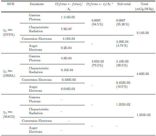
* i: organs of biokinetics
Table 5 shows dose values absorbed by the uterine wall and the fetus, considering the dose contributions for each of the maternal biokinetic organs (source organs).
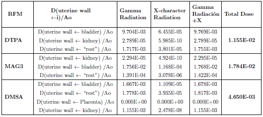
TABLE 5 Absorbed dose per unit of administered activity (mGy/MBq) of the radiopharmaceuticals: 99m Tc for DTPA, DMSA, and MAG3, in the uterin wall/fetus.
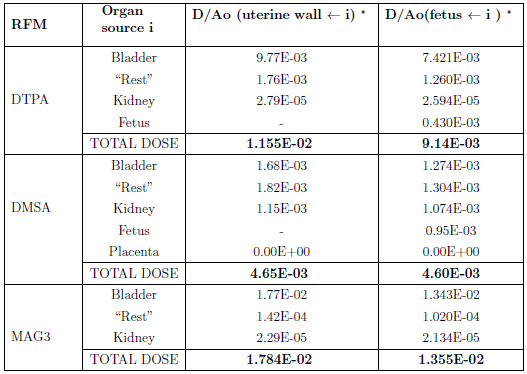
* i: organs of biokinetics
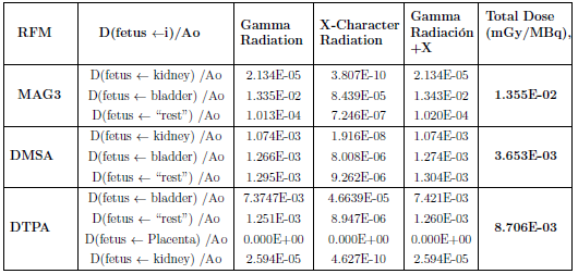
Tables 6 and 7 show the values of the dose absorbed by the fetus and the uterine wall, respectively, considering for their calculation, the dosimetric contributions of the gamma photons, and X-radiation characteristic generated by the organs of biokinetics.
TABLE 6 Absorbed dose of 99m Tc (DTPA, DMSA, and MAG3), in the fetus, due to gamma photons and characteristic X-radiation that come from organs of biokinetic.
Discussion
In renal function studies, the highest dose absorbed by the uterine wall and fetus of a woman at 03 months' gestation occurs when 99mTc-MAG3 is used, while the lowest dose is obtained when 99mTc-DMSA is used (Table 5).
When 99m Tc-MAG3 is used, the biokinetic source organ that contributes the highest dose to the uterine wall/fetus is the bladder, contributing 0.017 mGy/MBq / 0.01343 mGy/MBq, respectively (Tables 5-7). The dose in the uterine wall and fetus due to the activity of 99m Tc-MAG3 is the bladder, it constitutes the main source of irradiation to the fetus, this contribution can be decreased if the amount of the radiopharmaceutical in the bladder is reduced by hydration of the patient prior to radiopharmaceutical administration [11].
When 99m Tc-DMSA is used, the organ that contributes the highest dose to the uterine wall and fetus is the "rest" of organs, contributing 0.0018 mGy/MBq / 0.0013 mGy/MBq, respectively (Tables 5 - 7).
When 99m Tc-DTPA is used, the organ that contributes the highest dose to the uterine wall and fetus is the bladder, contributing 0.00977 mGy/MBq / 0.0074 mGy/MBq, respectively (Tables 5-7).
The gamma emitters that come from the "remainder" and the bladder are those that contribute most to the dose of the uterine wall and fetus. The contribution of X-characteristics is very insignificant (Tables 6-7).
The placental transfer produced by 99m Tc-DMSA to the fetus generates a dose (auto-dose) of 0.95E-03 mGy/MBq and represents approximately 20 % of its total dose. Only 10 % of this dose corresponds to the produced by the 99m Tc conversion electrons + Auger electrons deposited in the fetus, while the remaining 10 % is due to characteristic gamma photons and X-rays (Tables 4 - 5).
When DTPA is used, the placental transfer also occurs, increasing the dose to the fetus (auto dose) by just 5 % of its total. Electronic conversion + Auger electrons of the 99m Tc deposited in the fetus produce 4.47% of this dose, and the remaining 0.12% is due to characteristic gamma photons and X-rays. When MAG3 is used, no placental transfer occurs [8].
In general, the dosimetric contribution of the organs that are part of the biokinetics to the uterine wall and fetus for the 99mTc radiopharmaceuticals (DMSA, MAG3, and DTPA) are basically due to the gamma radiation emitted by the 99mTc (Tables 6-7).
During pregnancy, the risk due to the use of radiopharmaceuticals depends on the stage of pregnancy and the dose absorbed by the fetus. The risk is most relevant during organogenesis and the early fetal period [12].
Conclusions
The absorbed dose in the uterine wall and fetus of a woman with three months of gestation submitted to renal studies with 99m Tc-DTPA, 99m Tc-DMSA, and 99m Tc-MAG3 was calculated. For the dosimetric calculation, the MIRD methodology and the anthropomorphic representation of the maternal organs due to Stabin were used. The highest dose absorbed by the uterine wall and the fetus were obtained with the use of 99m Tc-MAG3. The organ of biokinetics that contributes to the highest dose is the bladder. The lowest dose absorbed by the uterine wall and the fetus were obtained with the use of 99m Tc-DMSA. The organ of biokinetics that contributes the highest dose is the "rest" of organs.

