


Servicios Personalizados
Revista
Articulo
 texto en
  Inglés (pdf)
 Articulo en XML
 Referencias del artículo
 Enviar articulo por email
Indicadores
-
 Citado por SciELO
 -
 Accesos
Links relacionados
-
 Citado por Google
 -
 Similares en
SciELO
Similares en
SciELO -
 Similares en Google
Compartir

 Permalink
Revista Colombiana de Reumatología
versión impresa ISSN 0121-8123
Rev.Colomb.Reumatol. vol.24 no.1 Bogotá ene./mar. 2017
https://doi.org/10.1016/j.rcreu.2016.10.005
Original Investigation
Semi-quantitative analysis of scintigraphic findings in the hands of adults without osteoarticular disease☆
a Servicio de Medicina Nuclear, Clínica Reina Sofía Colsánitas, Bogotá, Colombia
b Medicina Nuclear, Fundación Universitaria Sanitas, Bogotá, Colombia
c Epidemiología, Fundación Universitaria Sanitas, Bogotá, Colombia
Introduction:
Bone and joint disease has a high incidence and impact on the population. The bone scan is a diagnostic tool that provides important metabolic and clinical information; therefore the interpretation of the images by the nuclear medicine physician must be very precise. The isotopic distribution pattern in hands has not yet been described. For this reason a description is presented of common scintigraphic findings in adults without osteoarticular disease.
Materials and methods:
A prospective analysis was conducted on 156 hands of patients whose bone scans met inclusion criteria. There were delineated regions of interest in the carpal, metacarpal, proximal, and distal interphalangeal joints of the second and third fingers of both hands. An analysis was made, including the total counts, means, and standard deviations. The cut-offs were selected using the normal distribution, which was defined as the cut at the 99th percentile of each variable. A semi-quantitative analysis was made of the images.
Results:
The study included 36 men (23%) and 119 women (77%), and the mean age was 44.9 ± 13.9. The mean total counts gradually decreased from proximal to distal in all age groups and in both genders in the following proportions: the activity in carpus was 4.4 fold more than the metacarpus; the metacarpus was 1.7 fold more than proximal interphalangeal joint; proximal interphalangeal joint was 1.4 fold more than distal one.
Conclusions:
A scintigraphic pattern consisting of a gradual decrease from proximal to dis-tal joints (degradation) was found in the hands of adults without bone and joint disease, regardless of gender and age.
Keywords: Radionuclide imaging; Bone scan; Bones of hands
Introducción:
La gammagrafia ósea es uno de los estudios más frecuentemente utilizados para el abordaje diagnóstico de la patología osteoarticular, sin embargo, no se conocen patrones claros de normalidad para algunos sitios anatómicos, llevando a tasas altas de variabilidad interobservador, como es el caso de la evaluación de las manos. No se encuentra en la literatura una descripción del patrón gammagráfico normal sobre manos, por lo cual pretendemos describir el patrón de captación más frecuente en una población adulta sin enfermedad osteoarticular.
Materiales y métodos:
Se hizo un análisis prospectivo de 156 gammagrafías óseas sobre manos que cumplieron el criterio de inclusión. Se delinearon regiones de interés en el carpo, metacarpo, articulaciones interfalángicas proximales y distales de los dedos índice y medio de ambas manos; se tomaron las cuentas totales, se analizaron promedios y desviación estándar, y se hizo análisis semicuantitativo de la imagen.
Resultados:
Se incluyeron 36 hombres (23%) y 119 mujeres (77%), la edad media fue de 44,9 ± 13,9. Los promedios de las cuentas totales disminuyeron progresivamente deproximal a distal en todos los grupos de edad y en ambos géneros, siguiendo la siguiente proporción: carpo 4,4 veces más que metacarpo; metacarpo 1,7 veces más que interfalángica proximal; interfalángica proximal 1,4 veces más que distal.
Conclusiones:
En manos de pacientes adultos, sin enfermedad osteoarticular, sin distinción de género y edad, encontramos un patrón gammagráfico en «degradé» con mayor concentración isotópica en carpo, seguida del metacarpo y de las articulaciones interfalángicas proximales y distales.
Palabras clave: Medicina nuclear Gammagrafía ósea Articulaciones de las manos
Introduction
During the last decades, nuclear medicine studies have demonstrated an accelerated growth, due to the develop ment of new and better radiopharmaceuticals1-3; among the numerous isotopic studies, bone scintigraphy is the second most frequently performed procedure in the nuclear medicine services4,5 after myocardial perfusion; only in the United States, more than 3,450,000 of these procedures were carried out in the year 2005, being the inflammatory and osteoarticular pathology one of the most important indications.4,6
In the study of the osteoarticular pathology (degenera tive/inflammatory) of the hand, nonisotopic images play an important role, being conventional radiography the simplest, most economical and widely used method, especially in the initial assessment.7,8 Another important technique is the nuclear magnetic resonance which evaluates early changes in the articular cartilage before bone destruction occurs.9,10 On the other hand, ultrasonography is used to evaluate joint effusions and erosions, and currently there are stratification systems such as Power Doppler, which has demonstrated to be reproducible and accurate for synovitis of the hand.11-13
The bone scan is not part of the initial evaluation of osteoarticular disease, but is highly sensitive to detect changes previous to structural alterations, it is also reproducible and it has a negative predictive value higher than 90%14,15; it can help to discriminate the origin of the pain (soft tissues or bone), locate the most painful points in patients with complex symptoms16,17 and detect bone pathologies when other imag ing techniques have failed.18 The radiopharmaceutical most commonly used for bone imaging is methylene diphosphonate which, bound to Tc99m forms a radioactive compound which reaches the bone through the bloodstream and binds to hydroxyapatite crystal with high affinity, 19,20 allowing to evaluate indirectly the osteoblastic activity.
The identification and the familiarization with the normal pattern of radiotracer uptake in the hands are very important for the proper scintigraphic evaluation of this area; however, at the present time there is no recognized normal scintigraphic reference pattern. Making a review of the literature, it was found only one study related to the subject, conducted by Wilfrido et al., who in 1977 mentioned in a heterogeneous group of patients some normal trends of radiotracer concentration, divided into 2 groups, the first with a detailed image of the joints of the hand seen in adolescents, and the second, in a pattern called "washed-out" with less definition of the image, observed in older patients.21
Knowing the normal patterns of radiotracer concentration in the different bone structures allows to improve the diag nostic accuracy, avoiding overdiagnoses and underdiagnoses.
The purpose of this study is to describe the radiotracer distribution pattern in the bone scan of the hands of adult patients without osteoarticular pathology, under a semi quantitative analysis which can be reproducible and that allows and objective evaluation of the hands.
Materials and methods
Patients
In the study were included 156 hands of patients (36 men, 119 women) between 18 and 72 years old, referred between November 2012 and November 2013, to the Nuclear Medicine Service for a bone scan to study oncological or maxillofacial pathology, who to interrogation did not report antecedents of osteoarticular disease of degenerative or rheumatic inflam matory type, previous surgeries, metabolic bone disease, plegias or stress injuries; the images were taken from the records of patients of the Nuclear Medicine Service of the Colsanitas Clinics in Bogota (Colombia Clinic and Reina Sofia Clinic) and the antecedents were verified in the respective medical histories. The selection of the participants was made by convenience, based on the records of hands accomplished in the Nuclear Medicine Service.
Radiopharmaceutical
Tc99m-MDP was used; the administered dose was 20 mCi (740 MBq) and it was injected in a peripheral vein in the foot.
Scintigraphic images
The studies were acquired by a double-headed gamma cam era with a high resolution collimator, GE Infinia, coupled to an Odysey VP workstation, using the Xeleris Functional Imaging, bone evolution software. A 3-phase bone scan with special approach to the hands (in the 3 phases) was performed acquiring perfusion images during one minute (one frame/3 s) in a matrix of 64 x 64 on the hands, the image of the tis sue distribution was obtained 5 min after the injection of the radiopharmaceutical (tissue phase) in a matrix of 128 x 128, and 3 h post-injection were obtained images in a matrix of 128 x 128, with a number of 500,000 counts per image for the bone phase.22 Only the images of the bone phase were taken into account for the semi-quantitative analysis.
Analysis of the images
Regions of interest (ROI) of the same size were delineated on the carpus and the metacarpophalangeal and proximal and distal interphalangeal regions of the index (called finger 1 for the analysis) and middle (called finger 2 for the analysis) fin gers, obtaining total counts in order to analyze their trends and compare them each other (Fig. 1).
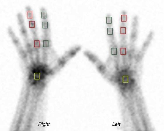
Fig. 1 Areas of interest. Bone scintigraphy of the hands; in green squares, areas of interest on the interphalangeal and metacarpophalangeal joints of the second finger; in red squares, areas of interest on the interphalangeal and metacarpophalangeal joints of the third finger; in yellow squares, areas of interest on the carpus.
Statistical analysis
The data were analyzed according to gender and age, for which the population was grouped as follows: 18-25, 26-35, 36-45, 46-55, 56-65 and older than 65 years.
The mean total counts, proportion ratios, mean and stan dard deviation were used for the statistical analysis, and their behavior in each variable was analyzed; p <0.05 was con sidered significant. The normality of each of the variables analyzed was evaluated using the Shapiro-Wilk test and the null normality hypothesis of normality was rejected with p<0.05. A bivariate analysis comparing the qualitative vari ables by gender was carried out using the Wilcoxon Rank sum test; and the analysis of each variable by age groups was per formed using the Kruskall-Wallis test.
Results
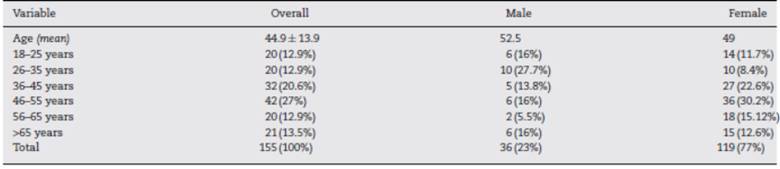
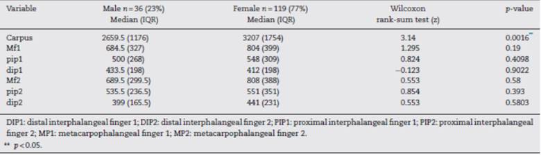
The hands analyzed corresponded to 155 patients, the overall average age was 44.9 years, 52.5 years for men and 49 years for women. The age group with the highest representation in this series of patients was the one of 46-55 years. The series is represented in 23% by men and 77% by women (Table 1).
The mean total counts from the carpus to the distal inter-phalangeal joints of the index and middle fingers, which were analyzed as a reference point, gradually decreased in all age groups; it was only found a difference in the counts of the car pus in the groups of 46-55 years and >65 years with regard to the other groups.
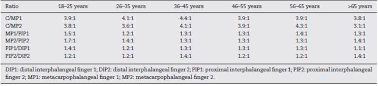
The proportion ratios between the different studied joints and in all age groups (carpus/metacarpophalangeal and proxi mal interphalangeal/distal interphalangeal in both hands) are shown in Table 2.
The trends of the means by age group in the different joints in which the ROIs were taken were similar (Figs. 2 and 3). A proximal to distal decrease of the counts was observed as mentioned above.
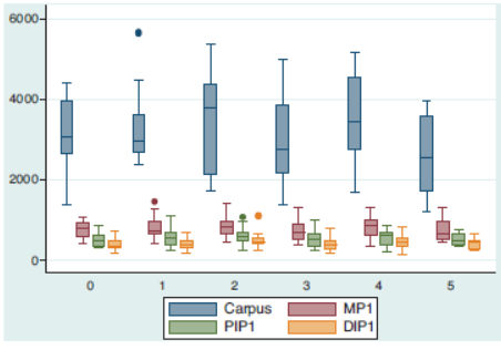
Fig. 2 Trend of the mean counts in each of the regions of interest for finger 1 according to age groups (0 = 18-25, 1 = 26-35, 2 = 36-45, 3 = 46-55, 4 = 56-65, 5 = >65 years).
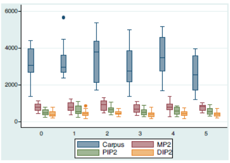
Fig. 3 Trend of the mean counts in each of the region of interest for finger 2 according to age groups (0 = 18-25, 1 = 26-35, 2 = 36-45, 3 = 46-55, 4 = 56-65, 5 = >65 years).
According to the gender, the median of the counts gradu ally decreased from proximal to distal in all joints, and without distinctions; the mean of the counts of the carpus was higher in women (Table 3).
Discussion
We observed in our series that the mean of total counts regis tered in each of the regions of interest showed a similar trend in all age groups and genders, finding the largest number of counts in carpi, followed by the metacarpophalangeal and the proximal and distal interphalangeal joints, respectively.
In the analysis of counts of the ROIs in the carpus, sta tistically significant differences were found in the groups of 35-45 years and older than 65 years, finding in the latter a smaller number of counts, explained probably by the physio logically decreased bone metabolic activity. Nevertheless, the trend and the relationship between the counts of the carpus and the metacarpophalangeal joints in these age groups was preserved; likewise, when the ROIs of the same joint were compared between genders, we found greater activity in the carpi of the hands of women with respect to men, a finding that could have been affected by a greater female population in the sample (77%).
The qualitative analysis of the characteristics of uptake was supported by the semi-quantitative analysis and we can say that the pattern is characterized by a progressive decrease in the concentration of the radiotracer that goes from proxi mal to distal and that we call "degradation". We also observed how the carpus was the region of interest of the hand with greater activity with an uptake of up to 4.4 folds more in rela tion with the metacarpophalangeal joint; the proportion ratios found demonstrate this behavior and also allow us to see how in the different age groups the metacarpophalangeal joint also shows and increase in the tracer uptake of up to 1.7 folds more than the proximal interphalangeal joint and this in turn concentrates up to 1.4 folds more than its adjacent distal inter phalangeal joint, being this finding another way to describe the degradation pattern previously described.
It should be mentioned that the osteoarticular pathology of the hand has received relatively little attention compared with other structures such as the hip of the knee, however, it causes significant disability and deterioration in the quality of life23,24; in osteoarticular disease the involvement of the hands is frequent and it can be made a clinical approach to the ori gin of the pathology depending on the location of the affected areas, for example, the commitment of the distal interphalangeal and the trapezium-metacarpal joints guides toward a degenerative origin,25 in rheumatoid arthritis the involvement is predominantly in the metacarpophalangeal joints with fre quent tenosynovitis of the flexor and extensor apparatus.26,27
The different diagnostic tools allow us to get closer to the understanding of these entities, the simple radiography is used both in the initial assessment and in the evalua tion of the severity and follow-up; it has the disadvantage that small variations in the position of the hand during its acquisition can simulate or mask lesions.7 Nuclear magnetic resonance is being increasingly used, especially with tech niques that use contrast media such as gadolinium, improving the specificity of the study; however, it requires a suitable and reproducible protocol, in addition to being limited in the pres ence of osteosynthesis materials.28,29
The bone scan of the hand is a very useful tool with great potential to provide accurate information to the clin ician, given its high sensitivity and reproducibility, but a clear normal pattern is not available; this situation demon strates why the interpretation of our studies must be based on a detailed analysis, in such a way that it will not gener ate wrong diagnoses. In the literature there are not studies that demonstrate the normal behavior of the joints of the hand under the nuclear medicine metabolic viewpoint, being this study, to our knowledge, the first of its kind; abnor mal scintigraphic patterns such as focal hyper-uptake in the distal interphalangeal, scaphotrapezial and the first carpometacarpal joints have been reported in patients with nodal osteoarthritis,30 as well as diffuse hyper-uptakes in the carpus of patients with psoriatic and rheumatoid arthri tis, however, without taking into account a basal normality pattern.31
Our study allowed us to know the scintigraphic pattern on the hands in terms of the relationship between each of the joints, however, we did not analyzed the maximum allowed ranges in each of them and we believe that it may be a subject for further studies. Likewise, it may be interesting to analyze in larger samples and with equal representative groups for each gender, is this trend is preserved and to present more accurately the model of "degradation" as a synonymous with normality.
Conclusions
Osteoarticular pathology is part of the most frequent reasons of consultation in the nuclear medicine units. This study is the first that analyzes in a detailed way the behavior of the hands in a bone scan from the qualitative and semi-quantitative viewpoints. Being the hands a frequent site of commitment in degenerative and inflammatory pathology, we believe that the description of a normal pattern called by us "degradation" becomes a tool of great usefulness for medical practice.
Study approved by the Ethics Committee of the Faculty of Medicine of the Sanitas University Foundation.
Ethical disclosures
Protection of human and animal subjects. The authors declare that no experiments were performed on humans or animals for this study.
Confidentiality of data. The authors declare that they have fol lowed the protocols of their work center on the publication of patient data.
Right to privacy and informed consent. The authors declare that no patient data appear in this article.
REFERENCES
1. Sopena Novales P, Plancha Mansanet MC, Martínez Carsi C, Sopena Monforte R. Nuclear medicine and radiopharmaceuticals. Radiologia. 2014;56 Suppl 1:29-37. [ Links ]
2. Clinical audit-ESR perspective. Insights into imaging. 2010;1:21-6. [ Links ]
3. Mirzaei S, Maffioli L, Hilson A. Clinical audit in nuclear medicine. Eur J Nucl Med Mol Imaging. 2011;38:3-4. [ Links ]
4. Mettler FA Jr, Bhargavan M, Thomadsen BR, Gilley DB, Lipoti JA, Mahesh M, et al. Semin Nucl Med. Seminars in nuclear medicine. 2008;38:384-91. [ Links ]
5. Agrawal K, Marafi F, Gnanasegaran G, van der Wall H, Fogelman I. Pitfalls and limitations of radionuclide planar and hybrid bone imaging. Semin Nucl Med. 2015;45:347-72. [ Links ]
6. Brenner AI, Koshy J, Morey J, Lin C, DiPoce J. The bone scan. Semin Nucl Med. 2012;42:11-26. [ Links ]
7. Hunter DJ, Arden N, Cicuttini F, Crema MD, Dardzinski B, Duryea J, et al. OARSI Clinical Trials Recommendations: Hand imaging in clinical trials in osteoarthritis. Osteoarthritis and Cartilage. 2015;23:732-46. [ Links ]
8. Feydy A, Pluot E, Guerini H, Drape JL. Role of imaging in spine, hand, and wrist osteoarthritis. Rheum Dis Clin N Am. 2009;35:605-49. [ Links ]
9. Haugen IK, Ostergaard M, Eshed I, McQueen FM, Bird P, Gandjbakhch F, et al. Iterative development and reliability of the OMERACT hand osteoarthritis MRI scoring system. J Rheumatol. 2014;41:386-91. [ Links ]
10. Haugen IK, Lillegraven S, Slatkowsky-Christensen B, Haavardsholm EA, Sesseng S, Kvien TK, et al. Hand osteoarthritis and MRI: development and first validation step of the proposed Oslo Hand Osteoarthritis MRI score. Ann Rheum Dis. 2011;70:1033-8. [ Links ]
11. Keen HI, Lavie F, Wakefield RJ, D'Agostino MA, Hammer HB, Hensor E, et al. The development of a preliminary ultrasonographic scoring system for features of hand osteoarthritis. Ann Rheum Dis. 2008;67:651-5. [ Links ]
12. Mathiessen A, Haugen IK, Slatkowsky-Christensen B, Boyesen P, Kvien TK, Hammer HB. Ultrasonographic assessment of osteophytes in 127 patients with hand osteoarthritis: exploring reliability and association with MRI, radiographs and clinical joint findings. Ann Rheum Dis. 2013;72:51-6. [ Links ]
13. Hammer HB, Bolton-King P, Bakkeheim V, Berg TH, Sundt E, Kongtorp AK, et al. Examination of intra and interrater reliability with a new ultrasonographic reference atlas for scoring of synovitis in patients with rheumatoid arthritis. Ann Rheum Dis. 2011;70:1995-8. [ Links ]
14. Jones AG, Francis MD, Davis MA. Bone scanning: radionuclidic reaction mechanisms. Semin Nucl Med. 1976;6:3-18. [ Links ]
15. Love C, Din AS, Tomas MB, Kalapparambath TP, Palestro CJ. Radionuclide bone imaging: an illustrative review. Radiographics. 2003;23:341-58. [ Links ]
16. Guermazi A, Hayashi D, Roemer FW, Felson DT. Osteoarthritis: a review of strengths and weaknesses of different imaging options. Rheum Dis Clin N Am. 2013;39:567-91. [ Links ]
17. Omoumi P, Mercier GA, Lecouvet F, Simoni P, Vande Berg BC. CT arthrography, MR, arthrography, PET, and scintigraphy in osteoarthritis. Radiol Clin N Am. 2009;47:595-615. [ Links ]
18. Huellner MW, Strobel K. Clinical applications of SPECT/CT in imaging the extremities. Eur J Nucl Med Mol Imaging. 2014;41 Suppl 1:S50-8. [ Links ]
19. Brown ML, O'Connor MK, Hung JC, Hayostek RJ. Technical aspects of bone scintigraphy. Radiol Clin N Am. 1993;31:721-30. [ Links ]
20. O’Connor MK, Brown ML, Hung JC, Hayostek RJ. The art of bone scintigraphy-technical aspects. J Nucl Med. 1991;32:2332-41. [ Links ]
21. Sy WM, Bay R, Camera A. Hand images: normal and abnormal. J Nucl Med. 1977;18:419-24. [ Links ]
22. Donohoe KJ, Henkin RE, Royal HD, Brown ML, Collier BD, O'Mara RE, et al. Procedure guideline for bone scintigraphy: 1.0. Society of Nuclear Medicine. Journal of nuclear medicine: official publication. J Nucl Med. 1996;37:1903-6. [ Links ]
23. Kloppenburg M, Maheu E, Kraus VB, Cicuttini F, Doherty M, Dreiser RL, et al. OARSI Clinical Trials Recommendations: Design and conduct of clinical trials for hand osteoarthritis. Osteoarthritis and cartilage/OARS. Osteoarthritis and Cartilage. 2015;23:772-86. [ Links ]
24. Michon M, Maheu E, Berenbaum F. Assessing health-related quality of life in hand osteoarthritis: a literature review. Ann Rheum Dis. 2011;70:921-8. [ Links ]
25. Punzi L, Frigato M, Frallonardo P, Ramonda R. Inflammatory osteoarthritis of the hand. Best practice & research Clinical rheumatology. 2010;24:301-12. [ Links ]
26. Longo UG, Petrillo S, Denaro V. Current Concepts in the Management of Rheumatoid Hand. International journal of rheumatology. 2015;2015:648073. [ Links ]
27. Rowbotham EL, Freeston JE, Emery P, Grainger AJ. The prevalence of tenosynovitis of the interosseous tendons of the hand in patients with rheumatoid arthritis. Eur Radiol. 2016;26:444-50. [ Links ]
28. Williams A, Shetty SK, Burstein D, Day CS, McKenzie C. Delayed gadolinium enhanced MRI of cartilage (dGEMRIC) of the first carpometacarpal (1CMC) joint: a feasibility study. Osteoarthritis and Cartilague 2008;16:530-2 [ Links ]
29. Miese F, Buchbender C, Scherer A, Wittsack HJ, Specker C, Schneider M, et al. Molecular imaging of cartilage damage of finger joints in early rheumatoid arthritis with delayed gadolinium-enhanced magnetic resonanc imaging. ArthritisRheum. 2012;64:394-9. [ Links ]
30. Hutton CW, Higgs ER, Jackson PC, Watt I, Dieppe PA. 99mTc HMDP bone scanning in generalised nodal osteoarthritis. I. Comparison of the standard radiograph and four hour bone scan image of the hand. Ann Rheum Dis. 1986;45: 617-21. [ Links ]
31. O'Sullivan MM, Powell N, French AP, Williams KE, Morgan JR, Williams BD. Inflammatory joint disease: a comparison of liposome scanning, bone scanning, and radiography. Ann Rheum Dis. 1988;47:485-91. [ Links ]
Received: June 24, 2016; Accepted: October 24, 2016
 Este es un artículo publicado en acceso abierto bajo una licencia Creative Commons
