SciELO - Scientific Electronic Library Online

 
vol.27 issue4Difficulties in establishing parameters for rheumatoid arthritis in Brazil author indexsubject indexarticles search
Home Pagealphabetic serial listing  

Services on Demand

Journal

Article

Indicators

Related links

  • On index processCited by Google
  • Have no similar articlesSimilars in SciELO
  • On index processSimilars in Google

Share


Revista Colombiana de Reumatología

Print version ISSN 0121-8123

Rev.Colomb.Reumatol. vol.27 no.4 Bogotá Oct./Dec. 2020  Epub Aug 16, 2021

https://doi.org/10.1016/j.rcreu.2019.10.008 

Letters to the editor

Immunological profile in biopsies of primary angiitis of the central nervous system

Perfil inmunológico en biopsias de vasculitis primaria del sistema nervioso central

Iván Posso-Osorioa 

Daniela Espinosa-Cárdenasb 

David Aguirre-Valenciaa 

Juan Naranjo-Escobara 

Luis-Alberto Escobarc 

Carlos A. Cañasa 

Gabriel J. Tobóna  d  * 

aG1RAT: Grupo de Investigación en Reumatología, Autoinmunidad y Medicina Traslacional, Fundación Valle del Lili and Univesidad Icesi, Cali, Colombia

bCentro de Investigaciones Clínicas, Fundación Valle del Lili, Cali, Colombia

cUnidad de Patología, Fundación Valle del Lili, Cali, Colombia

dLaboratorio de Inmunología, Fundación Valle del Lili, Cali, Colombia


Dear Editor,

Primary angiitis of the central nervous system (PACNS) is a rare autoimmune disease characterized by the inflammation of central nervous system arteries without compromising any other organ, and without a primary cause. This disease predominantly affects medium to small caliber arteries in the brain parenchyma, the spinal cord and the meninges, causing central nervous system (CNS) dysfunction.1 With an incidence rate of 2.4 cases per 1,000,000 habitants, it is a truly rare entity. Its pathophysiology is not yet elucidated, but it appears to be mediated by antigen presentation to the T lymphocytes in the arterial wall.2

PACNS is categorized into different subtypes according to pathological and histological findings: granulomatous angiitis of the central nervous system (GACNS), lymphocytic angiitis of the CNS, amyloid beta-related angiitis, mass-like lesions, and spinal cord vasculitis. With 59% of cases, GACNS is the most common of the subtypes.3

In 2013 our group reported 3 cases of patients with PACNS.4 This time, an immunohistochemical study was done in 2 of the patients' samples, revealing a leukocytic infiltratate (CD45+) constituted almost entirely of T lymphocytes (CD3+) with a clearly higher prevalence of CD4+ cells (Fig. 1A), with no evidence of plasmatic cells (CD138 and CD38 markers were negative) and an absence of Treg Lymphocytes (negativity for CD25). HLA was detected in 50% of the cells in both samples (Fig. 1B).
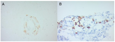
Fig. 1 Immunohistochemistry in biopsy samples of two patients with PACNS. (A) Leukocytic infiltrates mostly of T lymphocytes (CD3+) with higher prevalence of CD4+ cells. (B) HLA expression in PACNS (50% of the cells). 

In a report of 4 biopsies of Japanese patients, there was a T lymphocyte infiltrate, with a predominance of CD8+ cells in the brain parenchyma, and a B lymphocyte (CD20+) prevalence in the perivascular area.5 In another Japanese case report,6 the biopsy analysis showed an infiltrate of memory T cells (CD45RO+), suggesting that an activation by cytokines such as IFN-ү leads to an enhanced antigen presentation by perivascular macrophages, endothelial cells, smooth muscle cells, and astrocytes. These data, along with our results, suggests a T lymphocyte (with CD4 predominance), Th1 response (with IFN-ү and IL-12 cytokines) mediated disease. The negligible presence of B lymphocytes and the absence of plasmatic cells imply that this is not a humoral immunity mediated entity.

Nevertheless, more complex studies are required to understand the pathophysiology of this disease, the cytokine expression pattern, and to be able to establish a difference between the leukocyte infiltrate of the brain parenchyma and the perivascular space. In the future, this information could lead to targeted therapy against T lymphocyte co-stimulation.

REFERENCES

1. Mandal J, Chung SA. Primary angiitis of the central nervous system. Rheum Dis Clin [Internet]. Elsevier; 2017;43:503-18. doi:10.1016/j.rdc.2017.06.001. [ Links ]

2. Salvarani C, Brown RDJ, Hunder GG. Adult primary central nervous system vasculitis. Lancet. 2012;380:767-77. [ Links ]

3. Salvarani C, Brown RD, Christianson TJH, Huston J, Giannini C, Miller DV, et al. Adult primary central nervous system vasculitis treatment and course: analysis of one hundred sixty-three patients. Arthritis Rheumatol [Internet]. 2015;67:1637-45, http://dx.doi.org/10.1002/art.39068. [ Links ]

4. Coronel-Restrepo N, Bonilla-Abadia F, Cortes OA, Izquierdo JH, Shinchi AM, Bravo JC, et al. Primary angiitis of the centralnervous system: a report of three cases from a single colombian center. Case Rep Neurol Med. 2013;2013:940438. [ Links ]

5. Myung J, Kim B, Yoon B-W, Lee SK, Sung J-J, Chung CK, et al. B-cell dominant lymphocytic primary angiitis of the central nervous system: four biopsy-proven cases. Neuropathology. 2010;30:123-30. [ Links ]

6. Iwase T, Ojika K, Mitake S, Katada E, Katano H, Mase M, et al. Involvement of CD45RO+ T lymphocyte infiltration in a patient with primary angiitis of the central nervous system restricted to small vessels. Eur Neurol. 2001;45:184-5. [ Links ]

The patients authorized the publication of this article and signed informed consent.

Conflict of interest The authors declare they have no conflicts of interest. No financial support was received.

* Corresponding author. E-mail address: gtobon1@yahoo.com (G.J. Tobón). 0121-8123/

Creative Commons License This is an open-access article distributed under the terms of the Creative Commons Attribution License