SciELO - Scientific Electronic Library Online

 
vol.27 suppl.1Infections and systemic sclerosis: an emerging challengePharmacological treatment of scleroderma renal crisis: A systematic literature review author indexsubject indexarticles search
Home Pagealphabetic serial listing  

Services on Demand

Journal

Article

Indicators

Related links

  • On index processCited by Google
  • Have no similar articlesSimilars in SciELO
  • On index processSimilars in Google

Share


Revista Colombiana de Reumatología

Print version ISSN 0121-8123

Rev.Colomb.Reumatol. vol.27  supl.1 Bogotá Dec. 2020  Epub Aug 22, 2021

https://doi.org/10.1016/j.rcreu.2019.11.006 

Review Papers

Osteoarticular manifestations of systemic sclerosis: a systematic review of the literature

Sebastian Molina-Riosa  * 

Eliana Ordoñez C.a 

Gerardo Quintana-Lopeza  b 

aDepartment of Internal Medicine, School of Medicine, Universidad Nacional de Colombia. Carrera 30 No. 45-03. Postal Code 111321, Bogotá DC, Colombia

bREUMAVANCE Group, Rheumatology Section, Department of Internal Medicine, Fundación Santa Fe de Bogotá University Hospital. Carrera 7 No. 117-15. Postal Code 220246, Bogotá DC, Colombia


ABSTRACT

Introduction:

Osteoarticular manifestations (OAM) are frequently present in patients with systemic sclerosis (SSc). These OAM are related to important functional disability and a severe impact on patient's life quality, therefore, they require special attention from clinicians.

Objective:

Determining the frequency of reported OAM in SSc patients, the general impact of osteoarticular manifestations in patient's condition and the tools available for diagnosis and treatment.

Methodology:

A systematic review of the literature was performed for information published between January 1970 and December 2018 in the medical research databases of: Medline, Embase, Lilacs, Scielo, Cochrane and clinicaltrials.gov.

Results and Conclusions:

A total of 116 articles were included in this review. The type and prevalence of the different OAM reported in the literature were determined. In addition to the evolution of their diagnostic methods, there are more validated methods to assess the impact of OAM in patients with SSc. Randomized clinical trials are required to establish the best treatment strategy for these patients.

Keywords: Scleroderma; Systemic; Joints; Bone and Bones/complications; Quality of life; Systematic review

RESUMEN

Introducción:

Las manifestaciones osteoarticulares (MOA) están presentes de forma frecuente en pacientes con esclerosis sistémica (SSc). Generan gran discapacidad funcional con un impacto importante en la calidad de vida de los pacientes, por lo que requieren especial atención por parte de los clínicos.

Objetivos:

Determinar la frecuencia de las MOA dentro de la SSc, su impacto en la condición general de los pacientes y las diferentes herramientas disponibles de diagnóstico y tratamiento.

Metodología:

Se realizó una revisión sistemática de la literatura disponible desde enero de 1970 hasta diciembre de 2018 en las bases de datos de Medline, Embase, Lilacs, Scielo, Cochrane y clinicaltrials.gov.

Resultados y conclusiones:

Se incluyeron un total de 116 artículos en esta revisión. Se determinaron el tipo y la prevalencia de las diferentes MOA reportadas en la literatura. Además de la evolución en los métodos diagnósticos de las mismas, cada vez existen más métodos validados para evaluar el impacto de las MOA en los pacientes con SSC. Se requieren ensayos clínicos aleatorizados que permitan establecer la mejor estrategia de tratamiento para estos pacientes.

Palabras clave: Esclerosis sistémica; Articulaciones; Compromiso óseo; Calidad de vida Revisión sistemática

Introduction

Osteoarticular manifestations (OAM) are frequently present in patients with systemic sclerosis (SSc) with a reported prevalence between 40-80 % of cases.1,2 These OAM are related to important functional disability and a severe impact on patient's life quality. Clements et al. previously alerted on the knowledge gap regarding OAM in SSc and the need to further investigate this issue.3

During the past few years the importance of OAM in SSc has grown, increasing the number of published literature regarding this topic. Given the amount of new information available, we decided to do a systematic review of the literature with the purpose of determining the frequency of reported OAM in SSc patients, the general impact of OAM in patient's condition and the tools available for diagnosis and treatment

Methodology

A systematic review of the literature was performed for information published between January 1970 and December 2018. The search strategy was conducted in the medical research databases of: Medline, Embase, Lilacs, Scielo, Cochrane and clinicaltrials.gov. The terms used were: Scleroderma, Systemic,- "CREST syndrome", "scleroderma", "SSc", "systemic sclerosis", Joint diseases-,, Musculoskeletal Diseases-,- and "arthritis" (Supplementary material, Table S1) Search was limited by language to English and Spanish, and only articles regarding humans were included. The search included articles discussing research design, cohort, case control, and cross-sectional studies according to the objective of the review. Exclusion criteria considered were: pediatric population, individual case reports, genetic studies, narrative reviews, editor letters and opinion papers. Initially, 3613 articles were screened by title content, afterwards, two independent researchers reviewed the papers based on their abstracts. Duplicates were discarded (Fig. 1). In case of disagreement regarding the inclusion of an article, the article was reviewed by the two researchers. In case that no consensus was achieved the decision was made by the senior researcher. The first phase resulted in 130 articles to be selected by full text research (Fig. 1). After reading the studies related to musculoskeletal manifestations (21 articles), we considered that an independent systematic review focused on these manifestations should be performed. For this reason, we excluded these articles from the current revision. Fourteen additional articles were excluded based on their content (Table S2). Twenty-one papers were included after reviewing related articles and bibliography. Thus, a total of 116 articles were included (Table 1) in the present review. The information to be included was extracted independently by two independent researchers. In case of disagreement, the same methodology was used as in the selection of studies. The bias risk assessment was performed by two independent researchers (Table S3-S6).

Fig. 1 PRISMA flow diagram. 

Tabla 1 Articles included in the systematic review 

Results

SSc can have different levels of OAM affecting directly articular units or their adjacent tissue (Table 2). OAM are more frequently seen in the diffuse variety of SSc and their development is related to severity biomarkers of vascular, muscular and pulmonary compromise.2,4

Table 2 Prevalence of OAM in SSc. 

Inflammatory articular manifestations

Arthritis and synovitis are frequent manifestations during the course of the disease. Literature reports 9-70 % of OAM among patients with similar early and late disease stages2,4-11 (Fig. 2). The most frequent manifestation is mild arthritis with small and discrete erosions.11-13 However, the presence of erosive arthritis similar to that seen in rheumatoid arthritis (RA) patients has been reported (Fig. 3); which opens the debate regarding the presence of a clinical overlap between RA and SSc or an isolated manifestation of the latter.11,14,15 Overlap is highly suspected in patients who develop articular manifestations and Raynaud's phenomenon.16 Monoarthritis, oligoarthritis or polyarticular involvement17 are more frequently seen in diffuse SSc compared to the limited variety,2 although some studies report that deforming arthritis manifestations are more prevalent in SSc limited variety.11 OAM are also related to acute phase reactants with an OR of 2.1 (95 % CI 1.67-2.64).2 Furthermore, the presence of arthritis is a predictive factor for: general disease progression, reduced left ventricular function, development of digital ulcers and worsening in Rodnan score18; thus making it a highly valuable severity feature to evaluate in all SSc patients.18 Sacroiliac joint manifestations are not unusual, and the presence of high C-reactive protein (CRP) level or back pain should alert the clinician for the possibility of sacroiliitis.19

Fig. 2 Detection of inflammatory joint and tendon involvement with MRI (arrows). Adapted (with permission) from Avouac et al.1  

Joint contractures

Joint contractures are very frequent during the course of the disease with a prevalence of 30-80 % in all patients throughout all disease stages.2,4,9,10,20-22 The articular units involved are mainly the first and second metacarpophalangeal (MCP) joints with damage described in up to 82 % of patients with joint contracture. The involvement of wrists, shoulders and second and third proximal interphalangeal (PIP) joints is usually described20 (Fig. 4), while the hips, knees and elbows are seldom involved. Patients with: diffuse SSc, positive titers for anti-topoisomerase I, and pulmonary, cardiac and esophageal involvement are more prone to joint contractures.2,7,20,21 Additionally, the diffuse SSc variety is an independent predictor for contracture progression.23 Also, it has been described that patients with more than four joints involved per hand have a lower survival rate, thus, articular contractures can be interpreted as a disease severity marker.20

Fig. 3 Erosive radiographic arthritis characterized by erosions and joint space narrowing involving the PIP and DIP joints (arrows). Adapted (with permission) from: Avouac et al.1  

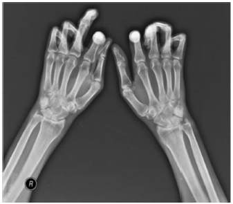
Fig. 4 Flexion contracture involving the hands. Adapted (with permission) from Tas et al.27Tendon friction rubs (TFR) 

TFR also appear in patients with SSc with a reported prevalence of 5-37 %, ocurring more often in patients with diffuse SSc.2,4,10,22,24 TFR manifest mainly in hands, elbows and ankles.24,25 They are found more frequently in young patients and during early stages of the disease (OR 2.58, 95 % CI 1.87-3.56), and their presence is related to a progression of the disease, and other severe manifestations such as pulmonary, renal and vascular compromise.2,18,24,26 Additionally, patients with early diffuse SSc who develop TFR have a lower five to ten-year survival, and more severe compromise of their life quality.24

In general, synovitis, articular contractures and TFR manifest together, thus, the presence of any one of the OAM is a risk factor for the development of other osteoarticular involvement.2

Acroosteolysis

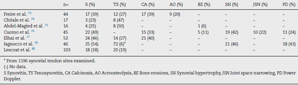
Distal phalanges bone resorption, or acroosteolysis, is another usual OAM, seen in up to 25 % of patients27 (Fig. 5). Some studies have described this manifestation related to more severe and systemic compromise of SSc.28 Furthermore, acroosteolysis is associated with digital ischemia severity,29,30 as well as with the presence of advanced videocapillaroscopy patterns, particularly capillary loss and angiogenesis.31 Similarly, the presence of digital ulcers is an independent predictor for acroosteolysis progression.23 Thus, distal bone resorption may correspond to an ischemic manifestation of SSc.

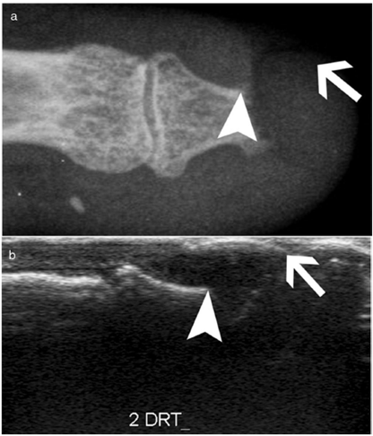
Fig. 5 Acroosteolysis of the index finger in a SSc patient. a. plain radiograph imaging. b. ultrasonography imaging. Adapted (with permission) from: Freire et al.73  

Calcinosis

Calcinosis, which has been described in up to 25 % of patients, is more frequently described in older patients and is related to the presence of osteoporosis and digital ulcers32,33 (Fig. 6). Additionally, digital ulcers are an independent progression factor for calcinosis.23 Some studies suggest a possible role for vascular damage in the pathogenesis of calcinosis since it is associated to advanced videocapillaroscopy patterns and digital ulcers.31

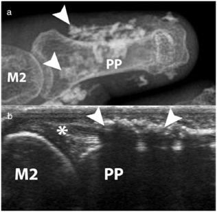
Fig. 6 Calcinosis of the index finger in a SSc patient. a. plain radiograph imaging. b. ultrasonography imaging. Adapted (with permission) from: Freire et al.73  

Avascular necrosis

Avascular necrosis in the femoral head has been reported as an infrequent OAM, although severe, in SSc patients. In early stages of the disease the patients maybe asymptomatic. However, as it progresses the cardinal symptom is pain in the groin, thigh and buttocks.35 Some rare cases involving the carpal bones have been reported, yet necrosis of the lunate bone might be underestimated and frequently misinterpreted as carpitis (erosive arthritis in the wrist).36,37

Feet involvement

Osteoarticular manifestations in the feet tend to appear later in the disease course and cause severe disability. At the time of SSc diagnosis, articular contractures, TFR and acroosteolysis involvement of the feet are far less common than these of the hands; nonetheless, feet arthralgia might be a common complaint.34 Moreover, feet involvement worsens during the course of the disease unlike hand involvement which tends to remain steady once it is manifested.34

Bone mineral density in SSc

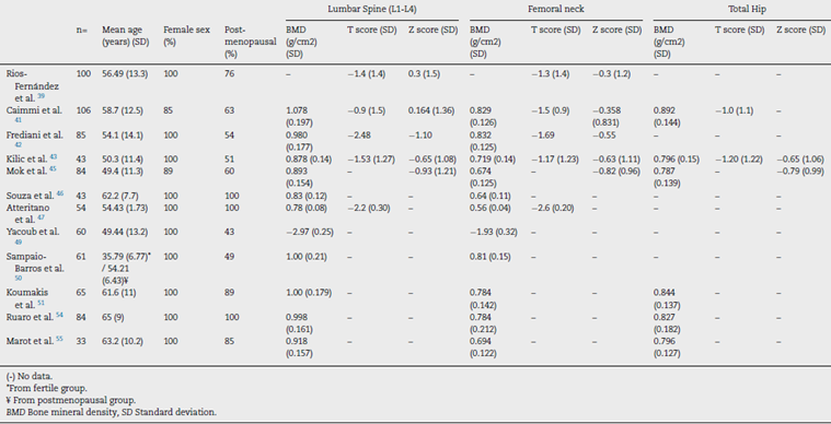
SSc patients have multiple known risk factors for the development of osteoporosis, i.e., chronic inflammation, steroids use, immobility and early menopause.38 Low bone mineral density (BMD) has been reported in around 27 %-62 % of SSc patients,38-45 while osteoporosis has been reported varying between 10 %-51 % of patients depending on the studied populations41-48 (Table 3). In SSc patients low BMD is more frequently seen in the femoral neck compared to the lumbar spine (0.829 + 0.126mg/cm2 vs. 1.078 + 0.197mg/cm2).41 The change in BMD seems to be influenced by the lower body mass index reported in this patients group compared to normal population.41,42,46

Table 3 Values of BMD, T scores and Z scores of patients with SSc. 

In a study in Moroccan population by Yacoub et al. the SSc population had a lower BMD, as well as lower vitamin D levels compared to healthy controls. In addition, there was an association between a low BMD and low vitamin D levels with the presence of severe joint involvement.49 Similarly, in two Spanish populations the presence of SSc and the longer duration of the disease were associated with a lower BMD when compared with a group of healthy women. However, vitamin D deficiency was not associated with lower BMD.39

Similar results were found in the femoral neck bone density in a group of 43 patients with SSc when compared with matched controls with RA43; however, a previous study with a larger number of patients did not find this difference.44 The cumulative dose of steroids is not a risk factor for the development of osteoporosis or fractures in SSc when evaluated with bone densitometry44,50; although patients receiving doses of prednisone of 5 mg or more per day have an altered bone microarchitecture when evaluated by tomography.51 In a prospective study of 50 Canadian SSc patients, those who were non-Caucasian had a lower BMD than Caucasians.40 The presence of calcinosis seems also to influence the appearance of osteoporosis32; nonetheless, this finding is not reproducible in all series.52 A low BMD in SSc patients is not associated with the severity of involvement of other organs.45

The presence of fragility fractures is not negligible. Omair and colleagues reported fragility fractures in up to 14 % of patients.40 In a previous study, up to 24 % of patients had a documented vertebral fracture, with a predominance of grade 1 fractures in the Genant classification.47 Avouac et al. found that 35 % had fragility fractures in a cohort of 71 patients with SSc, the number of fractures was similar to that found in patients with RA, and was higher than the reported in other SSc cohorts, probably due to the older age of patients in this cohort.44

Fauny et al. found that despite the increased risk of bone mineral density alteration, only 43 % of SSc patients are screened with Dual Energy X-ray Absorptiometry (DEXA).53 In addition, there are complementary strategies applied to tomography that allow to determine fracture risk in SSc patients, which seem to be more sensitive than DEXA in this specific population.53 The Trabecular Bone Score (TBS), a recent tool to evaluate bone microarchitecture, has been used in patients with SSc with good results.51,54 Ruaro et al. reported TBS as capable of discriminating patients with altered bone microstructure that increases the risk for fracture.54 In addition, TBS appears to be more altered in patients with early or active videocapillaroscopy patterns, suggesting a correlation between microvascular damage and bone involvement.54 In a previous study by Marot et al. an important compromise of trabecular bone was found, which seems to be related to the presence of digital ulcers.55 Bone microstructural characterization through TBS opens the possibility to its potential use as a complementary diagnostic tool for bone involvement secondary to corticosteroids, thus TBS could be used with DEXA to detect osteoporosis induced by corticosteroids.51

Serological markers

Anti-cyclic citrullinated peptide antibodies (anti-CCP) Several studies have looked at the relationship of anti-CCP with the development of joint manifestations in SSc. Anti-CCP positivity is reported in between 3 %-14 % of SSc patients according to the test generation and the cut-off point used.56-59 In Latin American population anti-CCP positivity has been described in up to 12 % of cases with SSc, however, their association with arthritis in this demographic group is not completely clear.56 Due to the high specificity of these antibodies for RA diagnosis, there is controversy regarding the usefulness of anti-CCP to diagnose the overlap of SSc with RA (SSc-RA). Santiago et al. found a significant association between positive titers of anti-CCP and the onset of arthritis, although there was no statistically significant association between the presence of these antibodies and the diagnosis of SSc-RA58; unlike other studies where anti-CCP were considered useful for identifying SSc-RA patients.57,60,61 Stamenkovic et al. also reported a significant relationship between anti-CCP and clinically evident arthritis.59 Similarly, a recent study found that the presence of anti-CCP is related to the appearance of bone erosions, and when associated with the presence of positive rheumatoid factor (RF) is related to the development of arthralgia in SSc patients.62 A meta-analysis by Laustriat et al. reported that in addition to conferring high risk of erosive arthritis (OR = 22.48 [95 % CI: 10.71-47.21]), anti-CCP positivity in SSc patients increases the risk for pulmonary fibrosis, as well as for esophageal and diffuse skin involvements.63

Other antibodies

The presence of antinuclear and double-stranded DNA antibodies (ANA and anti-dsDNA, respectively) in limited SSc patients seems tobe related to joint manifestations.11,64 Antibodies against SCL-70 (anti SCL-70) are higher among patients with low BMD.39,40,41 In addition, an association of anti SCL-70 with the development of articular contractures and deformity in hands has been reported (OR 7.01, 95 % CI 1.02-48.69), although it is not associated with the presence of arthralgia, or TFR.21,65 There is some debate about whether the presence of anti-topoisomerase I antibodies is associated with the presence of joint manifestations, especially joint contractures, since some of the studies that have established this association have selection biases.66

Cartilage oligomeric matrix protein (COMP)

COMP, a protein found in cartilage, and believed to be an important regulator of collagen fibers type I and II maintenance, has been studied as a possible marker of arthritis in SSc. A study of 40 SSc patients found that COMP levels were related to disease activity, presence of arthritis, anti-CCP and anti SCL-70 antibodies.67 However, a significant number of patients show positive COMP, so it does not seem to be very specific to identify arthritis.67

Platelet-activating factor acetylhydrolase (PAF-AH)

Normal levels of PAF-AH correspond to a risk factor for arthralgia or arthritis (OR 19.8, [95 % CI 2.4-164] and OR 1.86, [95% CI 1.33-2.59] respectively), which suggests that the presence of elevated PAF-AH has a possible protective role for joint involvement.68

YKL-40 or human cartilage glycoprotein 39 (HC gp-39)

YKL-40 was evaluated in a study of 40 SSc patients by La Montagna et al. reporting that YKL-40 levels are elevated in patients with arthralgia or arthritis, which suggests that it can be a possible serological marker of joint involvement in SSc.69

Radiological evaluation

Conventional radiography

Conventional radiography is a useful tool to address joint involvement in SSc used in conjunction with RF and anti-CCP, especially in cases where doubt exists of a possible overlap with RA.27 The findings can be divided into three patterns: articular degenerative damage, periarticular fibrotic changes, or articular inflammatory involvement; although this is a classification that is not reproducible in all studies.9

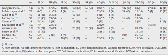
One of the most frequent findings described in simple X-rays is juxta-articular demineralization, reported in up to 42 % of cases with SSc. Decreased joint space and small and discrete joint erosions are common, up to 33 % and 31 % respectively12,27,33,70,71 (Fig. 7). Flexion deformities are present in a considerable number of patients.70 Extra-articular calcifications can appear in up to 18 % of cases12,27,30,33 (Table 4). Unlike RA, involvement of styloid process is uncommon, occurring only in 2 % of patients.12 In the distal phalangeal tuft (DPT) the most frequently described finding corresponds to DPT resorption reported in up to 28 % of the cases.12,30,70,72

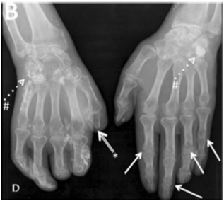
Fig. 7 Typical hand findings in simple radiography of a patient with SSc. (white arrow: yuxta articular erosions with joint space loss, asterix: acroosteolysis, hash: calcinosis). Adapted (with permission) from: Avouac et al.1  

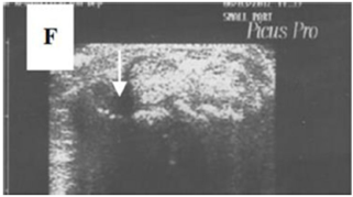
Fig. 8 A transverse ultrasonographic view of the common extensor tendon at the level of the wrist showing by grayscale tenosynovitis in the form of effusion (arrow). Adapted (with permission) from: Abdel-Magied et al.75  

Other findings reported are calcinosis in up to 12 %,30 as well as DPT proliferation or erosions, although with less frequency.72 When feet radiographs are evaluated, there is a lower frequency of acroosteolysis, erosions and calcinosis compared to hands. However, there are no significant differences in dem-ineralization, decreased joint space, or joint subluxations.34

Ultrasonography (US)

Due to the poor sensitivity of conventional radiography to detect early findings of joint involvement, the role of ultra-sonography in this scenario has been evaluated (Table 5). Freire et al. evaluated the US findings at the joint level in SSc patients with recent diagnosis.73 Synovitis was found in 39 % of patients, mostly mild. In addition, 27 % of patients presented tenosynovitis with greater involvement in extensor tendon sheaths than in flexors73 (Fig. 8). US is able to identify arthritis and tenosynovitis during the follow-up of the disease in SSc patients with arthralgias without clinically evident inflammatory changes, although it is not equally useful to identify erosions.74-76 Sclerosing tenosynovitis and soft tissue calcifications are specific findings of SSc that allow for differentiation from other pathologies such as RA.77 Likewise, US has an acceptable sensitivity to detect calcinosis and acroosteolysis, with a value of 89 % and 90 %, respectively,73 and is able to detect the presence of enthesopathy in SSc patients.78 Iagnocco et al. evaluated the prevalence of abnormal US findings combined with Power Doppler in carpal and hand joints from SSc patients.79 Similar to previously published studies, a high prevalence of abnormal findings was reported when compared to healthy population. The most frequent findings described were: synovial hypertrophy, joint effusion and tenosynovitis. In addition, the use of Power Doppler US improves the ability to recognize local inflammatory signs, often in the absence of clinically evident synovitis.79 Lescoat et al. in a recent study confirmed these results, reporting inflammatory synovitis in 17.5 % of patients, and sclerosing tenosynovitis in up to 18.5 %.80 These findings were associated with elevated CRP, clinically evident synovitis and pericarditis. In turn, sclerosing tenosynovitis was more frequently seen in men and was associated with pulmonary compromise, antibodies against RNA polymerase III, and diffuse type SSc.80

Table 4 Radiographic changes in hands of patients with SSc. 

Table 5 US changes in joints of patients with SSc. 

Table 6 MRI changes in hands of patients with SSc. 

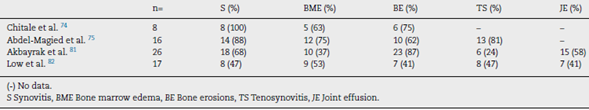
Magnetic resonance imaging (MRI)

Interest in MRI usefulness to detect joint involvement in SSc patients has increased in recent years. Synovitis with mild erosions and joint effusion associated with the presence of bone marrow edema are predominantly described (Table 6).81,82 Articular MRI is more sensitive than ultrasound to identify joint involvement in patients with arthralgia without clinically evident synovitis.74,75 Unlike US and radiography, MRI is able to identify early changes, especially at the level of the car pus with predominant involvement of the lunate bone, which seems to be a distinctive feature of the disease.81

MRI has a high yield to identify tenosynovitis involving the hands in patients with arthritis,83 as well as changes in SSc patients, in whom tenosynovitis is described in up to 88 % of those with arthralgia.74,75 Similarly, in patients with evidence of TFR, MRI identifies tenosynovitis in up to 50 % of cases.84

Clinimetry and impact on general functionality and quality of life

The quality of life of patients with SSc is affected at different levels,85 largely due to skin involvement86 and osteoarticular commitment that affects the development of daily activities in a moderate to severe degree.10,87,89 Moreover, psychological stress and the perception of disability negatively affect the quality of life in these patients.90 There are multiple factors that are associated with the loss of hand function, especially the presence of joint contractures, digital ulcers, and severe Raynaud's phenomenon.91

The Health Assessment Questionnaire (HAQ) disability index (DI), the Short Form 36 (SF-36) health survey physical component score (PCS) and the mental component score (MCS) have been validated in SSc.92 Wiese et al. documented that there is a correlation between the HAQ DI and SF36 PCS (r= -0.79), as well as between HAQDI and global health measurements given by physicians and patients with early diffuse SSc (r = 0.43 and r = 0.57, respectively).22 These measurements had an important association with the modified Rodnan score, although they did not have the same association with mus-culoskeletal manifestations or OAM.22 Additionally, using the same instruments, the disability generated by joint involvement is greater than that presented in PsA patients, with more intense pain than in RA patients.93 In a subsequent study of 326 patients with early diffuse SSc it was found that the loss of hand function, determined by the Cochin Hand Function Scale (CHFS), also contributes significantly to the disability generated by SSc.94

Kwakkenbos et al. validated in a cohort of 696 patients the PROMIS-29v2, a tool that evaluates seven domains related to quality of life in addition to the intensity of pain in SSc patients.89 They found that SSc negatively impacts quality of life largely due to joint contractures and gastrointestinal symptoms.89

The hand mobility test in SSc (HAMIS) was evaluated in a longitudinal study of 43 SSc patients with less than three years' duration of the disease.95 A follow-up of at least four years was carried out, reporting that, although it varied in 72 % of the patients, there were no significant changes in the mobility of the hand during the first years of the disease.95 In turn, in patients with longer duration of the disease, there is an important limitation to perform movements that involve handgrip or twisting.96 The delta finger-to-palm test has also been validated to evaluate the mobility of the hands in SSc patients,97 as well as the Michigan Hand Questionnaire (MHQ).98 Similarly, the hand anatomic index (HAI) corresponds to a useful tool to objectively assess hand deformity and secondary functional impairment in SSc patients.99

Erol et al., used the health-related quality of life (HRQoL) with the Duruöz Hand Index (DHI) to assess hand function in 44 SSc patients compared to RA patients.100 They described that the loss of hand function in SSc is not only similar to that of RA, although joint changes in both diseases are different, but it has an important influence on the quality of life in these patients.100 A previous study that used HAMIS and Cochin Scale (CS) reported similar findings.101

Furthermore, Lorand et al., validated the usefulness of the Disease Activity Score 28 (DAS 28) using PCR and ESR, as well as of the Simplified Disease Activity Index (SDAI) and the Clinical Disease Activity Index (CDAI), to assess joint involvement in SSc.102 They reported that, although the four instruments are valid to determine severity of joint involvement in SSc patients, DAS28-ESR showed the best performance regarding reliability and construct validity.102

The levels of fatigue reported in SSc patients, measured by means of the general fatigue index (IGF), are similar to the ones reported by patients with neoplasms, RA or systemic lupus erythematosus (SLE).103 In addition, fatigue has a negative impact at many different levels in patients with autoimmune diseases, including SSc.104 Fibromyalgia, a disease frequently associated with high rates of fatigue, is very common in SSc patients, occurring in between 57 %-72 %.105

Treatment

There are few studies on specific treatment of OAM in SSc. The first line treatments for the management of synovitis are adapted from the RA management schemes. A small retrospective study with patients from the EUSTAR database suggests a beneficial effect of hydroxychloroquine in the management of the inflammatory joint involvement in SSc.106 Patients who develop contractures in the first years of the disease could benefit from the use of cyclophosphamide, methotrexate or leflunomide.20 Immunoglobulin, used in a treatment scheme over six months, could be effective in reducing joint symptoms and secondary impact on quality of life in SSc patients with severe joint involvement refractory to first-line treatments.107

The possible role of anti TNF-α in the management of SSc has increased the interest of different groups worldwide. There are reports of studies that give a possible role for this group of treatments in the management of arthritis in the SSc population.108 In a study conducted by EUSTAR in 2011 in centers specialized in SSc management, it was evidenced that the majority of experts from this society recommended the use of anti TNF-α molecules only in randomized clinical trials since their potential adverse effects on the disease are unknown.109 However, due to the difficulty of conducting a randomized clinical trial with biological drugs, only observational studies have been published in this field.

Elhai et al. evaluated through an observational study the effect of tocilizumab, a humanized antibody against the IL-6 receptor, and abatacept, a recombinant fusion protein that modulates T-cell co-stimulation mediated by CTLA4, in the management of refractory polyarthritis or myopathy associated with SSc.110 DAS 28 was used to evaluate the response to treatment, and HAQ-DI to evaluate its impact on quality of life, suggesting that both drugs could improve polyarthritis secondary to SSc.110 A phase 3 study is currently underway to evaluate the response to rituximab compared with placebo in SSc patients with OAM who do not respond to first-line treatments.111

In SSc patients with painful hands of multicausal origin, including osteoarticular involvement, the carpal tunnel hydrodissection with lidocaine guided by US, associated to local corticoid injection reduces pain scores by up to 67 %, with a sustained duration of up to six months.112

Orthotic management for joint contractures can be ineffective in late stages of the disease, not only because it exacerbates symptoms such as Raynaud's phenomenon, but because it does not generate significant relief in the deformities generated by contractures.113 On the contrary, surgical management can be considered to reduce pain in patients with advanced disease refractory to other treatments.114 Procedures such as: sympathectomy, calcinosis resection, arthroplasties or capsulotomies have been practiced but the real benefits are few, so they have fall into disuse.115,116

In conclusion, OAM in SSc are frequent. They also have an important impact on patient's quality of life, especially due to the limitation in hand functionality. Treatment is difficult and requires a multidisciplinary approach. Due to the above, OAM in SSc require early identification and intervention. In addition, further research including good quality randomized clinical trials is required to establish the best treatment strategy for these patients.

REFERENCES

1. Avouac J, Clements PJ, Khanna D, Furst DE, Allanore Y. Articular involvement in systemic sclerosis. Rheumatol (Oxford). 2012;51(8):1347-56. [ Links ]

2. Avouac J, Walker U, Tyndall A, Kahan A, Matucci-Cerinic M, Allanore Y, et al. Characteristics of joint involvement and relationships with systemic inflammation in systemic sclerosis: Results from the EULAR Scleroderma Trial and Research Group (EUSTAR) database. J Rheumatol. 2010;37(7):1488-501. [ Links ]

3. Clements PJ, Allanore Y, Khanna D, Singh M, Furst DE. Arthritis in Systemic Sclerosis: Systematic Review of the Literature and Suggestions for the Performance of Future Clinical Trials in Systemic Sclerosis Arthritis. Semin Arthritis Rheum. 2012;41(6):801-14. [ Links ]

4. Ostojic P Damjanov N. Different clinical features in patients with limited and diffuse cutaneous systemic sclerosis. Clin Rheumatol. 2006;25(4):453-7. [ Links ]

5. Schmeiser T, Pons-Kühnemann J, Özden F, Müller-Ladner U, Dinser R. Arthritis in patients with systemic sclerosis. Eur J Intern Med. 2012;23(1), e25-9. [ Links ]

6. Tikly M. Clinical and laboratory manifestations of systemic sclerosis (scleroderma) in Black South Africans. Rheumatology. 1999;38(5):397-400. [ Links ]

7. Wangkaew S, Sivasomboon C, Leungwatthananon W, Kasitanon N, Louthrenoo W. Prevalence and predictors of hand involvement in Thai patients with systemic sclerosis. Int J Rheum Dis. 2018;21(1):240-8. [ Links ]

8. Arias-Nunez MC, Llorca J, Vazquez-Rodriguez TR, Gomez-Acebo I, Miranda-Filloy JA, Martin J, et al. Systemic sclerosis in northwestern Spain: A 19-year epidemiologic study. Medicine (Baltimore). 2008;87(5):272-80. [ Links ]

9. La Montagna G, Sodano A, Capurro V, Malesci D, Valentini G. The arthropathy of systemic sclerosis: A 12 month prospective clinical and imaging study. Skeletal Radiol. 2005;34(1):35-41. [ Links ]

10. Jaeger VK, Distler O, Maurer B, Czirják L, Lóránd V,Valentini G, et al. Functional disability and its predictors in systemic sclerosis: A study from the DeSScipher project within the EUSTAR group. Rheumatol (Oxford). 2018;57(3):441-50. [ Links ]

11. Misra R, Darton K, Jewkes RF, Black CM, Maim RN. Arthritis in scleroderma. Br J Rheumatol. 1995;34(9):831-7. [ Links ]

12. Allali F, Tahiri L, Senjari A, Abouqal R, Hajjaj-Hassouni N. Erosive arthropathy in systemic sclerosis. BMC Public Health. 2007;7:1-6. [ Links ]

13. Lovell CR, Jayson MIV. Joint involvement in systemic sclerosis. Scand J Rheumatol. 1979;8(3):154-60. [ Links ]

14. Armstrong RD, Gibson T. Scleroderma and erosive polyarthritis: a disease entity? Ann Rheum Dis. 1982;41(2):141-6. [ Links ]

15. Horimoto AMC, da Costa IP. Overlap between systemic sclerosis and rheumatoid arthritis: A distinct clinical entity? Rev Bras Reumatol. 2016;56(4):287-98. [ Links ]

16. Jinnin M, Ihn H, Yamane K, Asano Y, Yazawa N, Tamaki K. Clinical features of patients with systemic sclerosis accompanied by rheumatoid arthritis. Clin Exp Rheumatol. 2003;21(1):91-4. [ Links ]

17. Baron M, Lee P, Keystone EC. The articular manifestations of progressive systemic sclerosis (scleroderma). Ann Rheum Dis. 1982;41(2):147-52. [ Links ]

18. Avouac J, Walker UA, Hachulla E, Riemekasten G, Cuomo G, Carreira PE, et al. Joint and tendon involvement predict disease progression in systemic sclerosis: A EUSTAR prospective study. Ann Rheum Dis. 2016;75(1):103-9. [ Links ]

19. Arslan Tas D, Yildiz F, Sakalli H, Kelle B, Balli T, Erken E. Sacroiliac join involvement in systemic sclerosis. Int J Rheum Dis. 2015;18(1):84-90. [ Links ]

20. Bálint Z, Farkas H, Farkas N, Minier T, Kumánovics G, Horváth K, et al. A three-year follow-up study of the development of joint contractures in 131 patients with systemic sclerosis. Clin Exp Rheumatol. 2014;32(6). S-68-74. [ Links ]

21. Ashida R, Ihn H, Mimura Y, Jinnin M, Asano Y, Kubo M, et al. Clinical features of scleroderma patients with contracture of phalanges. Clin Rheumatol. 2007;26(8):1275-7. [ Links ]

22. Wiese AB, Berrocal VJ, Furst DE, Seibold JR, Merkel PA, Mayes MD, et al. Correlates and Responsiveness to Change of Measures of Skin and Musculoskeletal Disease in Early Diffuse Systemic Sclerosis. Arthritis Care Res. 2014;66(11):1731-9. [ Links ]

23. Avouac J, Mogavero G, Guerini H, Drapé JL, Mathieu A, Kahan A, et al. Predictive factors of hand radiographic lesions in systemic sclerosis: A prospective study. Ann Rheum Dis. 2011;70(4):630-3. [ Links ]

24. Khanna PP, Furst DE, Clements PJ, Maranian P, Indulkar L, Khanna D, et al. Tendon friction rubs in early diffuse systemic sclerosis: prevalence, characteristics and longitudinal changes in a randomized controlled trial. Rheumatology (Oxford). 2010;49(5):955-9. [ Links ]

25. Steen VD, Medsger TA. The palpable tendon friction rub: an important physical examination finding in patients with systemic sclerosis. Arthritis Rheum. 1997;40(6):1146-51. [ Links ]

26. Doré A, Lucas M, Ivanco D, Medsger TA, Domsic RT. Significance of palpable tendon friction rubs in early diffuse cutaneous systemic sclerosis. Arthritis Care Res. 2013;65(8):1385-9. [ Links ]

27. Tas DA, Erken E, Sakalli H, Yucel AE. Evaluating hand in systemic sclerosis. Rheumatol Int. 2012;32(11):3581-6. [ Links ]

28. Arana-Ruiz JC, Amezcua-Guerra LM. Acro-osteolysis as an indicator of severity in systemic sclerosis. Reumatol Clin. 2016;12(5):263-6. [ Links ]

29. Johnstone EM, Hutchinson CE, Vail A, Chevance A, Herrick AL. Acro-osteolysis in systemic sclerosis is associated with digital ischaemia and severe calcinosis. Rheumatol (Oxford). 2012;51(12):2234-8. [ Links ]

30. Koutaissoff S, Vanthuyne M, Smith V, De Langhe E, Depresseux G, Westhovens R, et al. Hand Radiological Damage in Systemic Sclerosis: Comparison with a Control Group and Clinical and Functional Correlations. Semin Arthritis Rheum. 2011;40(5):455-60. [ Links ]

31. Morardet L, Avouac J, Sammour M, Baron M, Kahan A, Feydy A, et al. Late Nailfold Videocapillaroscopy Pattern Associated with Hand Calcinosis and Acro-Osteolysis in Systemic Sclerosis. Arthritis Care Res. 2016;68(3):366-73. [ Links ]

32. Valenzuela A, Baron M, Herrick A, Proudman S, Stevens W. Calcinosis is associated with digital ulcers and osteoporosis in patients with systemic sclerosis: A Scleroderma Clinical Trials Consortium study. Semin Arthritis Rheum. 2016;46(3):344-9. [ Links ]

33. Avouac J, Guerini H, Wipff J, Assous N, Chevrot A, Kahan A, et al. Radiological hand involvement in systemic sclerosis. Ann Rheum Dis. 2006;65(8):1088-92. [ Links ]

34. La Montagna G, Baruffo A, Tirri R, Buono G, Valentini G. Foot Involvement in Systemic Sclerosis: A Longitudinal Study of 100 Patients. XXX. 2002;31(4):248-55. [ Links ]

35. Taccari E, Spadaro A, Riccieri V, Grandilli MA, Zoppini A. Avascular necrosis of the femoral head in long-term follow-up of systemic sclerosis: report of two cases. Clin Rheumatol. 1989;8(3):386-92. [ Links ]

36. Frerix M, Kröger K, Szalay G, Müller-Ladner U, Tarner IH. Is osteonecrosis of th lunate bone an underestimated feature of systemic sclerosis? A case series of nine patients and review of literature. Semin Arthritis Rheum. 2016;45(4):446-54. [ Links ]

37. Matsumoto AK, Moore R, Alli P, Wigley FM. Three cases of osteonecrosis of the lunate bone of the wrist in scleroderma. Clin Exp Rheumatol. 1999;17(6):730-2. [ Links ]

38. La Montagna G, Vatti M, Valentini G, Tirri G. Osteopenia in systemic sclerosis. Evidence of a participating role of earlier menopause. Clin Rheumatol. 1991;10(1):18-22. [ Links ]

39. Rios-Fernández R, Callejas-Rubio JL, Fernández-Roldán C, Simeón-Aznar CP, García-Hernández F, Castillo-Palma MJ, et al. Bone mass and vitamin D in patients with systemic sclerosis from two Spanish regions. Clin Exp Rheumatol. 2012;30(6):905-11. [ Links ]

40. Omair MA, McDonald-Blumer H, Johnson SR. Bone disease in systemic sclerosis: outcomes and associations. Clin Exp Rheumatol. 2014;32(6). [ Links ]

41. Caimmi C, Caramaschi P, Barausse G, Orsolini G, Idolazzi L, Gatti D, et al. Bone Metabolism in a Large Cohort of Patients with Systemic Sclerosis. Calcif Tissue Int. 2016;99(1):23-9. [ Links ]

42. Frediani B, Baldi F, Falsetti P, Acciai C, Filippou G, Spreafico A, et al. Clinical determinants of bone mass and bone ultrasonometry in patients with systemic sclerosis. Clin Exp Rheumatol. 2004;22(3):313-8. [ Links ]

43. Kilic G, Kilic E, Akgul O, Ozgocmen S. Increased risk for bone loss in women with systemic sclerosis: A comparative study with rheumatoid arthritis. Int J Rheum Dis. 2016;19(4):405-11. [ Links ]

44. Avouac J, Koumakis E, Toth E, Meunier M, Maury E, Kahan A, et al. Increased risk of osteoporosis and fracture in women with systemic sclerosis: A comparative study with rheumatoid arthritis. Arthritis Care Res. 2012;64(12):1871-8. [ Links ]

45. Mok CC, Chan PT, Chan KL, Ma KM. Prevalence and risk factors of low bone mineral density in Chinese patients with systemic sclerosis: A case-control study. Rheumatol (Oxford). 2013;52(2):296-303. [ Links ]

46. Souza RBC, Borges CTL, Takayama L, Aldrighi JM, Pereira RMR. Systemic sclerosis and bone loss: The role of the diseas and body composition. Scand J Rheumatol. 2006;35(5):384-7. [ Links ]

47. Atteritano M, Sorbara S, Bagnato G, Miceli G, Sangari D, Morgante S, et al. Bone Mineral Density, Bone Turnover Markers and Fractures in Patients with Systemic Sclerosis: A Case Control Study. PLoS One. 2013;8(6):6-11. [ Links ]

48. Omair MA, Pagnoux C, McDonald-Blumer H, Johnson SR. Low bone density in systemic sclerosis. a systematic review. Journal of Rheumatology. 2013;40:1881-90. [ Links ]

49. Yacoub Y, Amine B, Laatiris A, Wafki F, Znat F, Hajjaj-Hassouni N. Bone density in Moroccan women with systemic scleroderma and its relationships with disease-related parameters and vitamin D status. Rheumatol Int. 2012;32(10):3143-8. [ Links ]

50. Sampaio-Barros PD, Costa-Paiva L, Filardi S, Sachetto Z, Samara AM, Marques-Neto JF. Prognostic factors of low bone mineral density in systemic sclerosis. Clin Exp Rheumatol. 2005;23(2):180-4. [ Links ]

51. Koumakis E, Avouac J, Winzenrieth R, Toth E, Payet J, Kahan A, et al. Trabecular bone score in female patients with systemic sclerosis: Comparison with rheumatoid arthritis and influence of glucocorticoid exposure. Journal of Rheumatology. 2015;42:228-35. [ Links ]

52. Di Munno O, Mazzantini M, Massei P, Ferdeghini M, Pitaro N, Latorraca A, et al. Reduced bone mass and normal calcium metabolism in systemic sclerosis with and without calcinosis. Clin Rheumatol. 1995;14(4):407-12. [ Links ]

53. Fauny M, Bauer E, Albuisson E, Perrier-Cornet J, Deibener J, Chabot F, et al. Vertebral fracture prevalence and measurement of the scanographic bone attenuation coefficient on CT-scan in patients with systemic sclerosis. Rheumatol Int. 2018;38(10):1901-10. [ Links ]

54. Ruaro B, Casabella A, Paolino S, Pizzorni C, Alessandri E, Seriolo C, et al. Correlation between bone quality and microvascular damage in systemic sclerosis patients. Rheumatol (Oxford). 2018;57(9):1548-54. [ Links ]

55. Marot M, Valéry A, Esteve E, Bens G, Müller A, Rist S, et al. Prevalence and predictive factors of osteoporosis in systemic sclerosis patients: a case-control study. Oncotarget 2015;6(17):14865-73 [ Links ]

56. Polimeni M, Feniman D, Skare TS, Nisihara RM. Anti-cyclic citrullinated peptide antibodies in scleroderma patients. Clin Rheumatol. 2012;31(5):877-80. [ Links ]

57. Morita Y, Muro Y, Sigiur K, Tomita Y. Anti-cyclic citrullinated peptide antibody in systematic sclerosis. Clin Exp Rheumatol. 2008;26(4):542-7. [ Links ]

58. Santiago M, Baron M, Miyachi K, Fritzler MJ, Abu-Hakima M, Leclercq S, et al. A comparison of the frequency of antibodies to cyclic citrullinated peptides using a third generation anti-CCP assay (CCP3) in systemic sclerosis, primary biliary cirrhosis and rheumatoid arthritis. Clin Rheumatol. 2008;27(1):77-83. [ Links ]

59. Stamenkovic B, Stankovic A, Dimic A, Damjanov N, Nedovicc J, Stojanovic S, et al. The clinical significance of antibody determination to cyclic citrullinated peptides in systemic sclerosis. Srp Arh Celok Lek. 2012;140(5-6):350-4. [ Links ]

60. Ingegnoli F, Galbiati V, Zeni S, Meani L, Zahalkova L, Lubatti C, et al Use of antibodies recognizing cyclic citrullinated peptide in the differential diagnosis of joint involvement in systemic sclerosis. Clin Rheumatol. 2007;26(4):510-4. [ Links ]

61. Ueda-Hayakawa I, Hasegawa M, Kumada S, Tanaka C, Komura K, Hamaguchi Y, et al. Usefulness of anti-cyclic citrullinated peptide antibody and rheumatoid factor to detect rheumatoid arthritis in patients with systemic sclerosis. Rheumatology. 2010;49(11):2135-9. [ Links ]

62. Kamalaksha S, White DHN, Solanki KK. Significance of combined anti-CCP antibodies and rheumatoid factor in a New Zealand cohort of patients with systemic sclerosis. Int J Rheum Dis. 2018;21(7):1430-5. [ Links ]

63. Laustriat G, Ruyssen-Witrand A, Constantin A, Barnetche T, Adoue D, Cantagrel A, et al. Anti-citrullinated peptides antibodies in systemic sclerosis: Meta-analysis of frequency and meaning. Joint Bone Spine. 2018;85(2):147-53. [ Links ]

64. Serup J, Staun-Olsen P. Antinuclear antibodies and anti-DNA antibodies in scleroderma. A possible relationship between joint manifestations and increased antibodies in localized scleroderma. Allergy. 1986;41(6):452-6. [ Links ]

65. Foocharoen C, Suwannachat P, Netwijitpan S, Mahakkanukrauh A, Suwannaroj S, Nanagara R. Clinical differences between Thai systemic sclerosis patients with positive versus negative anti-topoisomerase I. Int J Rheum Dis. 2016;19(3):312-20. [ Links ]

66. Radic M, Kaliterna DM, Ljutic D. The level of anti-topoisomerase I antibodies highly correlates with metacarpophalangeal and proximal interphalangeal joints flexion contractures in patients with systemic sclerosis. Clin Exp Rheumatol. 2006;24(4):407-12. [ Links ]

67. Gheita TA, Hussein H. Cartilage Oligomeric Matrix Protein (COMP) in systemic sclerosis (SSc): Role in disease severity and subclinical rheumatoid arthritis overlap. Jt Bone Spine. 2012;79(1):51-6. [ Links ]

68. Yanaba K, Asano Y, Tada Y, Sugaya M, Kadono T, Sato S. Clinical significance of circulating platelet-activating factor acetylhydrolase levels in systemic sclerosis. Arch Dermatol Res. 2012;304(3):203-8. [ Links ]

69. La Montagna G, D'Angelo S, Valentini G. Cross-sectional evaluation of YKL-40 serum concentrations in patients with systemic sclerosis. Relationship with clinical and serological aspects of disease. J Rheumatol. 2003;30(10):2147-51. [ Links ]

70. Bassett LW, Blocka KLN, Furst DE, Clements PJ, Gold RH. Skeletal Findings in Progressive Systemic Sclerosis (Scleroderma). AJR Am J Roentgenol. 1981;136(6):1121-6. [ Links ]

71. Blocka KL, Bassett LW, Furst DE, Clements PJ, Paulus HE. The arthropathy of advanced progressive systemic sclerosis. A radiographic survey. Arthritis Rheum. 1981;24(7):874-84. [ Links ]

72. Izquierdo YE, Calvo Páramo E, Castañeda LM, Gómez SV, Zambrano FS. Radiographic changes of the distal phalangeal tuft of the hands in subjects with systemic sclerosis. Systematic review. Reumatol Clínica. 2016;14(1):20-6. [ Links ]

73. Freire V Bazeli R, Elhai M, Campagna R, Pessis É, Avouac J, et al. Hand and Wrist Involvement in Systemic Sclerosis: US Features. Radiology. 2013;269(3):824-30. [ Links ]

74. Chitale S, Ciapetti A, Hodgson R, Grainger A, O'connor P, Goodson NJ, et al. Magnetic resonance imaging and musculoskeletal ultrasonography detect and characterize covert inflammatory arthropathy in systemic sclerosis patients with arthralgia. Rheumatology. 2010;49(12):2357-61. [ Links ]

75. Abdel-Magied RA, Lotfi A, Abdelgawad EA. Magnetic resonance imaging versus musculoskeletal ultrasonography in detecting inflammatory arthropathy in systemic sclerosis patients with hand arthralgia. Rheumatol Int. 2013;33(8):1961-6. [ Links ]

76. Cuomo G, Zappia M, Abignano G, Iudici M, Rotondo A, Valentini G. Ultrasonographic features of the hand and wrist in systemic sclerosis. Rheumatology (Oxford). 2009;48(11):1414-7. [ Links ]

77. Elhai M, Guerini H, Bazeli R, Avouac J, Freire V, Drapé JL, et al. Ultrasonographic hand features in systemic sclerosis and correlates with clinical, biologic, and radiographic findings. Arthritis Care Res. 2012;64(8):1244-9. [ Links ]

78. Kilic E, Kilic G, Akgul O, Ozgocmen S. Presence of enthesopathy demonstrated with ultrasonography in systemic sclerosis. Mod Rheumatol. 2015;25(5):731-6. [ Links ]

79. Iagnocco A, Vavala C, Vasile M, Stefanantoni K, Valesini ProfG, Riccieri V. Power Doppler ultrasound of the hand and wrist joints in systemic sclerosis. Clin Exp Rheumatol. 2013;31 SUPPL.76. [ Links ]

80. Lescoat A, Ballerie A, Belhomme N, Cazalets C, de Carlan M, Droitcourt C, et al. Synovial involvement assessed by power Doppler ultra-sonography in systemic sclerosis: results of a cross-sectional study. Rheumatology. 2018;57(11):2012-21. [ Links ]

81. Akbayrak E, Dinser R, Müller-Ladner U, Tarner IH. Low-field magnetic resonance imaging study on carpal arthritis in systemic sclerosis - low-grade erosive arthritis of carpal bones is an unexpected and frequent disease manifestation. Arthritis Res Ther. 2013;15(1):2-9. [ Links ]

82. Low AHL, Lax M, Johnsoin SR, Lee P. Magnetic Resonance Imaging of the Hand in Systemic Sclerosis. J Rheumatol. 2009;36(5):961-4. [ Links ]

83. Tehranzadeh J, Ashikyan O, Anavim A, Tramma S. Enhanced MR imaging of tenosynovitis of hand and wrist in inflammatory arthritis. Skeletal Radiol. 2006;35(11):814-22. [ Links ]

84. Stoenoiu MS, Houssiau FA, Lecouvet FE. Tendon friction rubs in systemic sclerosis: A possible explanation-an ultrasound and magnetic resonance imaging study. Rheumatol (United Kingdom). 2013;52(3):529-33. [ Links ]

85. Stamm T, Hieblinge R, Bostrõm C, Mihai C, Birrell F, Thorstensson C, et al. Similar problem in the activities of daily living but different experience: A qualitative analysis in six rheumatic conditions and eight European countries. Musculoskeletal Care. 2014;12(1):22-33. [ Links ]

86. Malcarne VL, Hansdottir I, McKinney A, Upchurch R, Greenbergs HL, Henstorf GH, et al. Medical signs and symptoms associated with disability, pain, and psychosocial adjustment in systemic sclerosis. J Rheumatol. 2007;34(2):359-67. [ Links ]

87. Willems LM, Kwakkenbos L, Leite CC, Thombs BD, van den Hoogen FHJ, Maia AC, et al. Frequency and impact of disease symptoms experienced by patients with systemic sclerosis from five European countries. Clin Exp Rheumatol. 2014;32(6):5-9. [ Links ]

88. Bassel M, Hudson M, Taillefer SS, Schieir O, Baron M, Thombs BD. Frequency and impact of symptoms experienced by patients with systemic sclerosis: Results from a Canadian National Survey. Rheumatology. 2011;50(4):762-7. [ Links ]

89. Kwakkenbos L, Thombs BD, Khanna D, Carrier ME, Baron M, Furst DE, et al. Performance of the Patient-Reported Outcomes Measurement Information System-29 in scleroderma: A Scleroderma Patient-centered Intervention Network Cohort Study. Rheumatol (United Kingdom). 2017;56(8):1302-11. [ Links ]

90. Kwakkenbos L, Sanchez TA, Turner KA, Mouthon L, Carrier ME, Hudson M, et al. The association of sociodemographic and disease variables with hand function: a Scleroderma Patient-centered Intervention Network cohort study. Clin Exp Rheumatol. 2018;36(4):88-94. [ Links ]

91. Hyphantis TN, Tsifetaki N, Siafaka V, Voulgari PV, Pappa C, Bai M, et al. The Impact of Psychological Functioning upon Systemic Sclerosis Patients' Quality of Life. Semin Arthritis Rheum. 2007;37(2):81-92. [ Links ]

92. Cole JC, Khanna D, Clements PJ, Seibold JR, Tashkin DP, Paulus HE, et al. Single-factor scoring validation for the Health Assessment Questionnaire-Disability Index (HAQ-DI) in patients with systemic sclerosis and comparison with early rheumatoid arthritis patients. Qual Life Res. 2006;15(8):1383-94. [ Links ]

93. Johnson SR, Glaman DD, Schentag CT, Lee P, Johnson SR, Gladman DD, et al. Quality of Life and Functional Status in Systemic Sclerosis Compared to Other Rheumatic Diseases. J Rheumatol. 2006;33(6):1117-22. [ Links ]

94. Peytrignet S, Denton CP, Lunt M, Hesselstrand R, Mouthon L, Silman A, et al. Disability, fatigue, pain and their associates in early diffuse cutaneous systemic sclerosis: The European Scleroderma Observational Study. Rheumatol (Oxford). 2018;57(2):370-81. [ Links ]

95. Sandqvist G, Hesselstrand R, Eberhardt K. A longitudinal follow-up of hand involvement and activities of daily living in early systemic sclerosis. Scand J Rheumatol. 2009;38(4):304-10. [ Links ]

96. Cinar FI, Unver V, Cinar M, Yilmaz S, Simsek I, Tosun N, et al. Coping strategies for activities of daily living in women whose hands affected by systemic sclerosis. J Clin Nurs. 2014;23(11-12), 1630-8. [ Links ]

97. Torok KS, Baker NA, Lucas M, Domsic RT, Boudreau R, Medsger TA. Reliability and validity of the delta finger-to-palm (FTP), a new measure of finger range of motion in systemic sclerosis. Clin Exp Rheumatol. 2010;28 2 SUPPL. 58. [ Links ]

98. Schouffoer AA, Van Der Giesen FJ, Beaart-Van De Voorde LJJ, Wolterbeek R, Huizinga TWJ, Vliet Vlieland TPM. Validity and responsiveness of the Michigan Hand Questionnaire in patients with systemic sclerosis. Rheumatol (Oxford). 2016;55(8):1386-93. [ Links ]

99. Roberts-Thomson AJ, Massy-Westropp N, Smith MD, Ahern MJ, Highton J, Roberts-Thomson PJ. The use of the hand anatomic index to assess deformity and impaired function in systemic sclerosis. Rheumatol Int. 2006;26(5):439-44. [ Links ]

100. Erol K, Gok K, Cengiz G, Ozgocmen S. Hand functions in systemic sclerosis and rheumatoid arthritis and influence on clinical variables. Int J Rheum Dis. 2018;21(1):249-52. [ Links ]

101. Poole JL, Santhanam DD, Latham AL. Hand impairment and activity limitations in four chronic diseases. J Hand Ther. 2013;26(3):232-7. [ Links ]

102. Lóránd V, Bálint Z, Komjáti D, Németh B, Minier T, Kumánovics G, et al. Validation of disease activity indices using the 28 joint counts in systemic sclerosis. Rheumatol (Oxford). 2016;55(10):1849-58. [ Links ]

103. Choi HJ, Han JY, Seo MR, Ryu HJ, Baek HJ. Fibromyalgia with chronic rheumatic diseases in South Korea: a comparison of clinical and American College of Rheumatology criteria. Int J Rheum Dis. 2017;20(12):1922-6. [ Links ]

104. Overman CL, Kool MB, D Silva JAP, Geenen R. The prevalence of severe fatigue in rheumatic diseases: an international study. Clin Rheumatol. 2016;35(2):409-15. [ Links ]

105. Thombs BD, Bassel M, Mcguire L, Smith MT, Hudson M, Haythornthwaite JA. A systematic comparison of fatigue levels in systemic sclerosis with general population, cancer and rheumatic disease samples. Rheumatol (Oxford). 2008;47(10):1559-63. [ Links ]

106. Bruni C, Praino E, Guiducci S, Bellando-Randone S, Furst DE, Matucci-Cerinic M. Hydroxychloroquine and joint involvement in systemic sclerosis: Preliminary beneficial results from a retrospective case-control series of an EUSTAR center. Joint Bone Spine. 2017;84(6):747-8. [ Links ]

107. Nacci F, Righi A, Conforti ML, Miniati I, Fiori G, Martinovic D, et al. Intravenous immunoglobulins improv the function and ameliorate joint involvement in systemic sclerosis: A pilot study. Ann Rheum Dis. 2007;66(7):977-9. [ Links ]

108. Omair M, Phumethum V, Johnson SR. Long term safety and effectiveness of tumour necrosis factor inhibitors in systemic sclerosis patients with arthritis. Clin Exp Rheumatol. 2012;30 2 Suppl 71:S55-9. [ Links ]

109. Distler JHW, Jordan S, Airò P, Alegre-Sancho J, Allanore Y, Balbir Gurman A, et al. Is there a role for TNF-a antagonists in the treatment of SSc? EUSTAR expert consensus development using the Delphi technique. Clin Exp Rheumatol. 2011;29 2 SUPPL. 65:S40-5. [ Links ]

110. Elhai M, Meunier M, Matucci-Cerinic M, Maurer B, Riemekasten G, Leturcq T, et al. Outcomes of patients with systemic sclerosis-associated polyarthritis and myopathy treated with tocilizumab or abatacept: A EUSTAR observational study. Ann Rheum Dis. 2013;72(7):1217-20. [ Links ]

111. Allanore Y. Rituximab in Systemic Sclerosis (RECOVER). https://clinicaltrials.gov/ct2/archive/NCT01748084. [ Links ]

112. DeLea SL, Chavez-Chiang NR, Poole JL, Norton HE, Sibbitt WL, Bankhurst AD. Sonographically guided hydrodissection and corticosteroid injection for scleroderma hand. Clin Rheumatol. 2011;30(6):805-13. [ Links ]

113. Seeger MW, Furst DE. Effects of splinting in the treatment of hand contractures in progressive systemic sclerosis. Am J Occup Ther. 1987;41(2):118-21. [ Links ]

114. Norris RW, Brown HG. The proximal interphalangeal joint in systemic sclerosis and its surgical management. Br J Plast Surg. 1985;38(4):526-31. [ Links ]

115. Bogoch E, Gross D. Surgery of the hand in patients with systemic sclerosis: outcomes and considerations. J Rheumatol. 2005;32(4):642-8. [ Links ]

116. Gilbart MK, Jolles BM, Lee P, Bogoch ER. Surgery of the hand in severe systemic sclerosis. J Hand Surg Br. 2004;29(6):599-603. [ Links ]

Please cite this article as: Sebastian M-R, Eliana OC, Gerardo Q-L. Manifestaciones osteoarticulares de esclerosis sistêmica: una sistemática. Rev Colomb Reumatol. 2020. http://dx.doi.org./10.1016/j.rcreue.2020.01.001

Abbreviations: OAM, Osteoarticular Manifestations; SSc, Systemic Sclerosis; RA, Rheumatoid Arthritis; CRP, C-Reactive Protein; MCP, Metacarpophalangeal; PIP, Proximal Interphalangeal; TFR, Tendon Friction Rubs; BMD, Bone Mineral Density; DEXA, Dual Energy X-Ray Absorptiometry; TBS, Trabecular Bone Score; Anti-CCP, Anti-Cyclic Citrullinated Peptide Antibodies; ANA, Antinuclear Antibodies; Anti-dsDNA, Double-Stranded DNA Antibodies; Anti SCL-70, Antibodies Against SCL-70; COMP, Cartilage Oligomeric Matrix Protein; PAF-AH, Platelet-Activating Factor Acetylhydrolase; HC Gp-39, Human Cartilage Glycoprotein 39; SSc-RA, Overlap Of Ssc With RA; RF, Rheumatoid Factor; DPT, Distal Phalangeal Tuft; US, Ultrasonography; MRI, Magnetic Resonance Imaging; HAQ, Health Assessment Questionnaire; DI, Disability Index; SF-36, Short Form 36; PCS, Physical Component Score; MCS, Mental Component Score; CHFS, Cochin Hand Function Scale; HAMIS, Hand Mobility Test In Ssc; HRQoL, Health-Related Quality of Life; DHI, Duruöz Hand Index; DAS-28, Disease Activity Score 28; SDAI, Simplified Disease Activity Index; CDAI, Clinical Disease Activity Index; IGF, General Fatigue Index; SLE, Systemic Lupus Erythematosus.

Conflicts of interest None.

Received: July 30, 2019; Accepted: November 28, 2019

* Corresponding author. E-mail address: semolinar@gmail.com (M.-R. Sebastian). 2444-4405/© 2020 Published by Elsevier Espana, S.L.U. on behalf of Asociación Colombiana de Reumatología.

Creative Commons License This is an open-access article distributed under the terms of the Creative Commons Attribution License