


 text in
  English (pdf)
 Article in xml format
 Article references
 Send this article by e-mail
 Cited by SciELO
  Cited by Google
  Similars in
SciELO
Similars in
SciELO  Similars in Google
 Permalink
Introduction
Systemic lupus erythematosus (SLE) is a chronic multi-systemic autoimmune disease with high morbidity and mortality.1 It has a great variability in its clinical presenta tion and evolution, which is the result of the interaction of epigenetic, immunological, hormonal and environmental factors. (2 The clinical course of this condition is more severe in pediatric patients than in adults. The prevalence of SLE is 70-90 per 100,000 individuals, of whom 20% are diagnosed before the age of 18 years. (3 In Colombia, 50%-55% of the adults and 75% of the children with SLE suffer from lupus nephritis (LN) at some point in their evolution. (4 In infants, the female/male ratio is 4.5/1. (3,5 The mean age of presenta tion of LN varies between 12 and 17 years in children, being less frequent in those under five years of age. (3,6,7 Likewise, LN affects Black, Latino and Asian people more than Caucasian patients. (8
The diagnosis of LN represents a challenge for the clinical team. This disease can begin as a clinical picture simi lar to acute glomerulonephritis, or may be as severe as the rapidly progressive glomerulonephritis with renal failure. (9 The five-year renal survival of children with LN has improved markedly in the last decades and it currently ranges between 77% and 93%. However, compared to healthy infants, the mortality rate observed in this population is 19 times higher. (10
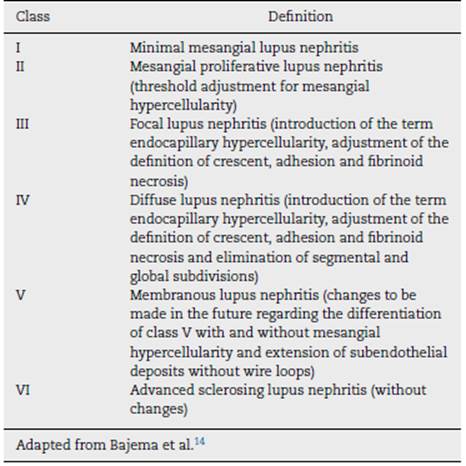
Taking into account that the clinical symptoms corre late with the patterns of glomerular injury, the renal biopsy (RB) is of great importance in the diagnostic approach of LN and, based on this, it is possible to guide the treatment and establish the prognosis. (11,12 In 1982, the World Health Organization (WHO) published the first classification of LN, which was used until 2004. Subsequently, the International Soci ety of Nephrology/Renal Pathology Society (ISN/RPS) classification arouse, which enhanced the standardization and reproducibility of the diagnosis to identify the established kidney injury. (13 In 2018, Bajema et al. conducted a revision of the ISN/RPS classification in order to reduce conflictive definitions and inter-observer variability. (14
Retaking the importance of the RB in the diagnosis of LN, different authors have tried to establish the relationship between the clinical, laboratory and histological findings in the adult and pediatric populations with contradictory results. Dasari et al., in a systematic review that included six stud ies, concluded that the ISN/RPS classification system and the activity or chronicity indices present a low level of agreement among pathologists, which makes difficult their use in clin ical practice. (15 On the other hand, Rijnink et al., in a cohort of 105 adult patients with LN, proposed the fibrinoid necro sis, the fibrous crescents and the interstitial fibrosis/tubular atrophy found in the biopsy as potential biomarkers that allow to assess the risk of progressive renal injury. (16 Currently, there is no consensus on the potential use of the RB find ings to predict the renal survival in pediatric patients with LN. Therefore, this study seeks to correlate the histopathological findings and the renal function of pediatric patients with LN.
Methods
Cali is the capital of the department of Valle del Cauca and is the third most populated city in Colombia. The Fundación Valle de Lili is a reference center in southwestern Colombia, with an approximate coverage of 10 million people. It has a service of pediatric nephrology that cares for 3000 patients per year. This study was conducted following the guidelines of the Declaration of Helsinki and was approved by the ethics committee of the institution (#1396).
Study groups and collection variables
An observational and retrospective study is presented in this document. All patients with histopathological diagnosis of lupus nephropathy were included. Those who were not followed-up by the institution or without availability of the blocks of renal tissue used for the diagnosis were excluded. The data were obtained from the medical records of the institution.
Exposure variables
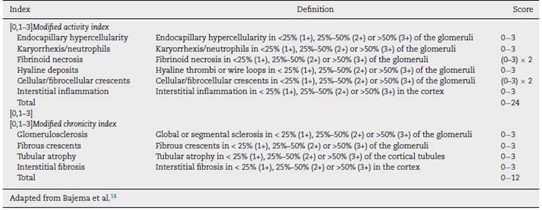
Demographic variables, clinical characteristics and histopathological results of patients with LN were collected. The blocks obtained in the RB were analyzed by a pathologist, who classified them according to the adjustments made to the classification (ISN/RPS) by Bajema et al. (Tables 1 and 2).v(14
Outcome variables
The main outcome variable was the glomerular filtration rate (GFR) at the time of diagnosis and at the last follow-up. The GFR was calculated using the Schwartz formula for people under 18 years of age. (17 Once measured, it was categorized according with the Kidney Disease: Improving Global Outcomes (KDIGO) international classification to assess the renal function of the patient. (18 Kidney failure was defined as a GFR < 60 mL/min/1.73 m2 or kidney failure classified as grade IV or V according to the KDIGO scale.
Arterial hypertension was defined as a systolic or diastolic blood pressure above the 95th percentile for age19 and pro teinuria in the nephrotic range (protein-creatinine ratio > 2.0). The need for renal replacement therapy, transplant and death were taken as secondary outcome variables.
Statistical analysis
Dichotomous variables were reported as percentages. Con tinuous variables were presented as medians, interquartile ranges or means and standard deviations, according to the normality of their distribution. The kappa coefficient was used to determine the level of agreement between renal failure and the presence or absence of each histopathological char acteristic of the ISN/RPS scale, revised by Bajema et al. The Mann-Whitney U test was used to determine differences in GFR at the last follow-up, according to the presence or absence of proteinuria at diagnosis. The P-value <.05 was considered significant. The analyses were conducted using the Stata® 14.0 statistical package (StataCorp, 2014, College Station TX, USA.).
Results
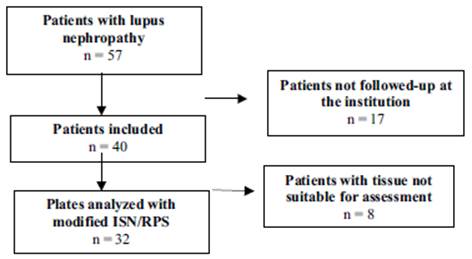
Between the years 2011 and 2018, 57 pediatric patients with lupus nephropathy were treated in the institution. Of them, 17 did not have histopathological tissue suitable for reading. The selection of patients is described in Fig. 1.
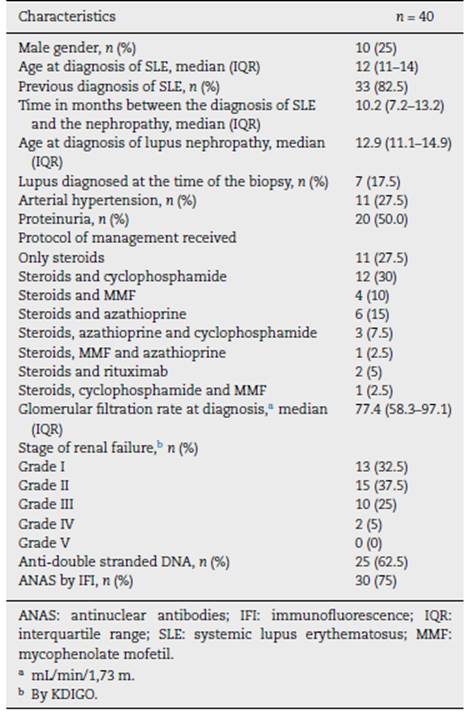
The description of the patients is found in Table 3. Of the 40 patients, 10 (25%) were male, the median age at the diagnosis of SLE was 12 years (IQR 11-14), 7 patients (17.5%) were diag nosed with SLE at the time of the RB. In the 33 patients (82.5%) with previous diagnosis, the time between the diagnosis of SLE and that of LN was 10.2 months (IQR 7.2-13.2). The median GFR at the time of the diagnosis was 77.4 mL/min/1.73 m2 (IQR 58.3-97.1). At the same time, 12 patients (30.0%) had a renal function below 60 mL/min/1.73 m2.
Level of agreement with the diagnosis
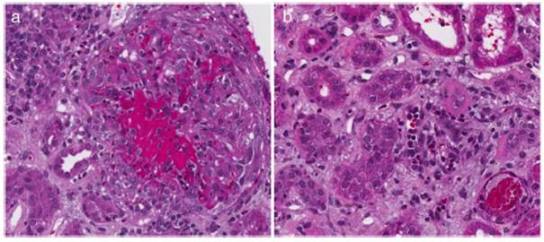
Table 4 describes the level of agreement between the histopathological findings and the GFR at the time of diagnosis. Thirty-two patients had histopathological blocks available and were classified by a pathologist (CJ). Nine of them (28.1%) presented GFR < 60 mL/min/1.73 m2. All the characteristics of the adjusted ISN/RPS scale presented low levels according to the renal failure, without statistical significance. In the activity index, karyorrhexis, with 34.48%, was the characteristic with the highest level according to the renal failure. Nevertheless, the kappa coefficient was insignificant (k = 0.1873, SE = 0.0759, P = .0068). In the chronicity index, tubular atrophy presented the highest percentage of agreement (27.59%) with insignif icant kappa coefficient (k = -0.1053, SE = 0.0952, P = .8655). Fig. 2 describes the findings of karyorrhexis and tubular atro phy seen in the biopsy.
Table 4 Level of agreement between the histopathological findings and the glomerular filtration rate at the time of diagnosis.
| Index | Number | Level of agreement | Expected agreement | kappa | Standard error | z-value | P-value |
|---|---|---|---|---|---|---|---|
| [0,1-8]Activity | |||||||
| Capillary hypercellularity | 32 | 27.59% | 20.57% | 0.0883 | 0.0794 | 1.11 | .1331 |
| Karyorrhexis | 32 | 34.48% | 19.38% | 0.1873 | 0.0759 | 2.47 | .0068 |
| Fibrinoid necrosis | 32 | 3.45% | 8.44% | -0.0545 | 0.0441 | -1.24 | .892 |
| Hyaline deposits | 32 | 27.59% | 23.54% | 0.0529 | 0.0775 | 0.68 | .2476 |
| Crescents | 32 | 6.90% | 11.65% | -0.0538 | 0.0475 | -1.13 | .8713 |
| Interstitial inflammation | 32 | 17.24% | 32.82% | -0.2319 | 0.1001 | -2.32 | .9897 |
| [0,1-8] | |||||||
| [0,1-8]Chronicity | |||||||
| Segmental global sclerosis | 32 | 13.79% | 15.58% | -0.0211 | 0.0605 | -0.35 | .6365 |
| Tubular atrophy | 32 | 27.59% | 34.48% | -0.1053 | 0.0952 | -1.11 | .8655 |
| Interstitial fibrosis | 32 | 20.69% | 26.28% | -0.0758 | 0.0878 | -0.86 | .806 |
| Fibrous crescents | 32 | 13.79% | 15.58% | -0.0211 | 0.0554 | -0.38 | .6485 |
Last follow-up
Data from the follow-up of the 40 patients were obtained. The median follow-up in them was 2.3 years (1.0-5.16). At the end of the follow-up, six individuals (15.0%) had arte rial hypertension and 15 (37.5%) proteinuria. Six patients (15.0%) required renal replacement therapy and one required renal transplantation (15.0%). The median GFR in all cases was 84.4 mL/min/1.73 m2 (67.4-97.0). Of them, 9 individ uals (22.5%) had GFR < 60 mL/min/1.73 m2. In the last follow-up, there was no difference in GFR between patients with presence and absence of proteinuria at diagnosis (P = .3936).
Level of agreement in the last follow-up
For the analysis in the last follow-up, the patients with renal failure at the time of diagnosis were excluded, calculating the levels according to the 23 remaining cases. Table 5 describes the level of agreement between the histopathological find ings and the GFR at the last follow-up. In the activity index, karyorrhexis was the characteristic with the highest level of agreement (26.1%), but with insignificant kappa (k = -0.044, SE = 0.0941, P = .3201). In the chronicity index, global seg-mental sclerosis (k = 0.1481, SE = 0.078, P = .0287) and tubular atrophy (k = -0.0395, SE = 0.1025, P = .6502) were the characteristics with the highest percentage of agreement (30.4% for both variables).
Table 5 Level of agreement between the histopathological findings and the glomerular filtration rate at the last follow-up.
| Index | Number | Level of agreement | Expected agreement | kappa | Standard error | z-value | P-value |
|---|---|---|---|---|---|---|---|
| [0,1-8]Activity | |||||||
| Capillary hypercellularity | 23 | 17.39% | 22.50% | -0.0659 | 0.0961 | -0.69 | .7534 |
| Karyorrhexis | 23 | 26.09% | 22.68% | 0.044 | 0.0941 | 0.47 | .3201 |
| Fibrinoid necrosis | 23 | 4.35% | 7.94% | -0.039 | 0.0484 | -0.81 | .7897 |
| Hyaline deposits | 23 | 13.04% | 16.45% | -0.0407 | 0.0752 | -0.54 | .7058 |
| Crescents | 23 | 4.35% | 9.64% | -0.0586 | 0.0483 | -1.21 | .8872 |
| Interstitial inflammation | 23 | 30.43% | 28.17% | 0.0316 | 0.1013 | 0.31 | .3776 |
| [0,1-8] | |||||||
| [0,1-8]Chronicity | |||||||
| Segmental global sclerosis | 23 | 30.43% | 18.34% | 0.1481 | 0.078 | 1.9 | .0287 |
| Tubular atrophy | 23 | 30.43% | 33.08% | -0.0395 | 0.1025 | -0.39 | .6502 |
| Interstitial fibrosis | 23 | 26.09% | 21.55% | 0.0578 | 0.0856 | 0.68 | .2496 |
| Fibrous c rescents | 23 | 13.04% | 14.56% | -0.0177 | 0.0587 | -0.3 | .6186 |
Table 6 describes the patients with GFR < 60 mL/min/1.73 m2 classified by the Bajema and the WHO scales. There were no significant differences between the initial classification according to both scales and the renal function at four years.
Table 6 Patients with GFR < 60 mL/min/1.73 m2 at four years of follow-up of agreement, according to the classification of the initial.
| Grade reported in the biopsy of diagnosis | [0,2-3]n = 9 | |
| Bajema | WHO | |
| Grade I | 0 | 0 |
| Grade II | 0 | 0 |
| Grade III | 1 | 1 |
| Grade IV | 5 | 5 |
| Grade V | 2 | 3 |
| Grade VI | 1 | 0 |
| WHO: World Health Organization. | ||
Discussion
In this retrospective study, which included 40 pediatric patients with LN from a middle-income country, the histopathological findings of RB showed a low level of agree ment with the renal function when the biopsy was taken and at the last follow-up. The ideal to base a biopsy-guided clin ical decision should be the precision in the evaluation of the renal histology with an interpathologist concordance. RB has an important implication to establish a possible prognosis and treatment of LN, in addition, is the idoneous method for rul ing out other alterations such as thrombotic microangiopathy, podocytopathies tubulointerstitial nephritis or drug-induced nephrotoxicity, entities that also compromise the kidney in patients with lupus. (20
Therefore, it should be considered that RB is the standard method for the diagnosis of LN and that renal involvement can be classified by the histopathological findings. The care guidelines in LN recommend to establish the prognosis and the management scheme according to the histopathological findings of the patients, taking into account the differences between the non-proliferative and the proliferative classes (class III, IV and III/IV + V). However, it is controversial whether it is an equally valid tool for the association with the renal prognosis. (20
Several investigations have tried to define clinical and sero logical variables as predictors of renal prognosis, some cohorts describe the presence of proteinuria <0.8 g/day at 12 months after diagnosis as a marker of renal survival at 7 years, regard less the ethnicity and the histological class in adult patients. To date, none of the clinical results is able to be a reliable marker of long-term renal survival. (21,22 Therefore, the possi bility of having other biomarkers (histological or molecular) that prematurely can predict renal survival in patients with LN is questioned.
In our study, at the time of diagnosis, 28% (n = 9) of the patients with histopathological block had a GFR < 60 mL/min/1.73 m2. A correlation with the components of the ISN/RPS scale was made and there was evidence of low level of agreement in all histopathological characteristics for renal failure. In the activity index, karyorrhexis was predominant (34%), and in chronicity, tubular atrophy (27%); the 2 variables with insignificant kappa coefficient. In a study of 203 patients with LN, that compared the factors associated with tubulointerstitial inflammation and interstitial fibrosis or tubular atrophy, it was found that 30% of the patients, had a GFR < 60 mL min 1.73 m2 at the time of the biopsy, 60% of them had moderate to severe tubulointerstitial inflam mation and 70% experienced moderate to severe tubular atrophy, which are related with a poor renal response in the follow-up. (23
At follow-up (median 2.3 years), 9 of 23 patients had a GFR < 60 mil min 1.73 m2. The correlation with the items of the ISN/RPS scale was also conducted and there was evi dence of low level of agreement in all the histopathological characteristics for renal failure. In the activity index, karyorrhexis was predominant (26%) and in chronicity, tubular atrophy and global segmental sclerosis (30.4%), the three vari ables with insignificant kappa coefficient. There are many efforts in the literature to find prognostic variables for renal survival, but with few satisfactory results. In cohorts of adult European patients with LN (n = 98), divided into proliferative and non-proliferative class, predictor histological markers for commitment were studied, according with the renal ISN/RPS classification, established as GFR < 60 mL/min/1.73 m2. The results regarding long-term renal survival were limited. (24 Tao et al. assessed the possible association of the presence of crescents with certain clinical characteristics in 288 adult patients with LN. Although the general presence of cellular or fibrocellular crescents (that is, without discriminating the percentage of glomerular involvement) was described in 50.7% of the patients, no positive correlations between these and the decrease >30% in the GFR, a GFR < 15 mL/min/1.73 m2, require ment of dialysis at least for 6 months or death at a follow-up median of 76.5 months were reported. The studies in pediatric population are even more limited, with no clear evidence of histopathological markers that identify a renal prognosis. (25
This study has several limitations: the first, due to its ret rospective nature, some of the data were not available. The second, due the convenience sample size, the results of the study cannot be extrapolated to other clinical environments. The third, the samples were evaluated by a single pathologist (CJ), therefore, it was not possible to estimate the interobserver variability for the diagnosis. Despite this, the study has strengths. The patients are classified with the current management scale (ISN/RPS) revised by Bajema et al., unlike previous studies that offer the classifications with previous scales. In Colombia, Beltrán-Avendaño et al. (25 described a cohort of 11 patients with lupus nephropathy and they found a renal involvement at the time of the RB in 74% of the cases and creatinine clearance reduced in 45%. The histopathological findings were classified according to the parameters of the WHO, but without the description of these in relation with renal survival. (25 As already mentioned, previous stud ies in pediatric patients in Colombia with LN had clinically characterized this population and in one study, the lesions observed in the RB were correlated with the clinical manifes tations and the treatment of the disease. Given the foregoing, this study has great strength, since it is the first to estimate the level of agreement between renal biopsies of pediatric patients with LN in Colombia and kidney function, evaluating it as a biomarker to predict kidney failure in our population.
Conclusion
The findings in the RB may be insufficient for assessing the prediction of the survival of the renal function, so the treat ment and the prognosis of the patients with LN should be carried out using other biomarkers and clinical signs. Prospec tive studies that can confirm this hypothesis should be carried out.

