


 text in
  English (pdf)
 Article in xml format
 Article references
 Send this article by e-mail
 Cited by SciELO
  Cited by Google
  Similars in
SciELO
Similars in
SciELO  Similars in Google
 Permalink
Introduction
Interosseous atrophy in the context of rheumatoid arthritis is a frequent finding, described for several years now, which is correlated with the loss of prehensile strength and functionality of the hand. This is due more to a compromise in the relationship of the muscle-joint system rather than to a compromise of the neuromuscular or nerve junction; however, there are other entities that present interosseous atrophy and in some cases they have unusual locations, different that those that are usually seen in rheumatoid arthritis.1-3
Clinical summary
A 66-year-old female patient, with a history of amyotrophic lateral sclerosis and bulbar involvement, in outpatient management with riluzole, who consults due to a clinical picture of 8 months of evolution consisting of dysphagia, initially for solids and later for liquids, associated with loss of approximately 22 kg in the mentioned period. Due to this clinical picture, the patient was in outpatient follow-up by internal medicine, which indicated a video swallowing study, in which severe dysphagia with involvement of the oral preparatory and pharyngeal phases of swallowing was found. During the study, aspiration occurred, for which the patient was referred to the emergency room for a gastrostomy. Within the family history, she stated that her mother died of respiratory failure in the context of amyotrophic lateral sclerosis of bulbar subtype.
At the physical examination on admission, the patient was hemodynamically stable, with no need for supplemental oxygen, cachectic, without cardiopulmonary or abdominal alterations; at the neurological level, she presented mutism, with fasciculations in the right hemitongue, decrease in both proximal and distal strength in the upper limbs (3/5), as well as loss of fine and gross motor skills in the hands that made it impossible for her to clamp or grasp objects. The foregoing was associated with hypotrophy of the interosseous and lumbrical muscles of both hands, asymmetric, more marked in the right hand, in which the sign of the double anatomical snuffbox or "split hand" was evident (Fig. 1); no involvement of the hypothenar region was found (there is no image of that).
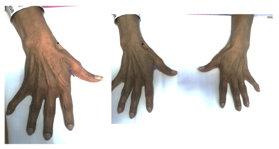
Figure 1 A) Atrophy of the first dorsal interosseous, right hand (arrows). B) Compared hands. Greater atrophy of the firstdorsal interosseous of the right hand is observed.
Due to severe dysphagia, the patient required hospitalization in order to perform a gastrostomy, which was carried out without complications, with adequate tolerance to enteral nutrition boluses, and after 2 days of hospitalization, she was discharged without complications.
Discussion
Amyotrophic lateral sclerosis is a heterogeneous neurodegenerative disease that affects both upper and lower motor neurons, with a wide spectrum of clinical motor and nonmotor manifestations.4 Motor involvement of the extremities is usually one of the initial manifestations in up to 60% of patients, with asymmetric involvement of the upper or lower limbs. On the other hand, bulbar involvement, which manifests itself with progressive dysarthria, followed by dysphagia and emotional lability, is also another frequent initial manifestation of this entity.4,5
In 1992, Wilbourn6 described the involvement of the hand, which was later named "split hand sign" or "double anatomic snuffbox sign" in patients with amyotrophic lateral sclerosis. Clinically, this finding corresponds to the atrophy of the first dorsal interosseous and the thenar region, but sparring the hypothenar region due to the compromise of the muscles innervated by the median nerve (abductor pollicis brevis and opponens muscle of the thumb) and the ulnar nerve (first dorsal interosseus, flexor pollicis brevis, adductor pollicis). The muscles of the hypothenar region (abductor digiti minimi) are not completely affected, which has its corresponding correlation with the evident compromise of these nerves by electrodiagnosis.
The finding described by Wilbourn6 was considered ominous in the context of the established amyotrophic lateral sclerosis; however, subsequent works highlight its importance for early diagnosis of the disease, being able to establish its phenotype and being part of the prognosis, according to other clinical and electrodiagnostic characteristics.5-8 Although it cannot be catalogued as a pathognomonic sign ofamyotrophic lateral sclerosis and there are other differential diagnoses, it has moderate sensitivity (52-74%) and good specificity (80-95%) in relation to other neurodegenerative entities with motor nerve involvement.5,6,9-11
It is of great importance to highlight that, even though it is not the case of our patient, the finding of the double anatomical snuffbox signis relevant in those patients with no history of this disease, since it can guide the clinician in the challenging diagnosis of amyotrophic lateral sclerosis. However, it should be emphasized, as established by Wilbourn, that not all atrophies of the first dorsal interosseous and thenar region result in amyotrophic lateral sclerosis; a correlation with electrodiagnosis is required to have a more accurate diagnosis.7
Conclusion
Physical examination and anamnesis continue to be fundamental tools in the clinical diagnosis of different entities, being essential in the area of rheumatology and in other areas of medicine. It must be remembered that the human body is the book in which diseases write their stories and it is up to us to read what they want to tell us.

