


 text in
  English (pdf)
 Article in xml format
 Article references
 Send this article by e-mail
 Cited by SciELO
  Cited by Google
  Similars in
SciELO
Similars in
SciELO  Similars in Google
 Permalink
Introduction
Takayasu arteritis (TA) is a large-vessel vasculitis, with variable worldwide incidence, with female predominance with a ratio of 6.9:1 compared to men,1 with a mean age of presentation of 25 years. A quarter of patients present the disease before age 20. Its etiology is not well defined, but genetic (associated with the major histocompatibility complex) and infectious factors are involved, mainly Mycobacterium tuberculosis; for example, in Mexican patients, genetic sequences of this mycobacterium have been found in biopsy samples.2 Its morbimortality is related to cardiovascular events and chronic kidney disease. In children it is the third general cause of vasculitis and the most frequent of large vessels; likewise, TA is the main cause of aortic stenosis and renovascular hypertension, with a peak incidence between 10 and 15 years.3
We present the case of a young patient with TA who developed nephritic syndrome in his evolution. The histopathological study is compatible with focal and segmental glomerulosclerosis with collapsing variety; secondary causes such as hemodynamic alterations of renal blood flow were ruled out; it is considered that there is an association with TA, probably through immune-mediated mechanisms.4
Case presentation
A 20-year-old Mexican male, with no relevant family history, began at age 16 with symptoms of pain and claudication in the lower extremities, fatigue, and elevated blood pressure levels. On physical examination, asymmetry of the popliteal and pedal pulses, abdominal bruit, and discrepancy of blood pressure values (>10mmHg) in upper limbs compared to lower limbs was observed. A secondary arterial hypertension approach was started; endocrinopathies were initially excluded. Hence, computed tomography angiography with three-dimensional reconstruction was performed (Fig. 1), in which 70% stenosis of the inframesenteric abdominal aorta involving both renal arteries at the level of the ostium was documented; supra-aortic trunks showed no lesions. An elevation of acute phase reactants (C-reactive protein 18.1mg/dl and erythrocyte sedimentation rate 64 mm/h) was found. With this information, TA classification criteria were met. Baseline renal function was preserved (serum creatinine: 0.58mg/dl and glomerular filtration rate: 146ml/min). Immunosuppressive treatment with steroids was started with prednisone at 50mg/day (1mg/kg/day), with a dose reduction to 20mg/day at 6 months follow-up and 5 mg/day after 12 months. In addition, methotrexate up to 15 mg/week, anticoagulation with rivaroxaban, and antihypertensive therapy with losartan and nifedipine were administered. In the third year of follow-up, there was an increase in serum creatinine; hematuria (20 red blood cells per high-power field), and albuminuria in the subnephrotic range (1441 mg in 24h) also were documented. A renal ultrasound is performed, evidencing normal morphology and size: right kidney 9.3 x 6.6 x 5 cm; left kidney 10.2 x 7.4 x 5.1cm in their longitudinal, anteroposterior, and transverse axes, respectively.
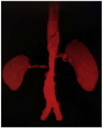
Figure 1 Abdominal aortic angiotomography with 3D reconstruction depicting stenosis of the adrenal portion at to the bifurcation, with stenosis of the renal arteries.
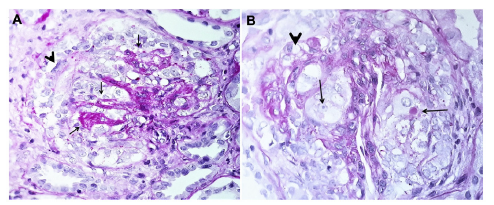
Given the suspicion of renovascular hypertension, aDoppler was practiced with the following findings (Fig. 2): maximum systolic velocity of 120 cm/s in the right renal arteryat the ostium and 120 cm/s at the hilum, with a resistanceindex of 0.60. The left renal artery evidenced a maximumsystolic velocity of 137 cm/s at the ostium and 164 cm/s atthe hilum, with a resistance index of 0.51. These normalvalues do not support the diagnosis of renovascular hypertension; therefore, to completely rule out this possibility, a99mTc-DMSA renal scintigram with captopril was performed, documenting baseline glomerular filtration rate in the leftkidney of 149.3 ml/min and the right kidney of 79.9 ml/min; postcaptopril: 232.6 ml/min and 156.4 ml/min, respectively. At follow-up, exclusive renal disease activity was foundwith normalization of acutephase reactants, yet a progressive drop in glomerular filtration rate. At the fourthyear of follow-up, creatinine increased up to 3.45 mg/dlwith a glomerular filtration rate of 34 ml/min and persis-tence of hematuria and albuminuria of 1840 mg in 24 h. Anephritic syndrome was diagnosed, and the following studies were conducted: anti-neutrophil cytoplasm antibodies,complement profile, systemic lupus erythematosus, and viralpanel, reported as normal. A renal biopsy was accomplished,demonstrating focal and segmental glomerulosclerosis with acollapsing variety (Fig. 3). High-dose glucocorticoid treatmentwas initiated (prednisone 50 mg/day-1 mg/kg/day-), switching methotrexate to azathioprine at 2 mg/kg/day, with noresponse: nitrogen compounds were elevated, with a serumcreatinine of 8.32 mg/dl. Renal replacement therapy was started with hemodialysis. The patient is under follow-up withno data on extrarenal vasculitis activity.
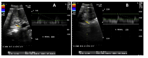
Figure 2 Doppler ultrasound of renal arteries. A) Right: maximum systolic velocity of 120 cm/s at the ostium level and 120cm/s at the hilum, with a resistance index of 0.60. B) Left: maximum systolic velocity of 137 cm/s at the ostium and 164 cm/s at the hilum, with a resistance index of 0.51.
Discussion
We present the case of a patient with a 4-year TA whose early signs and symptoms were claudication of the lower extremities, present in 10-35%3 of the cases, and arterial hypertension, which is detected in up to 80% of patients in this age group. Endocrinopathies were ruled out due to the presence of abdominal murmur and pulse asymmetry. A computed tomography angiography was performed, revealing abdominal aorta stenosis, with involvement of the renal arteries at the ostium. The case was classified as AT based on the EULAR/PRINTO/PRES criteria, which include as a mandatory criterion angiographic abnormality of the aorta or its main branches and pulmonary arteries, together with at least one of the following conditions: (a) absence of pulses or claudication in the peripheral artery induced by physical activity; (b) difference of at least 10 mmHg in systolic blood pressure in the extremities; (c) murmurs over large arteries; (d) hypertension; and (e) elevation of acute-phase reactants. Compliance with these criteria provides a sensitivity and specificity above 99%.5 The patient met all the above criteria. Differential diagnoses were also ruled out, such as fibromuscular dysplasia, present in young patients, which generally spares the aorta and affects the distal 2/3 of the renal arteries, with a characteristic tortuous morphology.6
Classification criteria for TA require evidence of angio-graphic abnormality, either by angiography, magnetic resonance angiography, or computed tomography angiography; the latter is a non-invasive method that offers excellent anatomical detail of both the lumen of the vessel and its wall. Along with magnetic resonance angiography, they have replaced angiography, historically considered the gold standard, for its invasive nature and the absence of information on the vessel wall.7 In this case, there was evidence of involvement limited to the abdominal aorta and renal arteries, classified as Numano IV, present in only 5.9% of patients diagnosed with TA.8
Doppler ultrasound is an inexpensive, non-invasive tool, without exposure to radiation or contrast media; it is an option when other procedures for diagnosis and monitoring of TA are not available. In this case, it was used to study renal artery stenosis. The best accepted criterion by different studies is a maximum systolic velocity above 180 cm/s, with a sensitivity that can reach up to 90%.9 A 99mTc-DMSA renal scintigram was also performed with captopril, which showed an increase in post-drug glomerular filtration rate, being negative for renovascular hypertension. This latter study is recommended for patients with low pre-test probability, with a high negative predictive value; in some series, it is reported up to 100%.10 Based on these data, renovascular hypertension was ruled out.
Renal manifestations of TA are related to renovascular hypertension and rarely to disorders that affect the renal parenchyma, although some cases have been reported. In the reviewed literature, a single previous report was found associated with focal and segmental glomerulosclerosis in a 29-year-old female patient who presented TA with multifocal disease, without evidence of renal flow alterations, with probable immune-mediated etiology.4 In a single-center series in China, six cases were reported, all female, with a mean age of 35 years. The clinical presentation in 2 cases was hematuria (10-15 red blood cells per field), in 5 significant proteinuria, of which 2 were in nephrotic range and one with nephrotic syndrome. Renal manifestations occurred in a mean of 60.7 months after the diagnosis of TA, and in no case alterations in renal blood flow were found. Histopathological studies reported IgA nephropathy in 2 cases, proliferative mesangial glomerulonephritis, membranoproliferative glomerulonephritis, minimal-change disease, and fibrillary glomerulonephritis (one case each).11
The initial response to treatment, when only the diagnosis of TA was available, was favorable; however, during follow-up, with the appearance of focal segmental glomerulosclerosis, the patient had proteinuria close to the nephrotic range, in addition to elevated nitrogen compounds, and a rapid decline in glomerular filtration rate, all poor prognosis criteria and response to steroids.12 Consequently, it was decided to initiate azathioprine instead of methotrexate based on recommendations drawn from the few existing studies. This combination, as primary treatment, achieves remission rates of up to 80%,13 but due to the baseline clinical features aforementioned and the histological variety, his evolution was poor, ending up on renal replacement therapy.
Conclusion
In this case report, one patient diagnosed with TA underwent a renal biopsy due to nephritic syndrome, with proteinuria in a subnephrotic range, without an evident cause. Changes consistent with focal segmental glomerulosclerosis were found; this clinicopathological entity is characterized by nephrotic or subnephrotic proteinuria, which may appear as a primary disease or be associated with multiple causes, including familial/genetic, viruses (HIV and cytomegalovirus), drugs, and structural and functional adaptive responses (such as hyperfiltration processes in obese patients, solitary kidneys, or reflux nephropathy.14 The collapsing variety also includes other etiologies allied to ischemia or marked reduction in renal flow.14 All these causes were ruled out in the patient.

