SciELO - Scientific Electronic Library Online

 
vol.29 suppl.1Pulmonary manifestations of rheumatoid arthritis, a review in SARS-CoV-2 pandemiaMonoarthritis induced by the Oxford-AstraZenecaR SARS-CoV-2 vaccine author indexsubject indexarticles search
Home Pagealphabetic serial listing  

Services on Demand

Journal

Article

Indicators

Related links

  • On index processCited by Google
  • Have no similar articlesSimilars in SciELO
  • On index processSimilars in Google

Share


Revista Colombiana de Reumatología

Print version ISSN 0121-8123

Rev.Colomb.Reumatol. vol.29  supl.1 Bogotá Dec. 2022  Epub Feb 03, 2024

https://doi.org/10.1016/j.rcreu.2020.11.004 

Revisión de Temas

Kawasaki disease in Colombia: A systematic review and contrast with multisystem inflammatory syndrome in children associated with COVID-19

Enfermedad de Kawasaki en Colombia: revisión sistemática y contraste con el síndrome inflamatorio multisistémico en niños asociado a COVID-19

Kevin Llinás-Caballero a  

Yhojan Rodriguez a  

Jaime Fernández-Sarmiento b   c   d  

Mónica Rodríguez-Jiménez a  

Juan-Manuel Anaya a   *  

a Center for Autoimmune Diseases Research (CREA), School of Medicine and Health Sciences, Universidad del Rosario, Bogotá, Colombia

b Department of Critical Care Medicine and Pediatrics, Universidad de La Sabana, Bogotá, Colombia

c Fundación Cardioinfantil-Instituto de Cardiología, Bogotá, Colombia

d Universidad CES Graduate School, Bogotá, Colombia


ABSTRACT

Introduction:

Kawasaki disease (KD) is an acute vasculitis with multisystem involvement. Recently, the increasing incidence of a condition that closely resembles KD in many cases, named multisystem inflammatory syndrome in children (MIS-C), has set off alarms amid the current worldwide coronavirus disease-19 (COVID-19) pandemic. Hence, the aim is to conduct a systematic review of the literature about KD in Colombia and contrast it with COVID-19-related MIS-C.

Materials and methods:

A search was carried out in both international and Latin American electronic databases for publications concerning patients with KD in the Colombian population. Records were then screened by titles and/or abstracts, assessed for eligibility, and reviewed. Preferred Reporting Items for Systematic Reviews and Meta-Analysis (PRISMA) guidelines were followed. The search included studies reporting MIS-C associated with COVID-19, and compared these patients with our findings of KD in Colombia. Results: Out of 36 publications retrieved, 17 were included, representing 120 individuals. Male to female ratio was 1.6, and most patients (90.4%) were aged 5 years or less. Among the main features of KD, fever was the most frequent (96.2% of the patients), while cervical lymphadenopathy was present in only 40.6%. Intravenous immunoglobulin was administered in 91.4% cases and 6.2% were resistant. Cardiac involvement was found in around 30%, and 20% had coronary artery lesions. Comparison between MIS-C associated with COVID-19 and KD in Colombia indicates that patients affected by MIS-C were older (72.2% of MIS-C patients > 5 years), had higher rates of cardiac involvement, and required critical care more often.

Conclusions:

Our findings of KD in Colombia are consistent with the available descriptions of KD in the scientific literature. Given the increasing rate of severe acute respiratory syndrome coronavirus-2 (SARS-CoV-2) infection in Colombia and Latin America, our study raises awareness about MIS-C in pediatric patients with COVID-19 and its relationship with KD.

Keywords: Kawasaki disease; Latin America; Colombia; Pediatric multisystem inflammatory disease; COVID-19 related; COVID-19; Cardiac involvement

RESUMEN

Introducción:

La enfermedad de Kawasaki (EK) es una vasculitis aguda con compromiso multisistémico. Recientemente, la incidencia creciente de una condición que se asemeja en forma considerable a la EK en muchos casos, denominada síndrome inflamatorio multisistémico (SIMS) en niños, ha encendido las alarmas en medio de la actual pandemia mundial de la enfermedad COVID-19. Por consiguiente, nos propusimos realizar una revisión sistemática de la literatura acerca de la EK en Colombia y contrastarla con el SIMS relacionado con COVID-19 en niños.

Materiales y métodos:

Buscamos publicaciones respecto a pacientes con EK en población colombiana, en bases de datos electrónicas tanto internacionales como latinoamericanas. Los registros hallados fueron tamizados por títulos o resúmenes, evaluados para elegibilidad y revisados. Se siguieron las guías Preferred Reporting Items for Systematic Reviews and Meta-Analysis (PRISMA). Posteriormente, buscamos estudios que reportaran SIMS temporalmente asociado con COVID-19 en niños y comparamos estos pacientes con nuestros hallazgos de EK en Colombia.

Resultados:

De 36 publicaciones encontradas se incluyeron 17, las cuales representaron 120 individuos. La razón hombre a mujer fue de 1,6 y la mayoría de los pacientes (90,4%) tenía 5 anos o menos. Entre las principales características de EK, la fiebre fue la más frecuente (96,2%), mientras que la linfadenopatía cervical estuvo presente solo en el 40,6%. La inmunoglobulina intravenosa se administró en el 91,4% de los casos y 6,2% presentaron resistencia. Se encontró compromiso cardiaco en alrededor del 30% de los pacientes, en tanto que el 20% tuvo lesiones de arterias coronarias. La comparación entre las características clínicas de la EK y el SIMS asociado a COVID-19 mostró que los individuos afectados por el SIMS eran mayores (72,2% con SIMS tenían más de cinco anos), tuvieron mayores índices de compromiso cardiaco y requirieron cuidado crítico con mayor frecuencia. Conclusiones: Nuestros hallazgos de EK en Colombia son consistentes con las descripciones disponibles de esta enfermedad en la literatura científica. Debido al aumento de infección por SARS-CoV-2 en Colombia y Latinoamérica, nuestro estudio busca crear conciencia sobre el SIMS en pacientes pediátricos con COVID-19 y su relación con la EK.

Palabras clave: Enfermedad de Kawasaki; América Latina; Colombia; Síndrome inflamatorio; multisistémico en niños; temporalmente asociado a COVID-19; COVID-19; Compromiso cardiaco

Introduction

Kawasaki disease (KD) is an acute, self-limited, systemic vasculitis, which primarily involves small and medium-sized vessels.1 It affects mostly males between 6 months and 5 years old, being the most common medium-sized-vessel vasculitis.2,3 Several theories have been proposed regarding the etiology of this condition, including pathogens, toxins, and autoimmune or autoinflammatory processes.4,5 Even though the current evidence points toward an infectious agent accompanied by a dysregulated immune response, a univocal agreement about the exact cause or causes of KD is lacking so far.2 Therefore, KD is nowadays considered to be a vasculitis of unknown origin, with a predilection for coronary arteries.6 Although acute manifestations usually resolve promptly upon adequate treatment, long-term cardiovascular sequelae, mainly coronary artery lesions (CALs), may develop in some patients. In fact, KD is the leading cause of acquired heart disease among children in developed countries.7 Nonetheless, if KD is identified and treated early, the risk of coronary artery aneurysms (CAAs) declines from 25% to 4%.8

In 1967, six years after seeing the first child presenting with a particular constellation of clinical manifestations, Japanese pediatrician Tomisaku Kawasaki described 50 patients that experienced an 'acute febrile mucocutaneous syndrome with lymphoid involvement with specific desquamation of the fingers and toes'.9,10 Subsequently, this syndrome was named mucocutaneous lymph node syndrome, and cases were reported outside Japan.11,12 Some years after that, KD was defined as equivalent to infantile periarteritis nodosa.13

Currently, the coronavirus-19 disease (COVID-19), caused by the severe acute respiratory syndrome coronavirus-2 (SARS-CoV-2) virus, has become a global public health issue. Interestingly, a link between SARS-CoV-2 infection and KD has been suggested following an increase of cases of KD, Kawasaki-like disease, and other inflammatory syndromes in multiple countries struck by the COVID-19.14-18 This condition has been termed multisystem inflammatory syndrome in children (MIS-C) temporally associated with COVID-19, among other names, and has posed several questions about the nature and the implications of this relationship.19 Since KD is considered not a very frequent condition, and the COVID-19 pandemic has been associated with MIS-C, we aimed to review the status of KD in Colombia and contrast the results with the clinical characteristics of MIS-C linked to COVID-19 in order to raise awareness of this association.

Materials and methods

Information sources and search strategy

A systematic review of the literature was done up to July 18th, 2020. The search was performed both in international and Latin American electronic databases. For this purpose, we searched using the terms: ("Kawasaki disease" OR "Kawasaki's disease" OR "Kawasaki syndrome" OR "Kawasaki's syndrome" OR "Kawasaki vasculitis" OR "Kawasaki's vasculitis" OR "mucocutaneous lymph node syndrome") AND "Colombia", or their Spanish language counterparts, in PubMed, LILACS, Embase and Web of Science. A more stringent search approach was also done in LILACS but yielded the same results (see Additional file 1). To identify potential additional studies for inclusion, we also hand-searched through other sources, including gray literature, and manually looked up the references of the articles found by the search strategy initially described above. No date, language, or population limits were applied.

Eligibility criteria

Articles included had to have been conducted in Colombian population and to describe methods implemented to study, diagnose, treat, or rehabilitate patients suffering from KD at any age. In addition to this, studies reporting the clinical outcomes of patients with KD were also eligible. Case reports, case series, cross-sectional, case control, cohort (either prospective or retrospective), or clinical trial studies were suitable for inclusion. Studies that were not carried in the population of interest, or that did not present relevant outcomes for the population of interest separately to those for other populations were excluded. We also ruled out records whose full text was not available to us, as well as systematic or narrative reviews and meta-analyses.

Study selection and data collection process

First, a primary reviewer screened all titles and/or abstracts of the publications found. Following this, eligibility assessment was performed. Retrieved articles were rejected if eligibility criteria were not met, and two secondary reviewers were consulted in cases in which eligibility criteria were unclear. After this, a single researcher collected the information regarding population, study design, clinical characteristics, treatment, and outcomes of all included studies; thereafter, a second reviewer verified the extracted information. Any discrepancies or missing information were resolved by consensus.

Preferred Reporting Items for Systematic Reviews and Meta-Analysis (PRISMA) guidelines were followed.20

Comparison with MIS-C associated with COVID-19

In order to find articles reporting cases of MIS-C related to COVID-19, we searched on PubMed on July 18th, 2020 using the following search string: (SARS-CoV-2 OR COVID-19) AND (multisystem inflammatory syndrome in children OR MIS-C OR pediatric multisystem inflammatory syndrome OR PIMS) and selected the articles corresponding to MIS-C related to COVID-19. No date, language, or population limits were applied. Some references found through hand-searching were also included. We then contrasted our results of KD in Colombia with the information on these studies. Statistical analysis was performed using x2 test and p-value < 0.05 was considered significant.

Results

Search and study selection results

A total of ten articles were identified through database searching, and 29 additional records from other resources were found. After duplicates removal, the titles and/or abstracts of 36 articles were screened. In the screening phase, ten articles were excluded. Twenty-six full-text articles were assessed for eligibility.

A study by Momenah et al.21 was excluded because it was not about Colombian population. Likewise, eight articles whose titles contained the search terms were also excluded because it was not possible to find out the full texts.

Ultimately, 17 publications were included in the qualitative synthesis for analysis (Fig. 1).

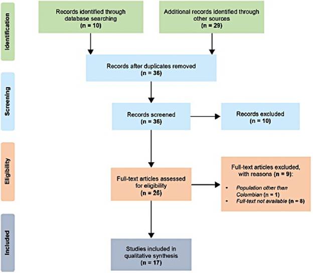
Fig. 1 Selection process. We followed the PRISMA guidelines for reporting in systematic reviews and meta-analyses.20  

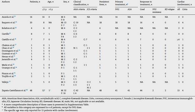
Table 1 summarizes the main features of these studies.22-38

Table 1 Description of KD cases in Colombiaa 

Demographic data

In the included studies, a total of 120 Colombian patients affected by KD were reported. 74 were males, with a male to female ratio of 1.6:1. Almost all of our patients (99.2%) were children or adolescents (<18 years). Among the 73 patients whose exact age was reported, 66 were <5 years. A single case was reported in an adult male aged 36 years.

Clinical classification and findings

From the reported cases, information about clinical presentation was available for 106 of them. The studies included were reviewed in the light of the American Heart Association (AHA)8 and the Japanese Circulation Society (JCS) criteria.39 All patients met at least one of these criteria sets for either complete or incomplete KD. Data regarding AHA and JCS criteria fulfillment are presented in Table 2.

Table 2-Fulfillment of AHA diagnostic criteria vs JCS diagnostic criteria among KD cases in Colombia. 

Concerning the principal symptoms of KD in these 106 patients, 102 of them presented with prolonged fever, 67 69 had peripheral extremity involvement (i.e., edema or desquamation of hands or feet), 74 76 had oral or pharyngeal mucosal changes (i.e., erythema, cracking of lips and/or strawberry tongue), 43 had cervical lymphadenopathy, 67 had conjunctival injection, and 78 had rash. From 58 patients, leukopenia was observed in 2 of them, lymphopenia in 3, thrombocytosis in 17, high C reactive protein (CRP) in 16, high erythrocyte sedimentation rate (ESR) in 14, abnormal liver tests in 7, hyponatremia in 2 and hypoalbuminemia in 2. Of these patients, pre- vs. post-treatment changes in platelets, CRP and ESR was reported in 20 of them. More data about other medical findings, including laboratory and imaging findings, are presented in Additional file 2.

Treatment and clinical response

Out of the 120 patients, the therapeutic approach was described for 116 of them. Among those, intravenous immunoglobulin (IVIG) was administered to 106 usually at doses of 2 g/kg, while 91 received acetylsalicylic acid (ASA). Eighteen patients were treated with antibiotics during the course of the illness (one of them also with an antiviral drug), 3 with paracetamol and/or metamizole, 2 with antihistamines, 4 with systemic corticosteroids (either as primary or secondary treatment), and 1 with inhaled short-acting 0-agonist and corticosteroids. One patient was also prescribed a topical ophthalmic drug because of secondary anterior uveitis. Besides this, the adult patient required intensive care unit (ICU) management with mechanic ventilation and renal replacement therapy.

Clinical response to treatment was reported in 97 cases. The majority of these (88, i.e., 90.7%) had a good clinical response, with resolution of fever and amelioration or full disappearance of the other complaints. IVIG resistance, defined as persistence or recrudescence of fever at least 36 h after the completion of the IVIG infusion,8 was observed in 6 patients (6.2%). On the other hand, 3 of them (3.1%) presented a relapse of the disease after 48 h of defervescence. IVIG resistant patients and those with relapses received additional doses of IVIG, were administered systemic corticosteroids, or both. Further treatment for these patients also included ASA in four of them.

Cardiac involvement and sequelae

Cardiac involvement was evidenced either clinically, by electrocardiography, or by cardiac imaging, in 36 38 patients. At least 48 individuals had an echocardiogram performed, and 21 or more showed abnormal results. Valvular heart disease was evidenced in 9 cases (regurgitation in all of them). Pericardial effusion or pericarditis was observed in 8 cases as well. Ejection fraction was reported as 25% in one patient, and below 70% in 2. Importantly, one had a previous history of a restrictive interventricular communication but also showed pericardial effusion in an echocardiogram performed during the workup. Two follow-up echocardiograms of this patient were reported as normal. CALs, including CAAs, were diagnosed in 24 cases. Ten patients had evidence of CAAs. No deaths were reported in the reviewed articles.

KD in Colombia vs. MIS-C associated with COVID-19

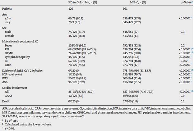
We collected 45 articles respecting MIS-C linked to COVID-19 and compared the patients reported there with those included in our review of KD in Colombia. Of note, all the latter were diagnosed with KD before the COVID-19 epidemic. MIS-C group comprehended 961 patients, three of them being adults (0.3%). Taking into account that the cases of KD in Colombia also included an adult cases, these three adult cases were not excluded. Information about age, sex, presence of main symptoms of KD, evidence of SARS-CoV-2 infection, ICU requirement, use of IVIG and ASA, cardiac involvement, and mortality for both groups is shown in Table 3.14,18,40-79

Table 3 Comparative analysis between KD in Colombia and reported MIS-C cases globally 

Discussion

KD occurs mainly in children and adolescents, and 85% of the patients are <5 years old.80 Therefore, the diagnosis of KD in adults is exceedingly rare, and very few cases have been reported worldwide.30 Furthermore, this condition is more frequent among male individuals, with a male to female ratio around 1.5 1.2 Consistent with these data, in the patients included in this study, 90.4% were aged 5 years or less, and most of them were male, with a male to female ratio of 1.6 1. However, in the articles reviewed, a case of a 36-years-old adult diagnosed with KD was reported.30

There are two main sets of diagnostic criteria used for KD, proposed by the AHA and the JCS separately.8,39 Despite being very similar to each other, the main difference between them is the fever being or not a requisite for the diagnosis. According to our analysis, the fulfillment of AHA and JCS criteria is very similar through the cases reported (Table 2).

In the AHA criteria, persistent fever (usually persisting > 5 days) is a sine qua non characteristic for the diagnosis of complete (or classic) KD and must be accompanied by at least four out of five main clinical findings.8 Conversely, the JCS criteria consider fever as one of six principal symptoms, and diagnosis is made when at least 5 of them are present.39 Differential diagnosis is key in KD, given that other illnesses must be ruled out to reach a correct diagnosis of KD.

Besides fever, changes of the oral cavity and lips are the commonest clinical feature in KD (96.5%), followed by polymorphous rash (96%), non-purulent bilateral conjunctivitis (89%), changes in the extremities (75.6%), and cervical lym (phadenopathy (62.7%).81 In our patients, fever was present in 96.2% of them, while cervical lymphadenopathy was the least frequent of the main symptoms, found in 40.6% of the cases.

Moreover, some patients that do not meet criteria to diagnose complete KD are thus evaluated for incomplete KD. AHA guidelines include an algorithm for the management of these patients according to C reactive protein values, other laboratory findings, and echocardiogram results.8 Table 2 shows how many patients were classified as incomplete KD according to each set of criteria. Special attention should be paid to the incomplete forms of the disease, which may be observed early during the course of the illness (i.e., the first days), as well as the forme frustre of this syndrome.28,31,34,35

The mainstay of the treatment for KD consists of IVIG (2 g/kg) within 10 days of the onset of symptoms, and ASA.4 The administration of IVIG must be done slowly, and pre-medication with antihistamines is often encouraged.4 Most patients resolve following this therapy; however, around 15% of the patients show IVIG resistance, requiring further doses of IVIG and/or use of systemic corticosteroids.2,4 The use of medications other than IVIG as adjunctive primary treatment, such as systemic corticosteroids, infliximab, or cyclosporin, has been studied, with mixed clinical results.4) Accordingly, most of the patients reviewed in this study received IVIG (91.4%) and ASA (89.2%) as part of their initial treatment. Systemic corticosteroids were used as primary treatment in two patients and two were treated with antihistamines, although not as premedication for IVIG in neither of both cases. None of the reviewed patients received infliximab or cyclosporin. IVIG resistance was present in 6.2% of the people and was treated with further IVIG doses and/or systemic corticosteroids.

The development of CALs, particularly CAAs, is the main determinant for the prognosis, long-term morbidity, and mortality of patients with KD. Thanks to the introduction of IVIG for the treatment of this syndrome, the rates of CAAs have dropped from 25% in untreated children to around 1 to 3% in children who received this medication.4 Fortunately, mortality in KD is very low.4,39 In this group of patients, altered coronary arteries were found in 20% of the cases, and CAAs were diagnosed in 8.3% (41.7% of the total CALs). Furthermore, cardiac involvement, in general, was identified in approximately 30% of the patients. There were no deaths reported. Our results in Colombian patients are similar to the data reported by the REKAMLATINA network, mainly those concerning sex distribution, clinical presentation, treatment, and CAAs prevalence.82

Finally, motivated by the recent rise in MIS-C cases and their association with COVID-19, we decided to compare the Colombian literature on KD included in this review with patients reported as suffering from MIS-C or related syndromes associated with COVID-19 (hereafter referred to only as MIS-C). It is well known that KD and MIS-C, despite sharing many similarities (not only clinically but also on a pathogenesis level), have important differences.83,84 Notwithstanding, given the undeniable resemblance in many cases and the paramount clinical importance, we made this contrast of KD in Colombia with global MIS-C cases since local ones are not available in the peer-reviewed literature at the time of writing. Of the MIS-C patients, at least 81% had laboratory evidence consistent with SARS-CoV-2 infection. Accordant with available descriptions,85 we found that 72.2% of MIS-C cases were in children older than 5 years, which is substantially different than our KD in Colombia findings. Furthermore, while KD has been associated with East Asian origin, MIS-C tends to affect more children of African American, Caribbean and Hispanic ancestry.85 This is of special interest to us given the triethnic origin of our population; withal, lack of consistency in reporting ethnic origin or ancestry among Colombian KD cases precluded further comparisons.

As previously described,85 the proportions of principal clinical findings of KD were lower in MIS-C patients compared to those included in our review, with statistically significant differences (except for fever). Correspondingly, as opposed to Colombian patients, IVIG was used less in the MIS-C group. These findings can be explained by the fact that not all the patients diagnosed with MIS-C had a presentation leading to KD diagnosis.

Besides traditionally measured acute phase reactants such as CRP and ESR, serum ferritin is very often elevated in MIS-C patients.84 Ferritin is a significant marker of M1 macrophage activation.86 Thus, it plays a key role in the workup of patients with suspected MIS-C.87 Nonetheless, we were not able to compare ferritin levels and other inflammatory parameters between global MIS-C versus Colombian KD patients because those variables are not systematically assessed in KD and were not included in Colombian reports.

Interestingly, we found a much greater rate of cardiac involvement in MIS-C patients than that found by us in Colombian KD patients. Likewise, more MIS-C patients required ICU management because of shock or other severe presentations. These results are consistent with the existing literature about this syndrome.19,88 Despite mortality being higher in the MISC group (1.8%), this difference was not statistically significant.

Nevertheless, some limitations should be noted. First, we collected a limited sample of patients, which may be in part due to the relative infrequency and/or underdiagnosis of the condition studied; but it is also because we could not retrieve the full text of all the articles reporting KD cases in Colombian population. The heterogeneous reporting of clinical characteristics and other attributes, along with the changing definition of MIS-C, may have hindered our analyses. We could not fully exclude a potential overlap within both groups. It is of note that we did not intend to perform a systematic review or a detailed description of the MIS-C cases here included, but to make a comparison between this cases and Colombian KD cases ascertained through a systematic review.

Conclusion and perspectives

KD is an idiopathic medium-sized-vessel vasculitis with multisystemic involvement that causes damage predominantly in extraparenchymal arteries, mostly coronary arteries. This syndrome is almost exclusive of pediatric patients but can seldom be found in adults. Even though the diagnosis is challenging, the timely administration of adequate treatment is a game-changer in the long-term outcomes of the disease. In this study, we found that KD Colombian patients exhibit similar characteristics with the international literature.

On the other hand, the rather recent outbreak of COVID-19 all over the world has brought forward several problems, including an association with an increase of cases of MISC, many of them presenting with features consistent with KD. This syndrome linked to COVID-19 seems to affect older children more, have higher rates of cardiac involvement, and entail more severe presentations requiring ICU management than the regular KD patients. Future descriptions of MISC cases related to COVID-19 in Colombia will broaden our understanding of how this entity behaves in our populations and will clarify the parallel between this condition and KD in our country. Consequently, given the high prevalence of SARS-CoV-2 infection, pediatric clinicians must maintain a high level of suspicion for the development of features of MIS-C in their patients. A guidance statement does not recommend immunomodulatory therapy for the majority of pediatric patients, who typically develop mild or moderate COVID-19. For children with severe or critical illness, the use of immunomodulatory agents may be beneficial.89

Follow up of these patients will allow us to know their real outcomes and sequelae. Also, the study of MIS-C offers a unique opportunity to explore the relationship between infection and autoimmune/autoinflammatory conditions such as KD.90

REFERENCES

1. Alexoudi I, Kanakis M, Kapsimali V, Vaiopoulos G. Kawasaki disease: current aspects on aetiopathogenesis and therapeutic management. Autoimmun Rev. 2011;10:544-7, http://dx.doi.org/10.1016/j.autrev.2011.04.005. [ Links ]

2. Dietz SM, van Stijn D, Burgner D, Levin M, Kuipers IM, Hutten BA, et al. Dissecting Kawasaki disease: a state-of-the-art review. Eur J Pediatr. 2017;176:995-1009, http://dx.doi.org/10.1007/s00431-017-2937-5. [ Links ]

3. Rowley AH. Is Kawasaki disease an infectious disorder? Int J Rheum Dis. 2018;21:20-5, http://dx.doi.org/10.1111/1756-185X.13213. [ Links ]

4. Son MBF, Newburger JW. Kawasaki disease. Pediatr Rev. 2018;39:78-90, http://dx.doi.org/10.1542/pir.2016-0182. [ Links ]

5. Marrani E, Burns JC, Cimaz R. How should we classify Kawasaki disease? Front Immunol. 2018;9:2974, http://dx.doi.org/10.3389/fimmu.2018.02974. [ Links ]

6. Dimitriades VR, Brown AG, Gedalia A. Kawasaki disease: pathophysiology, clinical manifestations, and management. Curr Rheumatol Rep. 2014;16, http://dx.doi.org/10.1007/s11926-014-0423-x. [ Links ]

7. Shulman ST, Rowley AH. Kawasaki disease: insights into pathogenesis and approaches to treatment. Nat Rev Rheumatol. 2015;11:475-82, http://dx.doi.org/10.1038/nrrheum.2015.54. [ Links ]

8. McCrindle BW, Rowley AH, Newburger JW, Burns JC, Bolger AF, Gewitz M, et al. Diagnosis, treatment, and long-term management of kawasaki disease: a scientific statement for health professionals from the American Heart Association. Circulation. 2017;135, http://dx.doi.org/10.1161/CIR.0000000000000484. [ Links ]

9. Kawasaki T. Acute febrile mucocutaneous syndrome with lymphoid involvement with specific desquamation of the fingers and toes in children. Arerugi. 1967;16:178-222. Japanese. [ Links ]

10. Burns JC. History of the worldwide emergence of Kawasaki disease. Int J Rheum Dis. 2018;21:13-5, http://dx.doi.org/10.1111/1756-185X.13214. [ Links ]

11. Kawasaki T, Kosaki F, Okawa S, Shigematsu I, Yanagawa I. A New infantile acute febrile mucocutaneous lymph node syndrome (MLNS) pevailing in Japan. Pediatrics. 1974;54:271-6. [ Links ]

12. Melish ME, Hicks RM, Larson EJ. Mucocutaneous lymph node syndrome in the United States. Arch Pediatr Adolesc Med. 1976;130:599, http://dx.doi.org/10.1001/archpedi.1976.02120070025006. [ Links ]

13. Landing BH, Larson EJ. Are infantile periarteritis nodosa with coronary artery involvement and fatal mucocutaneous lymph node syndrome the same? Comparison of 20 patients from North America with patients from Hawaii and Japan. Pediatrics. 1977;59:651-62. [ Links ]

14. Verdoni L, Mazza A, Gervasoni A, Martelli L, Ruggeri M, Ciuffreda M, et al. An outbreak of severe Kawasaki-like disease at the Italian epicentre of the SARS-CoV-2 epidemic: an observational cohort study. Lancet. 2020;6736:1-8, http://dx.doi.org/10.1016/S0140-6736(20)31103-X. [ Links ]

15. Riphagen S, Gomez X, Gonzalez-Martinez C, Wilkinson N, Theocharis P. Hyperinflammatory shock in children during COVID-19 pandemic. Lancet. 2020;395:1607-8, http://dx.doi.org/10.1016/S0140-6736(20)31094-1. [ Links ]

16. Belhadjer Z, Méot M, Bajolle F, Khraiche D, Legendre A, Abakka S, et al. Acute heart failure in multisystem inflammatory syndrome in children (MIS-C) in the context of global SARS-CoV-2 pandemic. Circulation. 2020, http://dx.doi.org/10.1161/CIRCULATIONAHA.120.048360. [ Links ]

17. Toubiana J, Poirault C, Corsia A, Bajolle F, Fourgeaud J, Angoulvant F, et al. Kawasaki-like multisystem inflammatory syndrome in children during the covid-19 pandemic in Paris, France: prospective observational study. BMJ. 2020:m2094, http://dx.doi.org/10.1136/bmj.m2094. [ Links ]

18. Feldstein LR, Rose EB, Horwitz SM, Horwitz SM, Collins JP, Newhams MM, et al. Multisystem inflammatory syndrome in U.S. children and adolescents. N Engl J Med. 2020:1-13, http://dx.doi.org/10.1056/NEJMoa2021680. [ Links ]

19. Son MBF. Pediatric inflammatory syndrome temporally related to covid-19. BMJ. 2020;369:m2123, http://dx.doi.org/10.1136/bmj.m2123. [ Links ]

20. Moher D, Liberati A, Tetzlaff J, Altman DG. Preferred reporting Items for systematic reviews and meta-analyses: the PRISMA statement. PLoS Med. 2009;6:e1000097, http://dx.doi.org/10.1371/journal.pmed.1000097. [ Links ]

21. Momenah T, Sanatani S, Potts J, Sandor GG, Human DG, Patterson MW. Kawasaki disease in the older child. Pediatrics. 1998;102:1-7, http://dx.doi.org/10.1542/peds.102.1.e7. [ Links ]

22. Anzola LK, Manrique MC, Bernal-Moreno MR. Hallazgos imagenológicos en la perfusión miocárdica con isonitrilos con SPECT en pacientes con diagnóstico de enfermedad de Kawasaki en las Clínicas Colsanitas de Bogotá. Rev Médica Sanitas. 2014;17:136-42. [ Links ]

23. Baquero R, Tuesca R, Muñoz C, Pérez J, Molina T, Bustamante MC. Enfermedad de Kawasaki en niños hospitalizados en cinco centros de Barranquilla, Colombia, 2002-2008. Infectio.2010;14:143-9, http://dx.doi.org/10.1016/s0123-9392(10)70103-x. [ Links ]

24. Bolaíios JM, Martínez PA, Calderón A, González A, Pérez C, Rincón C, et al. Enfermedad de Kawasaki: serie de casos en la Clínica Universitaria Colombia, 2007-2009. Pediatría. 2013;46:8-16, http://dx.doi.org/10.1016/S0120-4912(15)30026-4. [ Links ]

25. Carrillo NJ. Características clínicas y epidemiológicas de la enfermedad de Kawasaki en niños hospitalizados en el Hospital Napoleón Franco Pareja de la ciudad de Cartagena en el periodo comprendido entre enero del 2005 - diciembre del 2010. Published online 2011. [ Links ]

26. Castillo G, González LD, León HA, Botero VM, Muñoz C, Chávez M. Caracterización clínica de la enfermedad de Kawasaki en la Fundación Clínica Infantil Club Noel. Cienc Salud. 2014;2:33-9 [Internet]. Available from: http://revistas.usc.edu.co/index.php/CienciaySalud/article/view/374/332Links ]

27. Chalem P,Forero E, Tenorio A, Restrepo JF, Rondón F, Peña M, et al. Enfermedad de Kawasaki y fenómeno de Raynaud en un paciente de 15 años de edad. Acta Médica Colomb. 1995;20:241-4. [ Links ]

28. Chan L, Velasco M, Guerra G. Reporte de un caso clínico de Kawasaki apiréxico. Rev Colomb Salud Libr. 2008;3:202-6. [ Links ]

29. Domínguez AP, Cohen SA. Enfermedad de Kawasaki en femenina de tres años en la Unidad Materno Infantil. Sincelejo, abril de 2015. Biociencias. 2015;10:67-74, http://dx.doi.org/10.18041/2390-0512/bioc.1.2853. [ Links ]

30. Guzmán JC, Saldarriaga LM, Castro A, Henao CM. Kawasaki disease. Report of a rare case in an adult. Rev Colomb Reumatol. 2019;26:132-6, http://dx.doi.org/10.1016/j.rcreue.2018.03.005. [ Links ]

31. Henao AI, Eraso R, Aguirre C. Enfermead de Kawasaki incompleta en un adolescente. Informe de un caso. Iatreia. 2010;23:178-83. [ Links ]

32. Jaramillo JC, Aguirre CA. Enfermedad de Kawasaki, reporte de casos. Infectio. 2006;10:30-6. [ Links ]

33. Matiz S, Ariza C, Salinas C, Quiñones M, Sanguino R. Enfermedad de Kawasaki. Rev Colomb Cardiol. 2017;24, 307.e1-e6. [Internet]. Available from: http://www.scielo.org.co/pdf/rcca/v24n3/0120-5633-rcca-24-03-00307.pdfLinks ]

34. Ocampo DP, Santacoloma G, Jaramillo F. Enfermedad de Kawasaki. Rev Asoc Colomb Dermatol. 2010;18:48-50. [ Links ]

35. Pinzón JY, Pereira R, del PSuescún JM. Enfermedad de Kawasaki incompleta. Pediatría (Santiago). 2018;51:40-2. [ Links ]

36. Trujillo H, Mejía J. Enfermedad de Kawasaki: dificultades en el diagnóstico y tratamiento. Presentación de dos casos. Actual Pediátricas. 2003;13:167-70. [ Links ]

37. Vallejo LA. Enfermedad de Kawasaki: serie de casos clínicos enero 2006 - febrero del 2009 en Bogotá. Published online 2009. [ Links ]

38. Zapata-Castellanos AL, Eraso R, Pardo AL, Jaramillo JC, Aguirre C, Anaya JM, et al. Alta tasa de afectación cardiaca en pacientes colombianos con enfermedad de Kawasaki. Rev Colomb Reumatol. 2009;16:132-7, http://dx.doi.org/10.1016/S0121-8123(09)70110-0. [ Links ]

39. JCS Joint Working Group. Guidelines for diagnosis and management of cardiovascular sequelae in kawasaki disease (JCS 2013) - Digest version. Circ J. 2014;78:2521-62. doi:10.1253/circj.CJ-66-0096. [ Links ]

40. Abdel-Mannan O, Eyre M, Lobel U, Bamford A, Eltze C, Hameed B, et al. Neurologic and radiographic findings associated with COVID-19 infection in children. JAMA Neurol. 2020:1-6, http://dx.doi.org/10.1001/jamaneurol.2020.2687. [ Links ]

41. Bahrami A, Vafapour M, Moazzami B, Rezaei N. Hyperinflammatory shock related to COVID-19 in a patient presenting with multisystem inflammatory syndrome in children: first case from Iran. J Paediatr Child Health. 2020, http://dx.doi.org/10.1111/jpc.15048. [ Links ]

42. Bapst T, Romano F, Müller M, Rohr M. Special dermatological presentation of paediatric multisystem inflammatory syndrome related to COVID-19: erythema multiforme. BMJ Case Rep. 2020;13:e236986, http://dx.doi.org/10.1136/bcr-2020-236986. [ Links ]

43. Belot A, Antona D, Renolleau S, Javouhey E, Hentgen V, Angoulvant F, et al. SARS-CoV-2-related paediatric inflammatory multisystem syndrome, an epidemiological study, France, 1 March to 17 May 2020. Eurosurveillance. 2020;25:1-6, http://dx.doi.org/10.2807/1560-7917.ES.20202522.2001010. [ Links ]

44. Bettach E, Zadok D, Weill Y, Brosh K, Hanhart J. Bilateral anterior uveitis as a part of a multisystem inflammatory syndrome secondary to COVID-19 infection. J Med Virol. 2020, http://dx.doi.org/10.1002/jmv.26229, 0-2. [ Links ]

45. Blondiaux E, Parisot P, Redheuil A, Tzaroukian L, Levy Y, Sileo C, et al. Cardiac MRI of children with multisystem inflammatory syndrome (MIS-C) associated with COVID-19: case series. Radiology. 2020;78:202288, http://dx.doi.org/10.1148/radiol.2020202288. [ Links ]

46. Cant A, Bhujel N, Harrison M. Oral ulceration as presenting feature of paediatric inflammatory multisystem syndrome associated with COVID-19. Br J Oral Maxillofac Surg.2020:19-21, http://dx.doi.org/10.1016/j.bjoms.2020.06.037. [ Links ]

47. Capone CA, Subramony A, Sweberg T, Schneider J, Shah S, Rubin L, et al. Characteristics, cardiac involvement, and outcomes of multisystem inflammatory disease of childhood (MIS-C) associated with SARS-CoV-2 Infection. J Pediatr. 2020;21:1-5, http://dx.doi.org/10.1016/j.jpeds.2020.06.044. [ Links ]

48. Cheung EW, Zachariah P, Gorelik M, Boneparth A, Kernie SG, Orange JS, et al. Multisystem inflammatory syndrome related to COVID-19 in previously healthy children and adolescents in New York City. JAMA. 2020:8-10, http://dx.doi.org/10.1001/jama.2020.10374. [ Links ]

49. Chiotos K, Bassiri H, Behrens EM, Blatz AM, Chang J, Diorio C, et al. Multisystem inflammatory syndrome in children during the Coronavirus 2019 pandemic: a case series. J Pediatric Infect Dis Soc. 2020:3-8, http://dx.doi.org/10.1093/jpids/piaa069. [ Links ]

50. Dallan C, Romano F, Siebert J, Politi S, Lacroix L, Sahyoun C. Septic shock presentation in adolescents with COVID-19. Lancet Child Adolesc Heal. 2020;4:e21-3, http://dx.doi.org/10.1016/S2352-4642(20)30164-4. [ Links ]

51. Dasgupta K, Finch SE. A Case of pediatric multisystem inflammatory syndrome temporally associated with COVID-19 in South Dakota. S D Med. 2020;73:246-51 http://www.ncbi.nlm.nih.gov/pubmed/32580256Links ]

52. Davies P, Evans C, Kanthimathinathan HK, Lillie J, Brierley J, Waters G, et al. Intensive care admissions of children with paediatric inflammatory multisystem syndrome temporally associated with SARS-CoV-2 (PIMS-TS) in the UK: amulticentre observational study. Lancet Child Adolesc Heal. 2020:19-21, http://dx.doi.org/10.1016/S2352-4642(20)30215-7. [ Links ]

53. Derespina KR, Kaushik S, Plichta A, Conway EE Jr, Bercow A, Choi J, et al. Clinical manifestations and outcomes of critically Ill children and adolescents with COVID-19 in New York City. J Pediatr. 2020, http://dx.doi.org/10.1016/j.jpeds.2020.07.039. [ Links ]

54. Deza Leon MP, Redzepi A, McGrath E, Abdel-Haq N, Shawaqfeh A, Sethuraman U, et al. COVID-19-associated pediatric multisystem inflammatory syndrome. J Pediatric Infect Dis Soc. 2020;9:407-8, http://dx.doi.org/10.1093/jpids/piaa061. [ Links ]

55. Dolinger MT, Person H, Smith R, Jarchin L, Pittman N, Dubinsky MC, et al. Pediatric Crohn's disease and multisystem inflammatory syndrome in children (MIS-C) and COVID-19 treated with infliximab. J Pediatr GastroenterolNutr. 2020, http://dx.doi.org/10.1097/mpg.0000000000002809. [ Links ]

56. Dufort EM, Koumans EH, Chow EJ, Rosenthal EM, Muse A, Rowlands J, et al. Multisystem inflammatory syndrome in children in New York State. N Engl J Med. 2020, http://dx.doi.org/10.1056/NEJMoa2021756. [ Links ]

57. Greene AG, Saleh M, Roseman E, Sinert R. Toxic shock-like syndrome and COVID-19: a case report of multisystem inflammatory syndrome in children (MIS-C). Am J Emerg Med. 2020, http://dx.doi.org/10.1016/j.ajem.2020.05.117. [ Links ]

58. Grimaud M, Starck J, Levy M, Marais C, Chareyre J, Khraiche D, et al. Acute myocarditis and multisystem inflammatory emerging disease following SARS-CoV-2 infection in critically ill children. Ann Intensive Care. 2020;10:69, http://dx.doi.org/10.1186/s13613-020-00690-8. [ Links ]

59. Gruber C, Patel R, Trachman R, Lepow L, Amanat F, Krammer F, et al. Mapping systemic inflammation and antibody responses in multisystem inflammatory syndrome in Children (MIS-C). Cell. 2020;183:982-95, http://dx.doi.org/10.1016/j.cell.2020.09.034, e14. [ Links ]

60. Hameed S, Elbaaly H, Reid CEL, Santos RMF, Shivamurthy V, Wong J, et al. Spectrum of imaging findings on chest radiographs, US CT, and MRI images in multisystem inflammatory syndrome in children (MIS-C) associated withCOVID-19. Radiology. 2020;202543, http://dx.doi.org/10.1148/radiol.2020202543. [ Links ]

61. Jones VG, Mills M, Suarez D, Hogan CA, Yeh D, Segal JB, et al. COVID-19 and Kawasaki disease: novel virus and novel case. Hosp Pediatr. 2020, http://dx.doi.org/10.1542/hpeds.2020-0123. [ Links ]

62. Kaushik S, Aydin SI, Derespina KR, Bansal PB, Kowalsky S, Trachtman R, et al. Multisystem inflammatory syndrome in children associated with severe acute respiratory syndrome Coronavirus 2 infection: a multi-institutional study from New York City. J Pediatr. 2020:1-6, http://dx.doi.org/10.1016/j.jpeds.2020.06.045. [ Links ]

63. Licciardi F, Pruccoli G, Denina M, Parodi E, Taglietto M, Rosati S, et al. SARS-CoV-2-induced Kawasaki-like hyperinflammatory syndrome: a novel covid phenotype in children. Pediatrics. 2020, http://dx.doi.org/10.1542/peds.2020-171,e20201711. [ Links ]

64. Miller J, Cantor A, Zachariah P, Ahn D, Martinez M, Margolis K. Gastrointestinal symptoms as a major presentation component of a novel multisystem inflammatory syndrome in children (MIS-C) that is related to COVID-19: a single center experience of 44 cases. Gastroenterology. 2020, http://dx.doi.org/10.1053/j.gastro.2020.05.079. [ Links ]

65. Ng KF, Kothari T, Bandi S, Bird PW, Goyal K, Zoha M, et al. COVID-19 multisystem inflammatory syndrome in three teenagers with confirmed SARS-CoV-2 infection. J Med Virol. 2020, http://dx.doi.org/10.1002/jmv.26206. [ Links ]

66. Perez-Toledo M, Faustini SE, Jossi SE, Shields AM, Kanthimathinathan HK, Allen JD, et al. Serology confirms SARS-CoV-2 infection in PCR-negative children presenting with paediatric inflammatory multi-system syndrome. medRxiv. 2020, http://dx.doi.org/10.1101/2020.06.05.20123117,2020.06.05.20123117. [ Links ]

67. Pouletty M, Borocco C, Ouldali N, Caseris M, Basmaci R, Lachaume N, et al. Paediatric multisystem inflammatory syndrome temporally associated with SARS-CoV-2 mimicking Kawasaki disease (Kawa-COVID-19): a multicentre cohort. Ann Rheum Dis. 2020, http://dx.doi.org/10.1136/annrheumdis-2020-217960,annrheumdis-2020-217960. [ Links ]

68. Ramcharan T, Nolan O, Lai CY, Prabhu N, Krishnamurthy R, Richter AG, et al. Paediatric inflammatory multisystem syndrome: temporally associated with SARS-CoV-2 (PIMS-TS): cardiac features, management and short-term outcomes at a UK tertiary paediatric hospital. Pediatr Cardiol. 2020;2, http://dx.doi.org/10.1007/s00246-020-02391-2. [ Links ]

69. Rauf A, Vijayan A, John ST, Krishnan R, Latheef A. Multisystem inflammatory syndrome with features of atypical Kawasaki disease during COVID-19 Pandemic. Indian J Pediatr. 2020:2-4, http://dx.doi.org/10.1007/s12098-020-03357-1. [ Links ]

70. Regev T, Antebi M, Eytan D, Shachor-Meyouhas Y, Ilivitzki A, Aviel YB, et al. Pediatric inflammatory multisystem syndrome with central nervous system involvement and hypocomplementemia following sars-cov-2 Infection. Pediatr Infect Dis J. 2020:2-3,http://dx.doi.org/10.1097/INF.0000000000002804. [ Links ]

71. Riollano-Cruz M, Akkoyun E, Briceno-Brito E, Kowalsky S, Reed J, Posada R, et al. Multisystem inflammatory syndrome in children (MIS-C) related to COVID-19: a New York City experience. J Med Virol. 2020, http://dx.doi.org/10.1002/jmv.26224,0-3. [ Links ]

72. Rivera-Figueroa EI, Santos R, Simpson S, Garg P. Incomplete Kawasaki disease in a child with Covid-19. Indian Pediatr. 2020;19:1-4 [Internet]. Available from: http://www.ncbi.nlm.nih.gov/pubmed/32393680Links ]

73 Rodriguez-Gonzalez M, Rodríguez-Campoy P, Sánchez-Códez M, Gutiérrez-Rosa I, Castellano-Martinez A, Rodríguez-Benítez A. New onset severe right ventricular failure associated with COVID-19 in a young infant without previous heart disease. Cardiol Young. 2020:1-10, http://dx.doi.org/10.1017/s1047951120001857. [ Links ]

74. Schupper AJ, Yaeger KA, Morgenstern PF. Neurological manifestations of pediatric multi-system inflammatory syndrome potentially associated with COVID-19. Child's Nerv Syst. 2020, http://dx.doi.org/10.1007/s00381-020-04755-8. [ Links ]

75. Shaigany S, Gnirke M, Guttmann A, et al. An adult with Kawasaki-like multisystem inflammatory syndrome associated with COVID-19. Lancet. 2020, http://dx.doi.org/10.1016/S0140-6736(20)31526-9. [ Links ]

76. Sokolovsky S, Soni P, Hoffman T. Since January 2020 Elsevier has created a COVID-19 resource centre with free information in English and Mandarin on the novel coronavirus COVID-19. The COVID-19 resource centre is hosted on Elsevier Connect, the company âDTM s public news and information; 2020. [ Links ]

77. Waltuch T, Gill P, Zinns LE, et al. Since January 2020 Elsevier has created a COVID-19 resource centre with free information in English and Mandarin on the novel coronavirus COVID- 19. The COVID-19 resource centre is hosted on Elsevier Connect, the company âDTM s public news and information; 2020, January. [ Links ]

78. Whittaker E, Bamford A, Kenny J, Kaforou M, Jones CE, Shah P, et al. Clinical characteristics of 58 Children with a pediatric inflammatory multisystem syndrome temporally associated with SARS-CoV-2. JAMA. 2020:1-11, http://dx.doi.org/10.1001/jama.2020.10369. [ Links ]

79. Yozgat CY, Uzuner S, Duramaz BB, Yozgat Y, Erenberk U, Iscan A, et al. Dermatological manifestation of pediatrics multisystem inflammatory syndrome associated with COVID-19 in a 3-year-old girl. Dermatol Ther. 2020;33:e13770, https://doi.org/10.1111/dth.13770. [ Links ]

80. Burns JC, Glodé MP. Kawasaki syndrome. Lancet. 2004;364:533-44, http://dx.doi.org/10.1016/S0140-6736(04)16814-1. [ Links ]

81. Saguil A, Fargo M, Grogan S, Dwight D. Diagnosis and management of kawasaki disease. Am Fam Physician. 2015;91:365-71, http://dx.doi.org/10.1001/jama.265.20.2699. [ Links ]

82. Moreno E, Garcia SD, Bainto E, Salgado AP, Parish A, Rosellini BD, et al. Presentation and outcomes of Kawasaki disease in Latin American infants younger than 6 months of age: a multinational multicenter study of the REKAMLATINA Network. Front Pediatr. 2020;8:1-7, http://dx.doi.org/10.3389/fped.2020.00384. [ Links ]

83. Consiglio CR, Cotugno N, Sardh F, Sardh F, Pou C, Amodio D, et al. The immunology of multisystem inflammatory syndrome in children with COVID-19. Cell. 2020, http://dx.doi.org/10.1016/j.cell.2020.09.016. [ Links ]

84. Nakra NA, Blumberg DA, Herrera-Guerra A, Lakshminrusimha S. Multi-system inflammatory syndrome in children (MIS-C) following SARS-CoV-2 infection: review of clinical presentation, hypothetical pathogenesis, and proposed management. Children. 2020;7:69, http://dx.doi.org/10.3390/children7070069. [ Links ]

85. McCrindle BW, Manlhiot C. SARS-CoV-2-related inflammatory multisystem syndrome in children. JAMA. 2020, http://dx.doi.org/10.1001/jama.2020.10370. [ Links ]

86. Recalcati S, Locati M, Marini A, Santambrogio P, Zaninotto F, De Pizzol M, et al. Differential regulation of iron homeostasis during human macrophage polarized activation. Eur J Immunol. 2010;40:824-35, http://dx.doi.org/10.1002/eji.200939889. [ Links ]

87. Alunno A, Carubbi F, Rodríguez-Carrio J. Storm, typhoon, cyclone or hurricane in patients with COVID-19? Beware of the same storm that has a different origin. RMD Open.2020;6:1-4, http://dx.doi.org/10.1136/rmdopen-2020-001295. [ Links ]

88. Ebina-Shibuya R, Namkoong H, Shibuya Y, Horita N. Multisystem inflammatory syndrome in children (MIS-C) with COVID-19: insights from simultaneous familial Kawasaki Disease cases. Int J Infect Dis. 2020;97:371-3, http://dx.doi.org/10.1016/j.ijid.2020.06.014. [ Links ]

89. Dulek DE, Fuhlbrigge RC, Tribble AC, Connelly JA, Loi MM, El Chebib H, et al. Multidisciplinary guidance regarding the use of immunomodulatory therapies for acute COVID-19 in pediatric patients. J Pediatric Infect Dis Soc. 2020;piaa098, http://dx.doi.org/10.1093/jpids/piaa098. [ Links ]

90. Rodríguez Y, Novelli L, Rojas M, De Santis M, Acosta-Ampudia Y, Monsalve DM, et al. Autoinflammatory and autoimmune conditions at the crossroad of COVID-19. J Autoimmun. 2020;102506, http://dx.doi.org/10.1016/j.jaut.2020.102506. [ Links ]

Abbreviations: AHA, American Heart Association; ASA, acetylsalicylic acid; C, complete Kawasaki disease; CALs, coronary artery lesions; CAAs, coronary artery aneurysms; CI, conjunctival injection; COVID-19, coronavirus disease-19; CRP, C reactive protein; ESR, erythrocyte sedimentation rate; F, female; I, incomplete Kawasaki disease; ICU, intensive care unit; IVIG, intravenous immunoglobulin; JCS, Japanese Circulation Society; KD, Kawasaki disease; M, male; MIS-C, multisystem inflammatory syndrome in children; N, did not meet these criteria; OPMC, oral and pharyngeal mucosal changes; PEI, peripheral extremities involvement; PRISMA, Preferred Reporting Items for Systematic Reviews and Meta-Analysis; SARS-CoV-2, severe acute respiratory syndrome coronavirus-2.

Authors' contributions JMA conceived the study, contributed to the literature search, was the secondary reviewer for eligibility criteria, and drafted the article. KLC did the literature search, carried out the data analysis and drafted the manuscript. YR contributed to the literature search and helped to draft the manuscript. JFS contributed to the literature search. MRJ contributed to the literature search and helped to draft the manuscript. All authors read and approved the final version of the manuscript.

Ethics approval and consent to participate Not applicable.

Consent for publication Not applicable.

Availability of data and materials Data generated or analyzed during this study are included in this published article (and its supplementary information files). Further datasets are available from the corresponding author on reasonable request.

Funding This work was supported by Universidad del Rosario, Bogota, Colombia (ABN-011).

Appendix A. Supplementary data

Supplementary data associated with this article can be found, in the online version, at doi:10.1016/j.rcreu.2020.11.004.

Received: September 11, 2020; Accepted: November 23, 2020; Published: January 28, 2021

* Corresponding author. E-mail address: juan.anaya@urosario.edu.co (J.-M. Anaya). https://doi.org/10.1016/j.rcreu.2020.11.004

Conflict of interests

The authors declare that they have no competing interests

Creative Commons License This is an open-access article distributed under the terms of the Creative Commons Attribution License