


Services on Demand
Journal
Article
 Spanish (pdf)
 Article in xml format
 Article references
 Send this article by e-mail
Indicators
-
 Cited by SciELO
 -
 Access statistics
Related links
-
 Cited by Google
 -
 Similars in
SciELO
Similars in
SciELO -
 Similars in Google
Share

 Permalink
Revista MVZ Córdoba
Print version ISSN 0122-0268
Rev.MVZ Cordoba vol.19 no.3 Córdoba Sept./Dec. 2014
CLINICAL CASE
Diagnosis of contagious ecthyma in goats in a quarantine station in Panama
Diagnóstico de ectima contagioso en caprinos dentro de una estación de cuarentena en Panamá
Angie Magaña Ch,1* MMVZ, Nathaniel Kadoch Z,2 M.Sc, Anarvik Sánchez P,3 M.Sc, César Maure M,2 MV, Rolando Tello J,2 M.S.
1Ministry of Agricultural Development, National Directorate of Animal Health, Laboratory of Diagnosis and Veterinary Research Dr. Gerardino Medina H., Río Tapia, Tocumen, Edificio LADIV, Panamá, Panamá.
2Ministry of Agricultural Development, Executive Management of Agricultural Quarantine, Altos de Curundu, Calle River, Building 577, Panamá, Panamá.
3Ministry of Agricultural Development, Laboratory of Vesicular Disease Diagnosis, Río Tapia, Tocumen, LADIVES Building, Panamá, Panamá.
*Correspondence: patovetpanama@gmail.com
Received: September 2013; Accepted: April 2014.
ABSTRACT
We report an outbreak of contagious ecthyma (CE) in a herd of goats at Paso Canoas quarantine station, Panama. The goats were adult intact females. Visible clinical signs became apparent from day 13 after the start of quarantine. We performed clinical examination. Serum biopsy and scabs were collected from crusted lesions in the epithelium of the lips, nose and eyelid corners. Samples were studied by histopathology,complement fixation test, transmission electron microscopy (TEM), DAS-ELISA, viral isolationand nucleic acid amplification tests. Histopathology revealed ortho and parakeratotic hyperkeratosis, epithelial hyperplasia, viral inclusion bodies, keratinocytes with balonoid degeneration, vesicles with neutrophils and degenerated cells, in superficial dermis there is marked neovascularization. Complement fixation test, DAS-ELISA and nucleic acid amplification tests resulted positive for contagious ecthyma. TEM showed viral particles, consistent with Parapoxvirus. Clinical and laboratory findings were consistent with poxvirus infection in the quarantine goat herd.
Key words: Disease prevention, goats, orf virus, parapoxvirus (Source: MeSH MEDLINE).RESUMEN
El presente reporte describe un brote de ectima contagioso (EC) en un rebaño de cabras. Este caso tuvo lugar en la estación cuarentenaria de Paso Canoas, Panamá. Las cabras eran hembras, adultas, enteras y los signos clínicos fueron observados 13 días después de dar inicio al período de cuarentena. Se practicó el examen clínico, se colectaron fragmentos de costras y suero, además se realizaron biopsias de lesiones costrosas en el epitelio de los labios, nariz y comisuras palpebrales. Las muestras fueron analizadas por histopatología, prueba de fijación de complemento, microscopía electrónica de transmisión (MET), DAS-ELISA, aislamiento viral y amplificación de ácidos nucleicos. La histopatología reveló hiperqueratosis orto y paraqueratósica, hiperplasia epitelial, cuerpos de inclusión viral, eosinofílicos intracitoplasmáticos, degeneración balonoide de los queratinocitos, así como vesículas que contenían neutrófilos y células degeneradas; además, en la dermis superficial se observó una marcada neovascularización y edema. Las pruebas de fijación de complemento, DAS-ELISA y amplificación de ácidos nucléicos resultaron positivas para EC. El resultado de MET reveló partículas virales consistentes con Parapoxvirus. Los hallazgos clínicos y los resultados de laboratorio confirmaron el brote infeccioso de Parapoxvirus, agente etiológico de EC, en el hato en cuarentena.
Palabras clave: Cabras, orfvirus, parapoxvirus, prevención de enfermedades (Fuente: MeSH MEDLINE).
INTRODUCTION
Contagious Ecthyma (CE) is an acute viral disease, with a benign, self-limiting course, affecting epithelia. It is not systemic, is important in public health, and is also related to high economic losses (1-3). This disease has not been reported in the Republic of Panama or in any of the groups of animals previously received at the Paso Canoas quarantine station.
The CE etiologic agent is a DNA virus, epitheliotropic (2-4,6,7) of the Parapoxvirus genus, Poxviridae family (Orf virus) (1,2,6,7). The Poxvirus is oval and the surrounding area is distinguished by a spiral tubule, characteristic of this family. The dimensions of the Poxvirus range from 220 to 300 nm in length x 140 to 170 nm in width (4).
CE affects ruminants, particularly sheep, and less frequently goats, and has been reported in humans (1-8), dogs as well as in several wild species (1.4).
The infection in both animals and humans is transmitted by direct contact with the virus from infected animals, fomites, or grass; immunosuppressed animals and those with persistent infection keep the virus in the herd. The incubation period is four to eight days (1). The virus enters the body through broken skin and replicates in the cytoplasm of the host epithelial cell (1-3,6,7). The first cellular changes observed are epithelial hyperplasia, balanoid degeneration of the stratum spinosum cells, hyperplasia of the basal membrane of the epidermis, edema and granulomatous inflammation and in a few days the lesions become pustules (1).
The lesions spontaneously disappear over a period that can last up to six weeks (2,4,7-9), but there have been cases of CE that persist for a longer period in goats and sheep (7).
In goats, signs of CE are located in the epithelium of the lips, gingiva, tongue, nose, eyelid corners, inter-digital region, nipples, vulva and scrotum (1,4,6,7). The most frequent clinical lesions include: erythematous macules (1,2), papules (1, 2, 7), vesicles (1,2,4,10) and pustules (1,2,4,7,10). These lesions progress to scabs (1,2,7,8,10) and ulcers (7). CE is also known as sore mouth, contagious pustular dermatitis (1,3,8,9), scabby mouth (1,4,9), Orf (3-5,9) or cutaneous pustular dermatitis (3,4).
CE morbidity may reach 100% of the animals during an outbreak. However, mortality is low, and is usually linked to the presence of bacterial infections (1, 2, 4, 7-10) and secondary fungal infections (7).
Clinical observation of lesions may suggest the presence of CE in the herd. However, the characteristic CE lesions can be similar to those observed in foot and mouth disease, goat pox, vesicular stomatitis, Staphylococcal dermatitis, dermatophilosis, ulcerative dermatosis (1, 7) and blue tongue (7). Definitive diagnosis of CE requires laboratory tests that allow confirmation of the virus (2, 7). This report describes an outbreak of Contagious Ecthyma (CE) in a herd of goats that took place at the Paso Canoas quarantine station in Panama.
CLINICAL CASE
In October 2012, an import of 214 intact female goats was received at the Paso Canoas Ministry of Agricultural Development quarantine station, located 200 meters from the border between Panama and Costa Rica. This herd of goats was moved by land and entered Panama through the Paso Canoas border and would fulfill a quarantine period of fifteen days at the quarantine station before being delivered to the owner. Upon arrival, all animals were inspected and a serum sample was collected from each one. Four (1.9%) animals died during the first 24 hours after arrival in Panama. The technical staff at the quarantine station determined that these deaths were associated with bloat. Four (1.9%) animals were serologically positive for Caprine Arthritis Encephalitis (CAE). Additionally, the animals showed some of these conditions or a combination thereof: poor body condition, pregnancy, mucopurulent nasal discharge, frequent coughing and overgrown hooves.
Clinical History. After 13 days of quarantine, 11 (5.2%) animals had erythematous lesions and were devoid of hair on the lips, nose and palpebral fissures. At day 18, a total of 105 (50%) animals were affected with lesions similar to those described above. The characteristics of the lesions and the morbidity were suggestive of a viral process. The team considered ruling out the following differential diagnoses: CE, goat pox, foot and mouth disease, vesicular stomatitis, staph dermatitis, dermatophilosis, bluetongue and ulcerative dermatosis.
Quarantine facilities were disinfected daily with Virkon S (Antec International Ldt., England) diluted to 100 ppm and insect control was performed with Nuvan EC 1000 (Chemical Formulations SA, Costa Rica) at a dilution of 1.5 ml / L water.
The initial diet was based on concentrate and was replaced with fresh grass to avoid aggravating lesions on the lips.
RESULTS
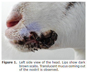
Clinical examination findings. In general, the physiological variables were within the normal range for the species. The animals were maintained a good appetite but with poor body condition, mucopurulent nasal discharge, coughing and overgrown hooves. On the lips, nose and palpebral fissures, shiny and hairless erythematous macules were observed, as well as vesicles and pustules, these lesions bleed easily when touched. At 11 days after the onset of clinical signs, the lesions evolved into scabs, multifocal and coalescing ulcers as a result of the detachment of the crusts (Figure 1).
Samples of skin lesions (10 animals), serum (5 animals) and cobalt (20 animals) were collected. The samples obtained were analyzed by histopathology, virus isolation, DAS-ELISA and nucleic acid amplification, following the standard procedures (11,12). Also, scab and serum samples were sent to the National Laboratory of Veterinary Services APHIS-USA (Ames, Iowa, USA) in order to perform the complement fixation test and transmission electron microscopy (TEM).
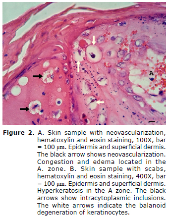
Conventional histopathological analysis of skin samples. The stratum corneum of the epidermis shows hyperkeratosis (ortho-and parakeratotic). In some sections of the lucid and granular stratum, the keratinocytes are degenerated (balanoid degeneration) with pyknotic nuclei and eosinophilic and refractile intracytoplasmic inclusions. Degenerated keratinocytes arise, leading to the formation of vesicles occupied by neutrophils and pus cells. The latter extend to the superficial dermis. The dermis showed neovascularization with congestion, edema and hemorrhage (Figure 2).
Viral isolation and DAS-ELISA. The DAS-ELISA test was performed on scabs for the detection of vesicular stomatitis and foot and mouth disease. The scabs were also used to inoculate Vero cells and in a period of less than 24 hours, the cytopathic effect was apparent. The DAS-ELISA technique was applied again, but this time the average with viable viral particles were isolated. Both scabs and viral particles were negative for vesicular stomatitis and foot and mouth disease.
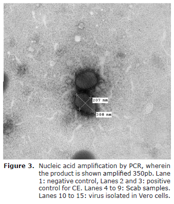
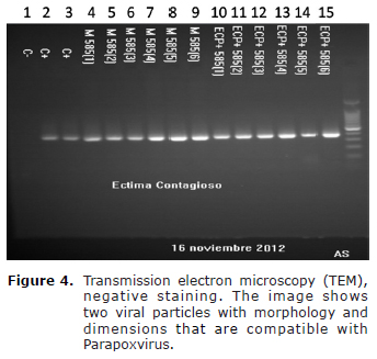
Nucleic acid amplification test. DNA was extracted from the scabs, and from the viral particles isolated in Vero cells. Individual amplification of nucleic acids was performed for vesicular stomatitis, foot and mouth disease and contagious ecthyma. Viral amplification was only observed in all samples processed for the diagnosis of contagious ecthyma (Figure 3).
Serology. The complement fixation test was positive for CE for four of the five animals tested.
Transmission electron microscopy (TEM). TEM results showed electron-dense, ovoid, viral structures, with a crosslinked membrane pattern. The size of these structures was 308nm x 207nm and they were consistent with the Parapoxvirus genus (Figure 4).
DISCUSSION
This message reports the diagnosis of a CE outbreak in the Paso Canoas quarantine station in Panama. This outbreak occurred in a herd of imported goats at 13 days into their quarantine, the animals were not released in Panamanian territory.
This disease has not been reported in the Republic of Panama, or in any of the groups of imported animals received by the quarantine station. Also, in the nearest border country, Costa Rica, there are no written reports of CE.
In this outbreak, erythematous macules were apparent after 13 days of quarantine, which differs from literature that mentions a 4-8 day incubation period (1); Also, according to consulted literature, CE mainly affects young animals and sheep (1-3, 7, 8), at this point it is important to mention that the quality and intensity of the infection could be related to the immune system's ability to control virus multiplication (1-3); the trip and other stressors may have played an important role in the timing of the disease. Also, one cannot dismiss the presence of persistently infected animals, which keep the virus circulating in the herd.
These lesions were exclusively distributed on the lips, nose and palpebral fissures. The distribution pattern of the lesions differs with that reported in literature where lesions can occur on the rest of the skin, genitals, udders and even on digestive epithelia (1, 2, 4, 8, 10).
Usually, acute erythematous lesions progress to scabs in about six weeks (1). However, in this case, the goats showed scabbing on day 11 after the start of acute macroscopic lesions.
In the histopathology, skin samples showed hyperkeratosis, balanoid degeneration of keratinocytes, neutrophilic infiltrated, intracytoplasmic, eosinophilic inclusion bodies, neovascularization with congestion, edema and hemorrhage. These lesions are characteristic of CE (1,7,10).
Clinical, histopathological and TEM diagnosis of CE were confirmed. The presence of other vesicular viral diseases was ruled out by means of the complement fixation test, DAS-ELISA and nucleic acid amplification tests.
Viral diseases such as foot and mouth disease, vesicular stomatitis, blue tongue and goat pox are differential diagnoses of CE, and are also considered important diseases for quarantine. For this reason, and upon reaching the etiologic agent of the disease described in this report, it was necessary to discard the presence of other diseases.
Biosecurity and disinfection measures were based on the physicochemical properties of the virus of the Poxvirus genus. These viruses are sensitive to high temperatures, and to benzene (5) which contains Virkon S.
CE in ruminants affects the development of the livestock industry (8), since CE outbreaks cause economic losses related to decreased food intake and lower weight gain (1, 2, 6, 7). Furthermore, CE has an impact on animal welfare due to pain caused by lesions on the animal (2) and is of importance in public health as zoonosis (1-8).
Effective therapy against Parapoxvirus infection remains contentious. (1,2,7). The goats also didn't have a severe bacterial infection. Therefore, animals affected with CE remained without drug treatment. Only feed was improved with fresh grass as a palliative treatment. After confirmation of CE in the herd, all animals were returned by land to their country of origin.
Quarantine stations are points of disease contention of vital importance to the country. Disease outbreaks in these facilities should be contained through strict biosecurity measures and diagnostic tests that establish the specific etiologic agent in the shortest possible period of time. For all imported animals, clinical inspection and alertness must be maintained in the event of any signs of disease manifestation during quarantine.
Acknowledgements
The authors acknowledge the financial, technical and logistical support of the following people and institutions: Dr. Enrique Evans, Dra. Kirian Cerceño, Dr. José Barrera, Licda. Yiris Rovira, Dr. Enzo Rodríguez, Dr. Manuel González Cano, Dr. Bredio Velasco, Dr. Humberto Hernández, Dr. Francisco Pinilla, Dr. Franklin Clavel, Dr. Carlos Lorenzo, Licda. Cristella Martínez y Licda. Krislly Ramírez from the Ministry of Agricultural Development and Palo Alto Laboratory of the México-United States Commission for the prevention of foot and mouth disease and other exotic diseases (CPA).
REFERENCES
1. Nandi S, De U, Chowdhury S. Current status of contagious ecthyma or orf disease in goats and sheeps-a global perspective. Small Rumin Res 2011; 96:73-82. [ Links ]
2. Cea M, Reyes E, Bravo A, Rojas R. Estandarización de una técnica molecular para el diagnóstico de virus orf útil en la ganadería ovina. Ciencia y trabajo 2008; 28:39-46. [ Links ]
3. Wet C, Murie W. Lamb pays lip services: two cases of ecthyma contagiosum (Orf). Scott Med J 2011; 56(1):1-4. [ Links ]
4. De La Concha-Bermejillo A, Guo J, Zhang Z, Waldron D. Severe persistent orf in Young goats. J Vet Diagn Invest 2003; 15(5):423-431. [ Links ]
5. Gallina L, Scagliarini A. Virucidal efficacy of common disinfectants against orf virus. Vet Rec 2010; 166(23):725-726. [ Links ]
6. Musser J, Taylor C, Guo J, Tizard I, Walker J. Development of a contagious ecthyma vaccine for goats. Am J Vet Res 2008; 69(10):1367-1370. [ Links ]
7. Hosamani M, Scagliarini A, Bhanuprakash V, McInnes C, Singh R. Orf: an update on current research and future perspectives. Expert Rev Anti Infect Ther 2009; 7(7):879-893. [ Links ]
8. Hosie B. Orf: reasons to take an interest in control. Vet Rec 2012; 170:671-672. [ Links ]
9. Lovatt F, Barker W, Brown D, Spooner R. Case-control study of orf in preweaned lambs and an assessment of the financial impact of the disease. Vet Rec 2012; 170(26):673. [ Links ]
10. Sargison N, Scott P, Rhind S. Unusual outbreak of orf affecting the body of sheep associated with plunge dipping. Vet Rec 2007; 160:472-373. [ Links ]
11. Devi B, A.R. S, Parameaswari P, Parijathan B. Domestic microwave versus conventional tissue processing: a quantitative and qualitative analysis. J Clin Diagn Res 2013;7(5):835-839. [ Links ]
12. Kottaridi C, Nomikuo K, Lelli R, Markoulatos P, Mangana O. Laboratory diagnosis of contagious ecthyma: comparison of different PCR protocols with virus isolation in cell culture. J. Virol. Methods 2006; 134(1-2):119-124. [ Links ]
