


Services on Demand
Journal
Article
 English (pdf)
 Article in xml format
 Article references
 Send this article by e-mail
Indicators
-
 Cited by SciELO
 -
 Access statistics
Related links
-
 Cited by Google
 -
 Similars in
SciELO
Similars in
SciELO -
 Similars in Google
Share

 Permalink
Revista MVZ Córdoba
Print version ISSN 0122-0268
Rev.MVZ Cordoba vol.19 no.3 Córdoba Sept./Dec. 2014
CLINICAL CASE
Pulmonary adenocarcinoma in cattle
Adenocarcinoma pulmonar en bovinos
Diogo Sousa Z,1 MV, Luis Rivera C,2* MVZ, Didier Quevedo C,1 M.Sc, Ana Claudia Gorino,1 MV, Simone Biagio C,1 Ph.D, Renée Laufer A,1 Ph.D.
1São Paulo State University “Júlio de Mesquita Filho” (UNESP), Faculty of Veterinary and Zootechnical Medicine, Clinical Veterinary Department, Botucatu, São Paulo, Brasil.
2São Paulo State University “Júlio de Mesquita Filho” (UNESP), Faculty of Agricultural and Veterinary Sciences, Veterinary Pathology Department, Jaboticabal, São Paulo, Brasil.
*Correspondence: lgriveramvz@gmail.com
Received: November 2013; Accepted: April 2014.
ABSTRACT
The Macroscopic, histological and immunohistochemical aspects of lung acinar adenocarcinoma and the presence of nodules in the abdominal cavity of an adult female bovine are reported. In the necropsy analysis samples were collected from the: lung, heart, spleen, liver, pancreas, kidney, uterus, intestine, brain, and from nodules found in the lung and abdominal cavity, which were routinely processed to be stained by hematoxylin-eosin and for an immunohistochemistry exam with the antibodies: cytokeratin (dilution 1:200 µL) and vimentin (dilution 1:1000 µL). The histopathological examination revealed neoplastic epithelial cells with acini formation. The immunohistochemical examination of the tumor cells showed positive marking for cytokeratin and the absence of marking for vimentin. According to anatomical, morphological, and histopathological findings, as well as the result of the immunohistochemical examination, the tumor was characterized as lung acinar adenocarcinoma.
Key words: Histopathology, immunochemistry, lung, metastasis, neoplasia (Source: CAB).
RESUMEN
Se relatan los aspectos macroscópicos, histológicos e inmunohistoquímicos de un adenocarcinoma acinar pulmonar y la presencia de nódulos en la cavidad abdominal en una hembra bovina adulta. En el análisis necroscópico fueron colectados fragmentos de: pulmón, corazón, bazo, hígado, páncreas, riñón, útero, intestino, encéfalo y de los nódulos hallados en pulmón y cavidad abdominal, los cuales fueron procesados rutinariamente para ser teñidos mediante Hematoxilina-Eosina y para examen inmunohistoquímico con los anticuerpos: citoqueratina (con dilución 1:200 µL) y vimentina (dilución 1:1000 µL). El examen histopatológico reveló células epiteliales neoplásicas con formación de acinos. El examen inmunohistoquímico de las células neoplásicas demostró marcación positiva para citoqueratina y ausencia de marcación para vimentina. De acuerdo con los hallazgos anatómicos, morfológicos, histopatológico, y el resultado del examen inmunohistoquímico, se logró caracterizar el tumor como adenocarcinoma acinar pulmonar.
Palabras clave: Histopatología, inmunohistoquímica, metástasis, neoplasia, pulmón (Fuente: CAB).
INTRODUCTION
Primary lung tumors are rare in cattle, so it is an unusual finding in slaughterhouses. The incidence of these tumors is 2.8% among all neoplasms found in the species (1).
Malignant tumors of epithelial origin related to lung tissue in cattle may arise from the epithelium, bronchial mucous glands (goblet cells) and alveolar liner. These tumors can be subdivided into four different histological types: papillary, acinar, solid and mixed, the papillary and acinar types are the most common in domestic animals (2).
Metastasis of lung carcinomas occurs due to local invasion by a hematogenous or lymphatic route (1). Due to the rare frequency of this neoplasm in cattle, little is known about its biological behavior. In Brazil, in a retrospective study performed by Lucena et al (3), between 1964 and 2008, samples were obtained in paraffin blocks from bovine necropsies performed on 6706 cattle, stored in the Pathology Service archives, at St. Mary's University, Rio Grande do sul. Of these samples, 586 were neoplasms. It was determined that four adenocarcinomas were of pulmonary origin, including two papillary adenocarcinomas, one acinar and one of small anaplastic cells. However, Viott et al (4) described, in detail, the first case of pulmonary adenocarcinoma in this country.
The aim of this study was to report the occurrence and describe the macroscopic, histological and immunohistochemical aspects of pulmonary acinar adenocarinoma, and the presence of malignant nodules distributed in the abdominal cavity of an adult female bovine.
CLINICAL CASE
A female bovine, Nelore breed, age nine, was received by the Department of Veterinary Pathology at the UNESP-Veterinary Hospital, Botucatu (SP), Brazil, for post mortem analysis. In the clinical history, clinical signs of hyperoxia, ruminal hypotonia, and persistent tachycardia were described during the first clinical examination. These clinical signs progressively intensified and 15 days after the first examination, an exploratory laparotomy was done where 50 liters of pink-colored abdominal fluid were drained and a mass in the left sacral wing with suspected mesothelioma was also observed. Due to the precarious economic condition of the animal owner, complementary procedures and examinations were not performed. Finally, with the authorization of the owner, the euthanasia of the animal was performed for academic purposes in the Veterinary Pathology sector.
During the necropsy, the animal obtained a body condition 3/5 (5), with oculo-palpebral mucous, oral and external genital of normal color. During In situ analysis, the lung showed areas of atelectasis in the cranial lobe, and left caudal, associated with multifocal fibrosis (Figure 1A), nodules between 1 and 4 cm in diameter were also present, with a firm consistency, ranging in color from white to yellow, and diffusely distributed in this organ (Figure 1B).
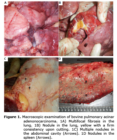
In the abdominal cavity, multi-sized nodules were observed, mainly in parietal and visceral serous, and diffusely distributed, measuring 0.2 to 2.0 cm in diameter (Figure 1C). A mass of 10.0 x 8.0 cm was also observed in the left sacral wing. The nodule in the sacral region was mainly composed of adipose tissue encapsulating a neoplastic mass of 2 cm in diameter. The uterus was found to be thickened, with a firm consistency, and upon cutting, viscous, translucent content was noted.
Samples were collected from the: Lung, heart, spleen, liver, pancreas, kidney, uterus, intestine, brain and from nodules found in the lung and abdominal cavity. All were set in 10% formalin, and routinely processed, using the hematoxylin-eosin method.
The histopathological examination of the nodules revealed neoplastic epithelial cells with acini formation. The cells exhibited moderately sized, eosinophilic cytoplasm, basophilic round nucleus with basal localization (Figure 2A). Little chromatin was found, identifying one or two nucleoli per cell; further observations included: anisokaryosis, anisocytosis, high pleomorphism and increased nucleus:cytoplasm ratio. On average, two mitotic figures per high magnification field (400x) were determined.
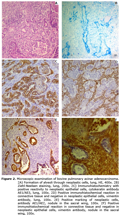
In some areas of the cut, multifocal and coalescing necrosis was found, mononuclear inflammatory cells predominantly infiltrated with lymphocytes, plasmocytes and discrete reactive macrophages. When performing Ziehl-Neelsen stain, the result was negative for Mycobacterium sp. (Figure 2B). Areas of mineralization and fibrosis, of moderate quantity, with multifocal and coalescent distribution were also identified. The spleen, sternal lymph node and abdominal nodes, showed infiltration of previously described neoplastic cells.
The Immunohistochemical technique by means of the endogenous peroxidase method was used for pan-cytokeratin (Mouse anti Cytokeratin (Pan) AE1/AE3, Invitrogen, Frederick, USA) (Dilution 1:200 ul) and Vimentin (Mouse anti Vimentin (V9), Invitrogen, Frederick, USA) (Dilution 1:1000 ul), both with cytoplasmic marking. For the revelation DAB was used (chromogen 3,3 âDiaminobenzidine, Dako, Glostrup, Denmark).
The immunohistochemistry test showed positive immunoreactivity for cytokeratin, being moderately cytoplasmic and diffused in neoplastic cells (Figure 2C, 2E). In malignant cells, no Vimentin marking was observed, this antibody was only positive for connective tissue (Figures 2D, 2F).
According to morphological, anatomical, and histopathological findings and to positive immunoreactivity for cytokeratin, the tumor was characterized as acinar adenocarcinoma in the lung and in the nodes identified in the abdominal cavity.
DISCUSSION
Pulmonary malignant neoplasms are classified according to their origin, epithelial cells in the lung originate lung adenocarcinomas (1,3,6), while the mesenchymal cells can produce osteosarcomas, chondrosarcomas, hemangiosarcomas, malignant histiocytosis, lymphomatoid granulomatosis, granular cell tumors and mesothelioma (7).
Pulmonary adenocarcinoma may be differentiated from mesothelioma by the presence of basal, uniform nuclei in cells that form acini, a feature which is absent in mesotheliomas, which have granular cells with irregular nuclei and primitive acini (8).
Additionally, immunohistochemistry is a useful tool in differentiating these tumors, since neoplastic cells in mesothelioma express epithelial cytokeratin and mesenchymal markers, such as Vimentin (9), whereas adenocarcinomas are specifically marked by cytokeratin.
Pulmonary adenocarcinomas, with esquirrosa, in cattle should be differentiated from other epithelial neoplasms, mainly primary neoplasms of the uterus and pancreas (3,9). Endometrial adenocarcinoma, besides being esquirroso and possessing large amounts of fibrous tissue, presents a disruption in glandular epithelial cells and high pleomorphism. This was not evident in uterine tissue and nodules scattered in the abdominal cavity of the animal under study (9). Moreover, pancreatic adenocarcinoma exhibits several cellular patterns: tubular, not delineated or solid. The cells may form acini or gross tubules, with a uniform, oval nucleus and dispersed chromatin. These tumors have areas of hemorrhage, mineralization or necrosis in the pancreas (7); the absence of macroscopic and microscopic alterations in this organ repudiate the emergence of this carcinoma
During the necropsy, the cheesy and calcareous appearance of the nodules suggested tuberculosis. However, histological sections subjected to the Ziehl-Neelsen stain were negative.
In humans, infection with Mycobacterium sp. was already associated with pulmonary adenocarcinoma (10,11). However, until now there has not been any report of animals associated with these diseases.
In conclusion, anatomical and histomorphological findings, and contributions described in the literature allow the tumor to be classified as lung acinar adenocarcinoma, this pattern of organization was also identified in the nodules collected from the abdominal cavity; suggesting that the primary focus of the neoplasm was from pulmonary origin. Positive immunoreactivity for AE1/AE3 and negative marking for vimentin were key determinants in completing the case.
REFERENCES
1. Ioan A, Câtoi C. Comparative Oncology. 1 Edition. Bucuresti: The Publishing House of the Romanian Academy; 2007. [ Links ]
2. Barley JP. Pulmonary papillary adenocarcinoma in an adult cow. Ir Vet J 2011; 1(12):674-675. [ Links ]
3. Lucena RB, Rissi DR, Kommers GD, Pierezan F, Oliveira-Filho JC, Macedo JTS. A retrospective study of 586 tumours in brazilian cattle. J Comp Path, 2011; 145:20-24. [ Links ]
4. Viott AM, Langohrii IM, Vannucci FA, Almeida AP, Leite RC, Ecco R. Adenocarcinoma pulmonar em um bovino. Ciênc Rural, 2010; 40(2):484-487. [ Links ]
5. Correa-Orozo A, Uribe-Velásquez LF, La condición corporal como herramienta para pronosticar el potencial reproductivo en hembras bovinas de carne. Rev Fac Nal Agr Medellín, 2010; 63(2):5607-5619. [ Links ]
6. Carminato A, Bozzato E, Trevisan L, Vascellari M, Catania S, Palma D, et al. Bovine pulmonary blastoma. Large Anim Rev, 2008; 14(1):3-5. [ Links ]
7. Santos, R. L.; Alessi, A.C. Patologia Veterinaria. Edición 1. São Paulo: Roca; 2011. [ Links ]
8. Merlo W, Rosciani A, Koscinczuk P, Ortega H, Insfrán RM, Macció OA. Mesotelioma peritoneal en un canino. Rev Vet 2007; 18(1):54-57. [ Links ]
9. Stilwell G, Peleteiro MC. Uterine adenocarcinoma with pulmonary, liver and mesentery metastasis in a hostein cow. Vet Med Int 2010; Article IDÂ 727856. http://dx.doi.org/10.4061/2010/727856. [ Links ]
10. Kobashi Y, Yoshida K, Miyashita N, Niki Y, Matsushima T. Pulmonary Mycobacterium avium disease with a solitary pulmonary nodule requiring differentiation from recurrence of pulmonary adenocarcinoma. Intern Med, 2004; 43(9):855-859. [ Links ]
11. Sawai T, Soda H, Kohno S. Mycobacterium intracellulare pulmonary infection which co-existed and mimicked lung cancer. Intern Med 2008; 47(5):459-462. [ Links ]
