


Services on Demand
Journal
Article
 Spanish (pdf)
 Article in xml format
 Article references
 Send this article by e-mail
Indicators
-
 Cited by SciELO
 -
 Access statistics
Related links
-
 Cited by Google
 -
 Similars in
SciELO
Similars in
SciELO -
 Similars in Google
Share

 Permalink
Revista MVZ Córdoba
Print version ISSN 0122-0268
Rev.MVZ Cordoba vol.20 no.1 Córdoba Jan./Apr. 2015
ORIGINAL
Blood gas analysis in Mangalarga Marchador horses with colic
Análisis de gases sanguíneos en caballos Mangalarga Marchador con cólico
Tiane F. Castro,1 M.Sc, Félix González,1* Ph.D.
1Universidade Federal do Rio Grande do Sul, Faculdade de Veterinária, Av. Bento Gonçalves, 9090, Porto Alegre, 94540-000, Brasil.
*Correspondence: felixgonzalez.ufrgs@gmail.com
Received: April 2014; Accepted: November 2014.
ABSTRACT
Objective. This study aims to distinguish blood gas changes in horses with colic syndrome in which small or large intestine is affected. Materials and methods. Thirty Mangalarga Marchador horses were assessed, divided into groups according to the affected intestinal segment in episodes of colic syndrome (ECS): a group (N=10) of horses suffering from ECS with lesions only in the small intestine, a group (N=10) of horses suffering from ECS with lesions only in the large intestine and a group (N=10) of healthy horses (control). All the animals with ECS were submitted to exploratory laparotomy in order to establish the intestinal segment affected. Blood samples were collected by venipuncture, before surgical procedure to determine sodium, potassium, chloride, urea, glucose, hematocrit, hemoglobin, pH, carbon dioxide partial pressure, total carbon dioxide concentration, bicarbonate, base excess and anion gap. Results. No significant changes were found in plasma levels of Na+, K+, Cl-, pCO2 and anion gap in any type of ECS. Horses with small intestine injuries presented higher levels of tCO2, urea and bicarbonate compared to those with large intestine injuries and to the control group, as well as higher levels of glucose and base excess than the control group. Conclusions. Horses with colic syndrome bearing small intestine injuries show wider variations in the blood gas parameters than horses with large bowel lesions.
Key words: Acid-base imbalance, electrolytes, large intestine, small intestine (Source:CAB).
RESUMEN
Objetivo. El presente estudio tuvo como objetivo diferenciar las alteraciones de gases sanguíneos que ocurren en casos de cólico equino con comprometimiento en intestino delgado o grueso. Material y métodos. Fueron evaluados 30 caballos Mangalarga Marchador con sindrome de colico equino (SCE) divididos en grupos según el segmento intestinal afectado con episodios de cólico: un grupo de animales (N=10) con SCE solo en el intestino delgado, un grupo (N=10) con SCE solo en el intestino grueso y un grupo (N=10) de animales sanos (control). Todos los animales fueron sometidos a laparotomía exploratoria para identificar el segmento intestinal afectado. Muestras de sangre fueron recogidas antes del procedimiento quirúrgico para determinar la concentración: sodio, potasio, cloro, urea, glucosa, hematocrito, hemoglobina, pH, presión parcial de dióxido de carbono, concentración total de dióxido de carbono bicarbonato, exceso de base y anión gap. Resultados. No fueron observadas alteraciones significativas en los niveles plasmáticos de Na+, K+, Cl-, pCO2 ni anión gap, independiente de la localización intestinal del SCE. Los equinos con lesiones en el intestino delgado presentaron niveles superiores de tCO2, urea y HCO3- con relación a los que tuvieron lesiones de intestino grueso y al control, así como niveles superiores de glucosa y de exceso de base con relación al control. Conclusiones. Equinos con síndrome de cólico con lesión de intestino delgado presentaron variaciones más amplias en los parámetros de gases sanguíneos que equinos con cólico afectados en el intestino grueso.
Palabras clave: Ácido-básico, desequilibrio, electrolitos, intestino delgado, intestino grueso (Fuente: CAB).
INTRODUCTION
Equine colic syndrome (ECS) or acute abdomen is a multifactorial syndrome that results in deep abdominal pain in horses, with a diversified etiology varying from excessive gas yielded from feed fermentation to obstruction and bowel torsion, commonly requiring surgical intervention (1). The diagnosis of ECS is relatively easy considering the features of the abdominal pain, which cause changes in animal behavior, but its etiology is yet a challenge for veterinarians, especially in the definition of the affected intestinal segment.
The use of supporting diagnosis tools, like hemogram, blood gas analysis and biochemical profile can give information concerning inflammatory focuses, dehydration or hipovolemia, toxemia, and electrolytic, acid-base or metabolic imbalances, providing a more precise diagnosis, an adequate treatment and a more accurate prognosis (2).
Practically all disorders affecting the gastrointestinal tract lead to imbalances in blood pH, bicarbonate, sodium, potassium and chloride that may be immediately corrected to avoid the risk of death. The acid-base and hydro-electrolytic evaluation, as well as the blood gas condition allow choosing the more appropriated treatment for the animal with colic syndrome. Blood gas analyzes the partial O2 and CO2 pressure as well as bicarbonate and pH (3), becoming the most appropriate method for evaluating the acid-base balance of organic fluids and their probable disturbances (4). Blood gas evaluation is a very useful parameter for determining the severity of the colic syndrome and also may helps in the identification of bowel lesions (1).
The present work had the aim of evaluating changes in blood gas parameters in Mangalarga Marchador horses suffering from colic syndrome, discriminating the intestinal bowel affected, in order to obtain additional tools for clinical and surgical assessment.
MATERIALS AND METHODS
Animals. All the horses in this work were selected following criteria of clinical signs of colic syndrome at a referred veterinary hospital. The animals were Mangalarga Marchador horses from 3 to 15 years old, from both sexes (18 males and 12 females). The location of the intestinal section affected was established through laparotomy procedure.
The animals were organized in three groups as follows: animals with lesions only in the small intestine (N=10); animals with lesions only in the large intestine (N=10); and healthy animals (control group) (N=10).
Sampling and analysis. Blood samples (2 mL), from the jugular vein, were collected anaerobically through heparinized tubes (Becton Dickinson, São Paulo, Brazil) for blood gas analysis before the surgical procedure. The analysis was done using a portable analyzer (I-Stat 1, Abbott, Princeton, USA), with specific cartridges following the manufacturer's instructions. The equipment was automatically calibrated before the samples analysis. The cartridge used was EC8+, which is able to determine sodium (Na+), potassium (K+), chloride (Cl-), urea, glucose, hematocrit, hemoglobin, pH, partial carbon dioxide pressure (pCO2), total carbon dioxide concentration (tCO2), bicarbonate (HCO3), base excess (EB) and anion gap (AG).
Statistical analysis. Statistical analysis was performed through Anova test, relating the intestinal bowel affected with the studied parameters. Quantitative variables (mean and standard deviation) were analyzed using the Tukey test, considering a significance level of 5%, using the Instat program.
RESULTS
The values obtained for hemogasometric analysis according to the affected intestinal section are showed in table 1.
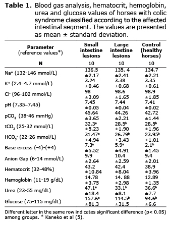
Plasma levels of Na+, K+, Cl-, pCO2 and anion gap did not show any significant alteration (p> 0.05) no matter which was the affected bowel segment. Glucose concentration was higher in 65% of the animals suffering from ECS, compared to the reference values (maximum reference value: 115 mg/dL). Glucose and urea concentrations were higher (p<0.05) in the group of horses with small intestine lesions compared to animals with large intestine lesions and to control animals. The values of hematocrit and hemoglobin did not show differences among all groups of horses (p>0.05).
Venous blood pH was higher in 40% of the ECS cases than in the control animals. However, no differences in blood pH were found between the two groups of horses with colic syndrome (p>0.05). The tCO2 was higher in horses with small intestine ECS compared to the other two groups of animals (p<0.05). The bicarbonate levels were increased in 70% of the horses with colic syndrome (maximum reference value: 26 mmol/L). It was observed that the highest the bicarbonate value, the more severe the clinical condition of the horses. The group of horses with small bowel lesions had the highest values of bicarbonate compared to the other groups of horses (p<0.05). In 60% of the animals with ECS the value of base excess was above +4 (reference value: -4 to +4), with higher values in the group with small bowel lesions (p<0.05).
DISCUSSION
This work shows evidences of blood gas alterations in horses with colic syndrome. The blood concentration of sodium was at the reference values in all the animals of this study, ECS and control, without any difference between the two groups of horses with colic syndrome. This was also the observation of Navarro et al (6) who studied electrolytic alterations in horses with gastrointestinal disorders and did not find changes in sodium concentrations in several types of intestinal lesions. Ihler et al (7) also did not found differences in blood sodium comparing horses that survived or deceased in cases of ECS. During a clinical situation of ECS, anorexia and adipsia due to the severe pain might cause a depletion of extracellular fluid with loss of body water, although with small loss of electrolytes (8). This might lead to hypernatremia by hemoconcentration, but such a situation did not occur.
As in the present study, Ferreira (8) did not find significant variations in potassium levels in horses submitted to temporary fistulization at the cecum. Di Filippo et al (9) found lesser serum potassium values in horses that survived to colic syndrome compared to those that died and to the control group. That work was done with animals observed at the moment of the clinical examination, same as our study. The reduction of potassium values in colic cases may be attributed to less feed intake or to gastrointestinal losses (10).
Chloride blood levels also were not affected by the clinical condition of ECS, as in the work of Navarro et al (6). Costa et al (11) observed significant decrease in serum chloride in horses with jejunum isquemia.
Glycemia was higher in horses with lesions in the small bowel. This result was also observed by Di Filippo et al (9) who found hyperglycemia in horses that survived to a colic syndrome. In another study (12) hyperglycemia was evident in animals with induced intestinal obstruction. Hyperglycemia is a frequent finding observed in cases of ECS and is generally associated with an unfavorable prognostic (13). This is related to the high secretion of catecholamines as a consequence of excitation, pain and stress, typical episodes of ECS (14).
Urea blood concentration is regulated by two events: synthesis rate in the liver and clearance rate by kidney. Synthesis rate depends on protein intake and catabolism. Clearance rate depends on glomerular filtration rate and reabsorption activity at the renal tubules (15). In dehydration conditions, as occur in colic syndrome, glomerular filtration rate is diminished resulting in pre-renal azotemia. In the present study, urea values were higher in horses with small bowel lesions, which suggest a greater compromise in that kind of lesions. Di Filippo et al (16) did not find variations in hepatic and renal blood markers in horses submitted to experimental intestinal obstruction. In agreement with those results, Granot et al (17) found that horses with colic impaction did not have different values of urea compare to animals that died or that survived. From our results it would be probable that horses with colic syndrome caused by lesions situated at the small intestine may be suffering from a more severe condition of water imbalance than horses with compromised large intestine.
In the present study, the hematocrit values were not different between individuals with colic syndrome and control animals. An increase in the hematocrit may occur in cases of equine colic syndrome as a result of hemoconcentration, dehydration and shock (14). Datt and Usenik (18) found significant hematocrit increase in horses after 12 hours of duodenum obstruction. It seems that hematocrit elevation in gastrointestinal disorders in horses may be more frequent in most severe cases. Concomitant with the result of the hematocrit, hemoglobin values were not significantly different among the groups of animals. However, horses with colic syndrome together had hemoglobin values greater than control horses. This result is in agreement with Silva (12), who found higher hemoglobin values in horses with colic syndrome than in healthy animals. It is plausible that hemoglobin could be a more sensitive indicator than hematocrit of gastrointestinal disorders of horses (18,19).
In our study, 40% of the horses with colic syndrome had increased blood pH in relation to the control group. Other authors also found elevated blood pH in horses with colon and jejunum obstructions (11,19), which was associated with increased bicarbonate concentration. In the present study, the rise in blood pH was linked not only with higher values of bicarbonate but also with more positive values of base excess in both groups of horses with colic syndrome, especially in animals with small intestine lesions. All together 60% of the colic affected horses had base excess values above +4, more severely when lesions occurred in the small intestine. Similar finding was observed in cases of horses with experimental intestinal obstruction (11). The tCO2 values had significant rise in horses with small bowel lesions. The tCO2 parameter expresses the alkaline reserve, with bicarbonate as its main component. A high tCO2 indicates the presence of metabolic alkalosis, and a low tCO2 indicates a metabolic acidosis (14). These increments in tCO2 and alkaline reserve in horses with small bowel lesions, were not expected in the situation of pain and stress in horses with colic, which should promote breath rate increment and decrease in pCO2 (respiratory alkalosis). In contrast with our findings, other authors found metabolic acidosis in horses suffering from colic syndrome (9), associated with a decline in HCO3- and base excess attributed to hipovolemia and generation of lactic acid.
The anion gap was within the reference values in 90% of the horses with colic syndrome. According to the anion gap equation [AG=(Na++K+) - (HCO3- + Cl-)] the AG values should be diminished, because the term HCO3- is augmented and sodium, potassium and chloride did not have alterations, as it was showed in our work. More research should be done to evaluate if there are other anion alterations in cases of equine colic syndrome.
In conclusion, horses with colic syndrome do not show significant alterations in plasma electrolytic parameters (Na+, K+, Cl-) or in the levels of pCO2 and anion gap, independently of the intestinal location of the lesion. Horses with small bowel lesions have higher values of tCO2, HCO3-, base excess, urea and glucose than horses with large bowel lesions, suggesting a condition of metabolic alkalosis and more severe stress.
REFERENCES
1. Nappert G, Johnson PJ. Determination of the acid-base status in 50 horses admitted with colic between December 1998 and May 1999. Canadian Vet J 2001; 42:703-707. [ Links ]
2. Nolen-Walston R, Paxson J, Ramey DW. Evidence-based gastrointestinal medicine in horses: It's not about your gut instincts. Vet Clin North Am Equine Pract 2007; 23:243-266. [ Links ]
3. Alves GES, Ribeiro Filho JD, Oliveira HP, Abreu JMG. Tratamento da compactação experimental do cólon maior em eqüinos: resultados de laboratório e exames bioquímicos. Arq Bras Med Vet Zootec 2005; 57:281-287. [ Links ]
4. Gokce G, Citil M, Gunes V, Atalan G. Effect of time delay and storage temperature on blood gas acid-base values of bovine venous blood. Res Vet Sci 2004; 76:121-127. [ Links ]
5. Kaneko JJ, Harvey JW, Bruss ML. (Eds.). Clinical Biochemistry of Domestic Animals. 6.ed. San Diego: Academic Press, 2008. [ Links ]
6. Navarro M, Monreal L, Segura D, Armengou L, Añor S. A comparison of traditional and quantitative analysis of acid-base and eletrolyte imbalances in horses with gastrointestinal disorders. J Vet Inter Med 2005; 19(6):871-877. [ Links ]
7. Ihler CF, Venger JL, Skjerve E. Evaluation of clinical and laboratory variables as prognostic indicators in hospitalised gastrointestinal colic horses. Acta Vet Scand 2004; 45(1-2):109-118. [ Links ]
8. Ferreira FPP. Fistulização temporária do ceco em eqüinos: estudo experimental da técnica cirúrgica e da viabilidade desta via para a administração de fluído enteral. [Tese de Doutorado]. Botucatu: Universidade Estadual Paulista. Programa de Pós-graduação em Cirurgia Veterinária, 2006. [ Links ]
9. Di Filippo PA, Santana AE, Pereira GT. Equilíbrio ácido-base e hidroeletrolítico em eqüinos com cólica. Ciência Rural 2008; 38(4):1003-1009. [ Links ]
10. Toribio RE. Essentials of equine renal and urinary tract physiology. Vet Clin North Am Equine Pract 2007; 23(3):533-561. [ Links ]
11. Costa NS, Ribeiro G, Dória RGS, Canola PA, Silva PC, Jorge RLN et al. Hemograma e hemogasometria de equinos submetidos a obstrução experimental de jejuno. Arq Bras Med Vet Zootec 2008; 60(6):1367-1374. [ Links ]
12. Silva CF. Valores hematológicos bioquímicos e exame do líquido peritoneal de eqüinos (Equus caballus, Linnaeus, 1758) durante síndrome cólica. [Dissertação de Mestrado]. Botucatu: Universidade Estadual Paulista. Faculdade de Medicina Veterinária e Zootecnia, 2005. [ Links ]
13. Marr CM. On the question of colic: are answers beginning to emerge?. Equine Vet J 2012; 44(4):384-386. [ Links ]
14. McGowan C. Clinical pathology in the racing horse: the role of clinical pathology in assessing fitness and performance in the racehorse. Vet Clin North Am Equine Pract, 2008; 24(2):405-421. [ Links ]
15. Stockham SL. Interpretation of equine serum biochemical profile results. Vet Clin North Am Equine Pract 1995; 11(3):391-414. [ Links ]
16. Di Filippo PA, Pereira RN, Perotta JH, Berlingieri MA, Freitas FC, Santana AE. Variações nas concentrações dos biomarcadores sanguíneos da função renal e hepática em equinos submetidos a obstrução experimental do duodeno, íleo e cólon maior. Ciência Animal Bras 2011; 12(1):178-186. [ Links ]
17. Granot N, Milgram J, Bdolah-Abram T, Shemesh I, Steinman A. Surgical management of sand colic impactions in horses: a retrospective study of 41 cases. Australian Vet J 2008; 86(10):404-407. [ Links ]
18. Datt SC, Usenik EA. Intestinal obstruction in the horse. Physical signs and blood chemistry. Cornell Vet 1975; 65:152-172. [ Links ]
19. Di Filippo PA. Obstrução intestinal experimental em equinos: parâmetros clínicos e laboratoriais. [Tese de Doutorado]. Jaboticabal: Universidade Estadual Paulista. Faculdade de Ciências Agrárias e Veterinárias, 2009. [ Links ]
