


Services on Demand
Journal
Article
 English (pdf)
 Article in xml format
 Article references
 Send this article by e-mail
Indicators
-
 Cited by SciELO
 -
 Access statistics
Related links
-
 Cited by Google
 -
 Similars in
SciELO
Similars in
SciELO -
 Similars in Google
Share

 Permalink
Revista MVZ Córdoba
Print version ISSN 0122-0268
Rev.MVZ Cordoba vol.20 no.3 Córdoba Sept./Dec. 2015
ORIGINAL
Concordance between otic cytology and culture in diagnosis of external otitis canine by Malassezia spp
Concordancia entre citología ótica y cultivo en el diagnóstico de otitis externa canina por Malassezia spp
Adriana Pulido-Villamarín,1* M.Sc, Rubiela Castañeda-Salazar,1 M.Sc, Melva Linares-Linares,1 M.Sc, Marcela Mercado-Reyes,2 M.Sc.
1Pontificia Universidad Javeriana, Facultad de Ciencias, Departamento de Microbiología, Unidad de Investigaciones Agropecuarias -UNIDIA-. Carrera 7 No. 43-82. Bogotá- Colombia.
2Instituto Nacional de Salud, Dirección de Vigilancia y Análisis de Riesgo en Salud Pública, Subdirección de Prevención y Análisis de Riesgo. Avenida Calle 26 #51-20. Bogotá- Colombia.
*Correspondence: adriana.pulido@javeriana.edu.co
Received: March 2014; Accepted: December 2014.
ABSTRACT
Objective. To determine the correlation between microbiological culture and otic cytology for diagnoses of external otitis by Malassezia in dogs. Materials and Methods. 158 ear swabs of dogs with clinical diagnosis of external otitis were analyzed by cytology, mycological culture and metabolic tests. Results. Were obtained a positive results by cytology of 62% and 75.3% by culture. The 31.1% of isolates were identified as M. pachydermatis, 12.6% as M. furfur and 56.3% were classified as Malassezia spp., because was not possible to define the species. We found a positive concordance between cytology and culture for Malassezia spp., of 0.76 with a kappa index of 0.448 (95% CI 0.30 to 0.60) which represents a moderate strength of concordance between the two techniques, without regard the identified species. Conclusions. The use of a diagnostic test is not enough to establish the participation of Malassezia spp., as a causal disease agent.
Key words: Canis familiaris, diagnostic techniques, Malassezia, otitis externa (Source: CAB).
RESUMEN
Objetivo. Determinar la concordancia entre el cultivo microbiológico y la citología ótica para el diagnóstico de otitis externa causada por Malassezia spp. en caninos. Materiales y Métodos. Se analizaron 158 muestras de hisopados de caninos con diagnóstico clínico de otitis externa, todas las muestras fueron analizadas mediante citología, cultivo micológico y pruebas metabólicas. Resultados. Se obtuvo una positividad mediante citología del 62% y por cultivo del 75.3%. El 31.1% de los aislamientos fueron identificados como M. pachydermatis, el 12.6% como M. furfur y un 56.3% se clasificó como Malassezia spp., dado que bioquímicamente no fue posible hallar su especie. Se determinó una concordancia observada entre técnicas positivas para Malassezia spp. de 0.76 con índice Kappa de 0.448 IC 95% (0.30 - 0.60) lo que representa una fuerza de concordancia moderada entre las dos técnicas, sin tener en cuenta la especie identificada. Conclusiones. El uso de una prueba diagnóstica no es suficiente para establecer la participación de Malassezia pachydermatis como agente causal de enfermedad.
Palabras clave: Canis familiaris, técnicas diagnósticas, Malassezia, otitis externa (Fuente: CAB).
INTRODUCTION
One of the most common causes of veterinary consult in dogs are ear diseases which must be diagnosed and treated. One of these conditions is external otitis whose reported prevalence is 10-20% (1,2), with a wide variety of causative agents. Malassezia spp., is the predominant organism in 50 - 83% of cases (3, 4), presenting themselves alone or in association with other infectious agents (5,6). Malassezia spp., has been recognized as commensal of the skin and mucous membranes on humans and animals (7), however it has been implicated in disease processes that go from either superficial infections to systemic processes, which are favored by exogenous host factors (environmental conditions) or endogenous (skin integrity, immunological conditions)(8-10).
Malassezia spp., are lipophilic yeasts; from which 14 species, have been recognized: Malassezia furfur, M. pachydermatis, M. globosa, M. restricta, M. obtusa, M. slooffiae, M. sympodialis, M. dermatis, M. yamatoensis, M. nana, M. caprae, M. cuniculi sp. nov, M. dermatis,M. japonica, M. equina (11-16) which have been identified by morphology, metabolic profile and molecular tests (3,4).
The laboratory diagnosis of external otitis is made by Wright staining cytology, Giemsa and Gram, that look for the presence of yeast structures whose ratio ≥5 cells per microscopic field (40X) is considered as indicator of pathology, while the presence of lesser amount of yeast indicates of commensal microorganism (2-4,8,17,18), on the other hand, the Malassezia cultivation is hard to do due to its strict nutritional requirements and morphological variability making difficult its isolation and identification, therefore, microbiological characterization is done only for epidemiological purposes or in cases of chronic external otitis and media otitis (1), but it should be noted that the identification of the etiologic agent by culture confirms the clinical suspicion and achieves therapeutic success (5).
Given that laboratory diagnosis is done by cytology and/or culture, the objective of this study was to determine the correlation of these techniques for the analyzed cases.
MATERIALS AND METHODS
Study population. 158 ear swabs of dogs with clinical diagnosis of external otitis were submitted by four veterinary clinics in Bogotá. All samples were analyzed by cytology and culture.
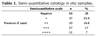
Cytology. Wright staining was performed to determine the presence of yeast, by semi-quantitative scale by crosses, thus: 1 to 5 cells/field (+), 6 to 10 cells/field (+ +), 10 to 19 cells/field (+++) and> 19 cells/field (++++)(1,3,4,18,19).
Culture and identification. Samples were cultured in Sabouraud agar (Oxoid, Wade Road, Basingstoke, UK. ) and Dixon's (Mycosell 36 g/L Malt extract 36 g/L, OxBil 20 g/L, Ac Oleic 2 ml/L, glycerol 2 ml/L, Tween 40 10 ml/L, Chloramphenicol 0.5 g/L) and incubated at 32°C for one week. Positive cultures were evaluated microscopically by Gram stain and identified by biochemical tests (urease activity, bile esculin hydrolysis, growth on agar Sabouraud assimilation of Tween 20, 40, 60, 80 (Merck KGaA, Darmstadt, Germany) and cremophor (Sigma-Aldrich, St Louis, MO, USA) (11,12), using reference strains: M. furfur CBS 7019, M. pachydermatis CBS 1879, M. symphodialis CBS 7222, M. restricta CBS 7877, M. slooffiae CBS 7956 and M. globosa CBS 7966.
Statistical Analysis. A prospective study was performed to determine the diagnostic value of cytology and culture for diagnosis of external otitis caused by Malassezia spp. Data were analyzed in SPSS 1.9; for quantitative variables descriptive statistics were done and for the ratio of diagnostic tests were analyzed for consistency of agreement by Kappa index.
RESULTS
Of the 158 samples analyzed by cytology, 62% (98/158) had the presence of yeast in different degrees as shown in table 1.
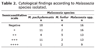
The microbiological culture for Malassezia was positive in 75.3% (119/158), of which by biochemical characterization, 31.1% (37/119) were classified as M. pachydermatis, 12.6% (15/119) as M. furfur and 56.3% (67/119) corresponded to Malassezia spp. In the corresponding smears were observed different cytological findings (Table 2).
The observed concordance between positive otic cytology for Malassezia Vs positive culture was 0.76 and the Kappa index was 0.448 (95% CI 0.30 to 0.60) which represents a moderate strength of agreement between the two techniques.
DISCUSSION
Of the 158 evaluated samples the detected cytology positivity was 62% and 75.3% for culture of all Malassezia species.
The semi-quantitative evaluation of otic cytology is controversial, especially regarding the amount of yeast per field associated with disease, some authors (19) suggest counts greater than 10 cells per field as indicative of disease, while others propose counts less than 5 cells per field (8,18,20,21), however other researchers indicate that the amount of yeast is directly related to the severity of the otitis (2). Given that the samples analyzed in this study were from animals clinically diagnosed with external otitis, these were considered positive if cytology showed 1 + and were correlated with positive cultures.
Although M. pachydermatis is frequently isolated from external otitis processes, there is not consensous regarding its prevalence as an etiologic agent of the same, being reported between 5.12% and 76.5% (5,19,20). In studies done by Boehriger (22) they reported a prevalence of 62% for cytology and 49% by culture (morphological macro/ microscopic and Sabouraud agar growth without lipid supplement), the difference between these data could be due to the methodology used in this study, since specific metabolic test were performed which allowed to identify M. pachydermatis by 31.1% and M. furfur in 12.6%.
Other Malassezia species reported in external otitis processes in dogs have been M. furfur with 3% and M. obtusa by 1.5% (8) which differs from the findings in this study where the prevalence of the first was 12.6%, while 56.3% of isolates could not be classified to species level because of atypical reactions in biochemical tests as stated by Crespo et al (9). It is important to note that most studies aimed to the identification of the etiologic agent of otitis processes were performed in dogs using culture media without lipidic supplements, limiting the grow of lipid-dependant species.
Concerning the two techniques, the results of the test showed that Kappa value and concordance test were similar to the ones reported by Boehringer (22), who found a concordance of 0.78, with a kappa of 0.56 (0.40-0.50), indicating that the tests have moderate agreement.
In conclusion, due to the complexity to identify the etiological agents involved in external otitis and considering that Malassezia is a commensal organism potentially pathogen, the use of a diagnostic test is not enough (9, 22) to establish its participation as a cause of disease, especially when there is no response to therapy (8).
Conflict of interest
The authors declare that they have no conflict of interest.
Acknowledgements
Authors would like to thank to research vice-rectory of the Pontificia Universidad Javeriana for funding.
REFERENCES
1. Dragonetti AM, Broglia G. Otitis externa canina aproximación al diagnóstico. Vet Cuyana 2007; 1-2:28-33. [ Links ]
2. Zur G, Lifshitz B, Bdolah-Abram T. The association between the signalment, common causes of canine otitis externa and pathogens. J Small Anim Pract 2011; 52:254-258. [ Links ]
3. Girão MD, Prado MR, Brilhante RSN, Cordeiro RA, Monteiro AJ, Sidrim JJC and Rocha MFG. Malassezia pachydermatis isolated from normal and diseased external ear canals in dogs: A comparative analysis. The Vet J 2006; 172:544-548. [ Links ]
4. Khosravi AR, Eid S, Ziglari T, Bayat M. Isolation and Differentiation of Malassezia Species Isolated from Healthy and Affected Small Animals, Ear and Skin. World J Zool 2008; 3(2):77-80. [ Links ]
5. Sarierler M, Kirkan S. Microbiological Diagnosis and Therapy of Canine Otitis Externa. Veteriner Cerrahi Dergisi 2004; 10(3-4):11-15 [ Links ]
6. Petrov V, Mihaylov G. Malassezia pachydermatis - etiology and clinical findings in canine external otitis - therapeutic approaches. Trakia J Sci (TJS) 2008; 6(1):123-126. [ Links ]
7. Brito EHS, Fontenelle ROS, Brilhante RSN, Cordeiro RA, Monteiro AJ, Sidrim JJC, Rocha MFG. The anatomical distribution and antimicrobial susceptibility of yeast species isolated from healthy dogs. Vet J 2009; 182:320-326. [ Links ]
8. Murphy KM. A review of techniques for the investigation of otitis externa and otitis media. Clin Tech Small Anim Pract 2001; 16(3):236-241. [ Links ]
9. Crespo MJ, Abarca ML, Cabañes FJ. Occurrence of Malassezia spp. in the external ear Canals of dogs and cats with and without otits externa. Med Mycol 2002; 40:115-121. [ Links ]
10. Rosser EJ. Causes of otitis externa. Vet Clin North Am Small Anim Pract 2004; 34:459-468. [ Links ]
11. Guého E, Midgley G, Guillot J. The genus Malassezia with description of four new species. Antonie van Leeuwenhoek 1996; 69:337-355. [ Links ]
12. Guillot J, Guého E, Lesourd M, Midgley G, Chévrier G, Dupont B. Identification of Malassezia species. J Mycol Med 1996; 6:103-110. [ Links ]
13. Sugita T, Takashima M, Shinoda T, Suto H, Unno T, Tsuboi R, Ogawa H, Nishikawa A. New yeast species Malassezia dermatis isolated from patients with atopic dermatitis. J Clinic Microbiol 2002; 41:4695-4699. [ Links ]
14. Hirai A, Kano R, Makimura E, Duarte R, Hamdam M, Lachance A, Yamaguchi A, Hasegawa A. Malassezia nana sp. nov., anovel lipid-dependent yeast species isolated from animals. Int J Syst Evol Microbiol 2004; 54:623-627. [ Links ]
15. Ashbee R, Bignell EM. Pathogenic yeast: The yeast handbook. Berlin: Springer; 2010. [ Links ]
16. Cabañes FJ, Vega S, Castellá G. Malassezia cuniculi sp. nov., a novel yeast species isolated from rabbit skin. Med Mycol 2011; 49:40-48. [ Links ]
17. Angus JC. Otic citology in health and disease. Vet Clin North Am Small Anim Pract 2004; 34:411-424. [ Links ]
18. Fernández G, Barboza G, Villalobos A, Parra O, Finol G, Ramírez RA. Isolation and identification of microorganisms present in 53 dogs suffering otitis externa. Rev Cient FCV-LUZ 2006; 16(1):23-30. [ Links ]
19. Nobre MO, Pötter De Castro A, Nascente PS, Ferreiro L, Meireles MCA. Occurrence of Malassezia pachydermatis and other infectious agents as cause of external otitis in dogs from Rio Grande do Sul state, Brazil (1996/1997). Braz J Mycrobiol 2001; 32:245-249. [ Links ]
20. Crespo MJ, Abarca ML, Cabañes FJ. Atypical lipid-dependent Malassezia species isolated from dogs with otitis externa. J Clin Microbiol 2000; 38(6):2383-2385. [ Links ]
21. Ginel PJ, Lucena R, Rodríguez JC, Ortega J. A semiquantitative cytological evlauation of normal and pathological samples from the ear canal of dogs and cats. Vet Dermatol 2002; 13:151-156. [ Links ]
22. Boehringer SI. Valor diagnóstico del examen citológico en las otitis externas de caninos. Rev Vet 2011; 22:38-42. [ Links ]
