


 English (pdf)
 Article in xml format
 Article references
 Send this article by e-mail
 Cited by SciELO
  Cited by Google
  Similars in
SciELO
Similars in
SciELO  Similars in Google
 Permalink
INTRODUCTION
In recent years, milk production has gained attention in the agribusiness scenario of Santa Catarina state, Brazil. The incidence of cattle diseases is also growing along with production increase, and tick fever caused by Babesia spp., and Anaplasma spp., is one of them. In Brazil, the main etiological agents of tick fever are Anaplasma marginale, Babesia bovis and B. bigemina1. The areas of enzootic stability are those in which there is a balance between immunity and disease, i.e. 75% of the animals older than nine months are carriers of these blood parasites. Therefore, we can infer that most of these animals are acquiring the infection when young 2,3, this situation also in western Santa Catarina, area that is inserted the studied herd. This infection is asymptomatic in older animals since reinfections occur due to the maintenance of the tick Rhipicephalus microplus, responsible for transmission of tick fever agents 4, which causes infestations in cattle all old. Thus, it results in a low mortality by blood parasites in adult animals. In endemic areas, where the vector population is high throughout the year, most young animals are infected, which leads to natural resistance to the disease 2,3.
Studies have shown that asymptomatic infections by infectious agents may alter the composition and quality of the milk, i.e. according to the literature in cases of subclinical mastitis caused by Staphylococcus epidermidis and Staphylococcus caprae in goats 5, where SCC significantly increased in cases of intra-mammary infections. Similar findings have been reported in cattle 6. Similarly, other infectious diseases can affect milk quality (SCC), e.g. bovine viral diarrhea in dairy cattle 7,8. According to researchers, these alterations may be related immune responses the host's against infectious agents, as agents of tick fever. Thus, the objective of this study was to evaluate the influence of subclinical infection by agents of tick fever in dairy cattle on milk production, chemical composition and quality.
MATERIALS AND METHODS
Local and animals. Blood and milk samples were initially collected from 75 Jersey cows with average body weight of 420 Kg and 2 to 4 years of age of a dairy farm located in Quilombo city (Western part of Santa Catarina state, Southern Brazil - Latitude: 26°43'34" S; Longitude: 52°43' 14" W). Lactating animals were kept in free-stall housing system with three milking each day. They were fed with 20% crude protein concentrate, Tifton 85 hay, whole-plant corn silage, vitamin and mineral supplements.
Blood samples were prepared for B. bovis, B. bigemina and A. marginale PCR analyses at the beginning and at the end of the experiment (months 0 and 6, respectively) 9-11. Based on PCR results, animals that were negative for all agents were grouped separated from those considered positive for at least one agent.
Milk samples were individually collected at intervals of 30 days for the assessment of quality (SCC) and composition (fat, protein, lactose, and total solids), resulting in a total of seven sampling times. Sampling was performed during midday after the California Mastitis Test (CMT). Animals CMT positives were excluded from the study. Milk samples were placed in plastic bottles provided by the laboratory that performed the analysis, identified with the earring number of each animal, stored in a styrofoam box with ice (2 to 8°C), and immediately transported to the laboratory.
Molecular analysis of tick fever etiological agents. For DNA extraction, 200 μL of blood were suspended in lysis buffer (10 mM Tris, pH 7.4/10 mM NaCl/25 mM EDTA/1% SDS) and Proteinase K (100 μg/mL), and incubated in a water bath at 42°C for 12 hours. DNA was purified using phenol, phenol-chloroform (1:1), and chloroform. A final centrifugation (14.000×g for 10 min) was carried out between each step, where the supernatant was recovered and forwarded to the next stage 9-11.
Purified DNA was then precipitated with isopropanol (60% solution) and washed with 70% solution of ethanol. The alcohol was evaporated in a Plus concentrator (Eppendorf) at 45°C for 10 min. The resulting DNA was then eluted in 50 mL of ultrapure DNAse free water. After the extraction, DNA concentration was measured using NanoDrop 2000 spectrophotometer (Thermo Scientific, NanoDrop 2000). DNA was diluted to a minimum concentration of 20 ng/μL 9-11.
For DNA amplification, Multiplex PCR was carried out in 0.2 mL microtubes on a final volume of 10 µL containing: 1U of Taq polymerase (GoTaq(r) Hot Start Polymerase, Promega), 84.5 pmol of each primer, 0.2 mM dNTPs, 1.5 mM magnesium chloride, 1 μL of 10x green buffer (Promega), 1 μL of DNA (20 ng/μL) and ultrapure water. A negative control was used to ensure the quality of sensitivity and specificity of the technique, using the same procedure before mentioned, replacing the genomic DNA by ultrapure DNAse free water. The following primers were used: BoF 5'CAC GAG GAA GGA ACT ACC GAT GTT GA3' and BoR 5'CCA AGG AGC TTC AAC GTA GCA GGT CA3', which resulted in an amplified fragment of 356 pb for Babesia bovis according Suarez et al 9; BiIA 5' CAT CTA ATT TCT CTC CAT ACC CCT CC 3' and BiIB 5' CCT CGG CTT CAA CTC TGA TGCCAA AG 3', resulting in an amplified fragment of 278pb for B. bigemina according to Figuero et al (10); and 1773F 5' TGT GCT TAT GGC AGA CAT TTC C 3' and 2957R 5' AAA CCT TGT AGC CCC AAC TTA TCC 3', which resulted in an amplified fragment of 1000pb for A. marginale according to Lew et al 11. Amplification involved a hot start of 5 min at 95˚C, followed by 35 cycles of 1 min at 95˚C, 1 min at 58˚C, 1 min at 72˚C, and a final extension step of 10 min at 72˚C. PCR products were separated by electrophoresis through a 2% agarose gel. Positive and negative controls for these etiological agents were added to the reaction, to validate the PCR to B. bovis, B. bigemina and A. marginale.
Experimental design. Dairy cows (n=37) were selected based on PCR results for the etiologic agents of tick fever. Animals on the same stage of lactation and with no diagnosis of mastitis were selected. In this group, 17 animals were positives for A. marginale, 13 for B. bovis, 15 for B. bigemina, considering that 11 of them were positives for two or three of these etiological agents. Seven cows were PCR-negatives for these etiological agents, and therefore they were used as negative control.
Milk analyses. Milk chemical composition (fat, protein, lactose, total solids) and urea were analyzed via infrared spectroscopy (IDF Standard 141C:2000). The SCC was determined using flow cytometry (IDF Standard 148-2:2006).
Statistical analysis. The data were analyzed using linear mixed model considering animals studied as replications. The parameters measured were: fat, protein, lactose, total solids and SCC. Milk production was considered a dependent variable and the tick fever agents analyzed were B. bovis, B. bigemina, and A. marginale. Two variables were considered regarding agent infection: one when all the agents were analyzed at the same time (1 - positive or 0 - negative) and a dichotomous variable considering simple infection or mixed infection. In all models the month of collection was considered a covariate for potential correction of the time effect. Graphical analysis was performed for checking the normality of residuals. Residual values and predicted values were analyzed in order to verify the normality and homoscedasticity conditions. There were no issues regarding the aforementioned premises. For all linear mixed models, the model adequacy was assessed by measuring the -2log likelihood, the Akaikes information criterion (AIC) and the Bayesian information criterion (BIC). Significance was set at 5% (P≤0.05). All analyses were performed in v.2.15.2 R (R development Core Team, 2012). A univariate model was built for each measured parameter: fat, protein, total solids and SCC in their gross values (continuous variables). The independent variables analyzes were the etiological agents: B. bovis, B. bigemina and Anaplasma. All response variables were dichotomized (responses were either 1 or 0). Finally, the measured parameters (fat, protein, lactose, total solids, and SCC) were correlated to milk production via Spearman correlation coefficient. Correlation between age of cows and SCC values was also done via Spearman correlation coefficient.
RESULTS
When comparing negative and positive animals for the three etiological agents of tick fever, a significant relationship (p=0.02) regarding SCC was observed. This means that infected animals showed higher values of SCC (Figure 1). This effect on SCC was mainly due to A. marginale infection (odds ratio 1:33, 95% CI, 1.04-1.69). There was no correlation between age of cows and SCC values (p>0.05). Milk protein, fat, total solids and lactose contents were not affected by the infection of tick fever agents in this statistical model. There was no significant relation between milk compositions and the presence or absence of mixed infection. Therefore, mixed infections did not induce changes in the quality of milk when compared to simple infection.
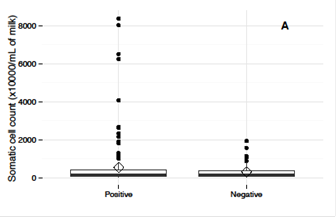
Figure 1 Somatic cell counts (SCC) in milk from cows positive for one or more etiological agent of tick fever (Babesia spp e Anaplasma marginale) when compared to negative cows.
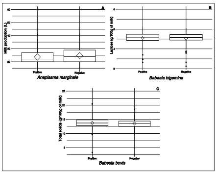
The three etiological agents of tick fever were individually evaluated regarding their effects on milk composition during the first six months of the study (Table 1, 2, and 3). A significant relation was observed between milk production and A. marginale infection, resulting on a 45% lower in this parameter (Table 3; Figure 2A). Also, positive animals for B. bigemina showed increase levels of lactose (23%, p=0.04, odds ratio 1:23, 95% CI, 1.03-1.50) (Table 1; Figure 2B), and milk total solids from B. bovis infected animals was increased by 40%, p=0.05 (odds ratio 1:40, 95% CI, 1.21-1.72) (Table 2; Figure 2C). All other evaluated parameters did not change, which may be related to the presence of babesiosis (p>0.05; Tables 1 and 2). No relation was observed between A. marginale infection and the chemical composition of milk (p>0.05, Table 3). Moreover, no significant correlation was observed when comparing the measured parameters and the milk production. Animal with single or mixed infections by agents of tick fever did not show differences on urea levels in milk (p>0.05) samples. There was also no correlation between urea levels and milk composition and quality (p>0.05).
Table 1 Effect of infection by Babesia bovis in dairy cattle over milk composition (protein, total solids, lactose and fat) and quality (SCC) compared to cows negative for this etiological agent of tick fever.
| Variable | groups | Median | Amplitude | Odds rate (CI 95%) | P |
|---|---|---|---|---|---|
| Protein | Positive | 3.51 | 5.18 | 1.37(0.32-1.48) | 0.79 |
| Negative | 3.49 | 1.63 | - | ||
| Total solids | Positive | 13.79 | 8.39 | 1.40(1.21-1.72) | 0.05 |
| Negative | 13.44 | 17.24 | - | ||
| Lactose | Positive | 4.58 | 1.93 | 0.93(0.81-4.02) | 0.30 |
| Negative | 4.73 | 8.83 | - | ||
| Fat | Positive | 4.59 | 1.80 | 1.78 (0.92-2.03) | 0.93 |
| Negative | 4.31 | 10.22 | - | ||
| Milk production | Positive | 23.5 | 19 | 1.45 (0.56-2.32) | 0.53 |
| Negative | 22 | 29 | - | ||
| SCC | Positive | 153 | 6501 | 0.99 (0.81-3.40) | 0.90 |
| Negative | 167 | 8376 | - |
TabLe 2 Effect of infection by Babesia bigemina in dairy cattle over milk composition (protein, total solids, lactose and fat) and quality (SCC) compared to cows negative for this etiological agent of tick fever.
| Variable | Groups | Median | Amplitude | Odds rate (CI 95%) | P |
|---|---|---|---|---|---|
| Protein | Positive | 3.46 | 1.60 | 1.14 (0.93-1.39) | 0.17 |
| Negative | 3.50 | 5.18 | - | ||
| Total solids | Positive | 13.71 | 17.06 | 0.80 (0.72-1.08) | 0.21 |
| Negative | 13.62 | 10.91 | - | ||
| Lactose | Positive | 4.65 | 1.65 | 1.23 (1.03-1.50) | 0.04 |
| Negative | 4.57 | 1.95 | - | ||
| Fat | Positive | 4.47 | 9.93 | 1.19 (0.98-1.35) | 0.25 |
| Negative | 4.59 | 10.88 | - | ||
| Milk production | Positive | 22 | 29 | 0.32 (0.12-3.41) | 0.31 |
| Negative | 23 | 27 | - | ||
| SCC | Positive | 172 | 4052 | 1.02 (0.75-1.39) | 0.70 |
| Negative | 153 | 8376 | - |
TabLe 3 Effect of infection by Anaplasma marginale in dairy cattle over milk composition (protein, total solids, lactose and fat) and quality (SCC) compared to cows negative for this etiological agent of tick fever.
| Variable | Groups | Median | Amplitude | Odds rate (CI 95%) | P |
|---|---|---|---|---|---|
| Protein | Positive | 3.50 | 2.24 | 0.57 (0.40-2.28) | 0.20 |
| Negative | 3.49 | 5.18 | - | ||
| Total solids | Positive | 13.62 | 8.48 | 1.00 (0.99-4.30) | 0.21 |
| Negative | 13.73 | 17.24 | - | ||
| Lactose | Positive | 4.60 | 1.80 | 0.93 (0.09-1.84) | 0.41 |
| Negative | 4.58 | 1.95 | - | ||
| Fat | Positive | 4.44 | 8.14 | 0.13(0.10-1.34) | 0.70 |
| Negative | 4.62 | 10.98 | - | ||
| Milk production | Positive | 21 | 30 | 6.31 (1.03-7.23) | 0.04 |
| Negative | 24 | 28 | - | ||
| SCC | Positive | 165 | 1911 | 0.96 (0.80-1.46) | 0.66 |
| Negative | 150 | 8376 | - |
DISCUSSION
The results show that cows infected by at least one of the etiological agents of tick fever had higher counts of somatic cells when compared to the negative animals. SCC from infected animals reached 8x10⁶ cells/mL, four times higher than non-infected cows, which shows that even subclinical infections can affect milk quality. As reported in the literature, infectious agents can influence SCC of dairy cows since the infection may stimulate an immune response and thus, increases the number of leukocytes. It should be noticed that all cells present in milk are somatic cells, including cells originated from the blood stream as leukocytes and peeling cells from the secretory epithelium (6-8), which may explain the increase in SCC observed. The same authors also stated that most leukocytes are mobilized from the bloodstream into the mammary tissue after changes in the capillary permeability. Milk from cows with intramammary infections may show increased numbers of immune cells, leading to a predomination of neutrophils, followed by macrophages and lymphocytes, while the number of epithelial cells remains unchanged 12,13. Previous study showed that bovine viral diarrhea affects SCC 8, the lack of information concerning infectious diseases motivated the present study on the relationship between milk composition and quality on dairy cattle with tick fever.
Milk from B. bigemina positive animals showed 23% increase on lactose levels, which disagrees with other authors findings who reported a decrease when mastitis and high SCC are present 14. The lactose increase evidenced by the current study was unexpected because, as mentioned above, lactose and fat contents are adversely affected in inflammatory processes, in addition to milk production reduction may be related indexes of carbohydrates consumed by animals, despite receiving the same diet, and the intake may vary between animals.
Total solids content has a tendency to decrease (3-12%) with increasing SCC, what is mainly related to mastitis cases 15. However, the assessment carried out on milk from B. bovis positive animals showed an increase only on the levels of total solids, but we have to remember that a specific infection (mastitis) have different behavior of a systemic infection, investigated in our herd.
Animals A. marginale positives produced lower amounts of milk when compared to non-infected animals, as previously observed by other authors 16,17, as well as in cases of tick fever 18. This lower milk production can be easily explained by the anemia and hyperthermia caused by the intracellular bacteria, since milk production is related to the volume of blood that passes through the mammary gland. In Argentina, it has been reported that dairy cows experimentally infected by an attenuated strain of A. marginale showed reduction of milk production, as well as fever, anorexia, adynamia, icterus, and death of the most severely affected 16. In the current study, animals were asymptomatic, but still produced lower volumes of milk.
Subclinical infection by agents of tick fever (B. bovis, B. bigemina, A. marginale) may affect milk quality, i.e. increase SCC due to stimulated immunity on infected animals. Lactose and total solids can also be increased in milk from B. bigemina and B. bovis positives cows. On the other hand, A. marginale negatively affects milk production.

