


 English (pdf)
 Article in xml format
 Article references
 Send this article by e-mail
 Cited by SciELO
  Cited by Google
  Similars in
SciELO
Similars in
SciELO  Similars in Google
 Permalink
INTRODUCTION
Pythiosis or Oomycosis is a granulomatous disease of cutaneous and subcutaneous tissue caused by Pythium insidiosum1. It is known in Colombia as "espundia equina or phycomycosis" 2-4; in Brazil, as "ferida brava, mal dos pântanos y ferida de moda" 5; in other parts of the world as "granular dermatitis, Florida leeches, Gulf Coast fungus, bursatee and hyphomycosis" 6.
Pythium insidiosum is a microorganism classified as belonging to the Stramenopila kingdom, Pseudofungi phylum, Oomycetes class, Pythiales order, Pythiaceae family and Pythium genus, so that the members of the Oomycetes class are phylogenetically distant from the kingdom of the fungi and closest to algae 7.
The geographic distribution is extensive, pythiosis has been reported in several tropical and subtropical countries, including Brazil, Venezuela, Colombia, Argentina, Costa Rica, Guatemala, Panama, Nicaragua, Haiti and the United States 7. The environmental conditions are determinant for the development of the organism in its ecosystem. Temperatures between 30 and 40 °C are necessary for the production of zoospores, in addition to the accumulation of water in flooded regions. The great majority of cases of pythiosis were observed during or after the rainy season 8.
Marcolongo-Pereira et al 9 stated that pythiosis, since there is no efficient antimycotic treatment, generates a negative economic impact due to the death or disability of the animals in most cases, since according to Cardona et al 10,11, disabling lesions are located particularly in the limbs, mouth and chest, because they are the areas of greatest exposure to the microorganism.
The lesion is characterized by the formation of serious granulomatous, granulocytic, prominent and raised ulceration with irregular edges and in the shape of a crater, the size of the lesions depends on the site and time of evolution of the infection and can measure between 12 and 50 cm in diameter, with the presence of fistulous paths or cavitations formed by the oomycete in its invasive process in the granular tissue.
In pythiosis lesions, dead cells behave as a foreign body, triggering an inflammatory response of the body in order to promote phagocytosis and allow for the subsequent repair of the tissue affected, so in the repair and healing process there are necrotic masses and calcifications of a white-yellowish color containing hyphae and eosinophil infiltrates, which dimensions vary from 2 to 10 mm in diameter, called "kunkers", which, together with the presence of fistulous trajectories and fibrinosanguinolent discharges, are unequivocal signs of pythiosis 3.
There are new therapies available every day to eliminate the etiologic agent of pythiosis, improve tissue quality and promote tissue repair. Despite the use of antimycotics as a treatment option, there are few studies reporting favorable results and do not show results with convincing evidence of the efficacy of the treatment in cutaneous granuloma due to pythiosis 7.
Triamcinolone acetonide has been used in the therapy of joint, respiratory and ocular diseases in horses; however, it has not been used in the treatment of dermopathies such as equine cutaneous pythiosis. The purpose of this study is to clinically and macroscopically characterize the healing process of cutaneous pythiosis in horses treated with triamcinolone acetonide.
MATERIALS AND METHODS
Ethical aspects. For the accomplishment of this descriptive, non-probabilistic study, animals of convenience were naturally infected with cutaneous pythiosis. The study was approved by the Ethics Commission on Animal Use (CEUA) of the Federal University of Viçosa (UFV), process No. 74/2012. The animals were not subjected to processes that developed unnecessary pain and/or stress. The animals were immobilized taking into account the technical standards of animal handling and restraint, in compliance with the Universal Declaration of the Rights of Animals, referring to the international ethical principles for biomedical animal research CIOMS (Council for International Organizations of Medical Sciences) established by the UNESCO (United Nations Educational, Scientific and Cultural Organization) and WHO (World Health Organization) of 1949 and Law 84 of October 27, 1989 (Colombian Code for Animal Protection) 12.
Location. The experiment was carried out on the Caribbean coast of Colombia, in the department of Cordoba, located at the coordinates 7° 23' 'and 9° 26' north latitude and 74° 52' and 76° 32' west longitude of the Greenwich meridian, at an altitude of 30 meters above sea level, with an annual average temperature of 28°C, relative humidity of 82%, annual rainfall of 1.400 mm and a tropical forest climate. There are two well-defined seasons (rainy season and dry season) 13.
Animals. Twenty-four horses with cutaneous pythiosis of both sexes, different ages and an average weight of 380 kg were used. The inclusion criterion was based on the presence of clinical cutaneous pythiosis, diagnosed by the general physical examination performed during the selection process and confirmed by histopathology in the staining of Hematoxylin Eosin (HE) and Grocott (GSM).
Methodology. Animals were fed with grass and water ad libitum. Animals were divided into two groups. One group received intramuscular treatment with 50 mg of triamcinolone acetonide every 15 days until completing three applications (treated group - TG). The second group did not undergo any treatment (control group - CG).
All animals (TG and CG) underwent clinical evaluation and macroscopic characterization of the lesion every four days until the complete recovery of the granuloma in the TG. The reduction of the fibrinosanguinolent exudate, pruritus, presence of fistulous trajectories and exit of kunkers was verified in that evaluation; as well as the reduction of the granuloma, leveling with healthy skin and the formation of scabs.
The clinical and macroscopic characteristics of the lesions were classified from 0 to 4 taking into account the degree of clinical expression (Table 1). In addition, the animals with lesion in the distal part of the members were subject to radiological projections to evaluate the degree of bone and tissue fibrosis involvement. High-definition photographs were taken for further analysis (Sony DSC-HX10V (r) , China).
Table 1 Clinical characteristics of granulomatous lesions in horses with pythiosis subject or not to treatment with triamcinolone acetonide according to the time of the study.
| Day | Degree* | Treated Group | Degree *2 | Control Group |
|---|---|---|---|---|
| 0 | 4 | Marked fibrinosanguinolent exudation intense pruritus Large number of fistulous trajectories Marked presence of Kunkers Exuberant ulcerative lesion overlying skin | 4 | Marked fibrinosanguinolent exudation intense pruritus Large number of fistulous trajectories Marked presence of Kunkers Exuberant ulcerative lesion overlying skin |
| 16 | 3 | Absence or mild fibrinosanguinolent exudation No pruritus Moderate number of fistulous trajectories Moderate presence of Kunkers Less prominent skin lesion Moderate formation of scabs at the edges of the wound Contraction of the lesion by 64.7% | 4 | Marked fibrinosanguinolent exudation intense pruritus Large number of fistulous trajectories Marked presence of Kunkers Exuberant ulcerative lesion overlying skin |
| 32 | 2 | Slight presence of Kunkers and fistulous trajectories Marked decrease in granuloma and leveling with normal skin Increased formation of scabs covering the wound Contraction of the lesion by 86.3%. | 4 | Marked fibrinosanguinolent exudation intense pruritus Large number of fistulous trajectories Marked presence of Kunkers Exuberant ulcerative lesion overlying skin |
| 48 | 1 | Formation of scabs covering the entire wound Contraction of the lesion by 97.3% | 4 | Marked fibrinosanguinolent exudation intense pruritus Large number of fistulous trajectories Marked presence of Kunkers Exuberant ulcerative lesion overlying skin Increased wound size Presence of pus |
| 60 | 0 | Absence of clinical manifestations Contraction of the lesion by 100% | 4 | Marked fibrinosanguinolent exudation intense pruritus Large number of fistulous trajectories Marked presence of Kunkers Exuberant ulcerative lesion overlying skin Increased wound size Presence of pus Mutilation |
| * Degrees from 0 to 4 of lesions, where 4 is the highest expression of the disease and 0 is the total recovery of the lesion and absence of clinical signs. Source: authors |
Wound retraction was assessed from day zero until the complete closure of the granuloma by measuring the area in cm2, contoured with a permanent marker on a transparent plastic sheet of an overhead projector, to be then digitized, and the area was determined with the use of the BioEstat 5.0 software 14.
Descriptive analyzes were performed for the macroscopic evaluation of the wound (wound retraction, reduction of fibrinosanguinolent exudate, reduction of pruritus and scab formation). The variance analysis (ANOVA) was used for the comparison of the data generated for comparisons between two or more means. The ANOVA was used to verify if there were any significant differences between the two groups (CG and TG) over time. The minimum level of statistical significance used in all tests applied was 95% (p≤0.05). All statistical tests were performed with the help of the computer program S.A.S. 9.1.3. 15.
RESULTS
The clinical evaluation and the anatomopathological characterization of the lesions in the horses of the study, together with the histopathological results, confirmed the diagnosis of cutaneous pythiosis according to what was reported as a diagnostic method for equine pythiosis 3,5,10,16-19.
All the animals in the study (CG and TT) showed the five clinical manifestations characteristic of the disease, according to Cardona et al 2-4, such as granulomatous ulceration with irregular surface, fibrinosanguinolent exudate, pruritus, fistulous trajectories with exit of purulent and caseific necrotic material called "Kunkers" (Figure 1).

Figure 1 Clinical characterization of pythiosis on horses under study. a.) Granulomatous ulceration, prominent and elevated with irregular and crater-like edges. b.) Outflow of water from the fistulas formed by the oomycete in its invasive process in the granular tissue. c.) Fibrinosanguinolent secretion of the granulomatous lesion. D.) Exit of Kunkers or necrotic masses and calcifications that easily detach with white-yellowish coloration (black arrows).
The clinical and macroscopic evaluation of the lesion and its clinical evolution (wound contraction, presence of fibrinosanguinolent exudate, pruritus, fistulous trajectories, exit of Kunkers, and scab formation) can be observed in table 2. It was found that all TG animals showed a marked decrease in clinical characteristics, with the last observation of pruritus at 11.83±1.47, exit of Kunkers at 13.33±1.03, fibrionosanguinolent secretion at 14.2±1.47 and presence of fistulous trails at 14.83±1.33; similarly, at 16±1.4 days after treatment, clinical signs were absent and the formation of scabs in the lesion began (Figures 2 and 3). None of the CG animals showed any improvement in the clinical characteristics of pythiosis and remained the same throughout the study.
Table 2 Graduation of the clinical characteristics of the granulomatous lesion in horses with pythiosis subject or not to treatment with triamcinolone acetonide according to the time of experiment.
| Time (days) | |||||||||||||
|---|---|---|---|---|---|---|---|---|---|---|---|---|---|
| Lesion (Degree ± DE) | D0 | D4 | D8 | D12 | D16 | D20 | |||||||
| GC | GT | GC | GT | GC | GT | GC | GT | GC | GT | GC | GT | ||
| Exit of kunkers | 4.0±0.0 | 4.0±0.0 | 4.0±0.0 | 3.5±0.2 | 4.0±0.0 | 1.7±0.5 | 4.0±0.0 | 1.0±0.0 | 4.0±0.0 | 0.0±0.0 | 4.0±0.0 | 0.0±0.0 | |
| Fistulous trajectories | 4.0±0.0 | 4.0±0.0 | 4.0±0.0 | 2.8±0.4 | 4.0±0.0 | 2.3±0.5 | 4.0±0.0 | 1.3±0.5 | 4.0±0.0 | 0.5±0.5 | 4.0±0.0 | 0.2±0.4 | |
| Secretion | 4.0±0.0 | 4.0±0.0 | 4.0±0.0 | 3.3±0.3 | 4.0±0.0 | 2.2±0.3 | 4.0±0.0 | 1.2±0.4 | 4.0±0.0 | 0.3±0.5 | 4.0±0.0 | 0.3±0.5 | |
| Pruritus | 4.0±0.0 | 4.0±0.0 | 4.0±0.0 | 3.0±0.0 | 4.0±0.0 | 1.7±0.5 | 4.0±0.0 | 0.7±0.5 | 4.0±0.0 | 0.0±0.0 | 4.0±0.0 | 0.0±0.0 | |
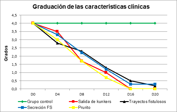
Figure 2 Proportion of the clinical characteristics of the granulomatous lesion in horses with pythiosis subject or not to treatment with triamcinolone acetonide according to the time of experiment.
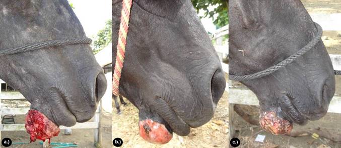
Figure 3 Clinical and macroscopic characterization of the lesion. a.) Decreased fibrinosanguinolent secretion at 4 days after treatment. b.) Decreased pruritus and onset of scab formation at 16 days after treatment. Note the absence of mutilation injuries. c.) Complete scab formation at 20 days after application of triamcinolone acetonide.
Table 3 and figure 4 describe the distribution of mean values and standard deviation of the area (cm2) and percentage of contraction of the granulomatous lesion in horses with pythiosis subject or not to treatment with triamcinolone acetonide according to the time of the experiment. It was observed that all TG granulomas recovered in 100%. So there was a significant reduction in the granuloma area at 16 days of 64.7% (p<0.0032), at 32 days of 86.3% (p<0.0004), at 48 days of 97.3% (p<0.0001) and completely closed at 60±3.4 days (p<0.0001) after the application of the treatment (Figure 5 and 6), as all the wounds of CG animals showed an increase of 1.9% in the area and a contraction percentage of 0,0%. The Student's t test was used to determine highly significant differences (p=0.0001≤0.05) between the two groups (TG and CF). Considering as a response variable to the reduction in the affected area in a period of 60±3.4 days in the treated group.
Table 3 Distribution of mean values and standard deviation of the area (cm2), contraction percentage of granulomatous lesion and significance level in horses with pythiosis subject or not to treatment with triamcinolone acetonide according to the time of experiment.
| Time (days) | Application of Medicine | TG Area (cm2±DE) | Contraction of the area (%) | Significance Level | ||||
|---|---|---|---|---|---|---|---|---|
| GT | GC | GT | GC | GT | GC | P- Valor | ||
| 0 | 1 dose AT | S/n saline | 95.2±55.1 | 93.4±54.8 | 0.0 | 0.0 | 0.9336 | |
| 4 | --- | --- | 60.5±32.5 | 93.4±54.8 | 36.4 | 0.0 | 0.0875 | |
| 8 | --- | 49.1±29.3 | 93.5±54.8 | 48.4 | 0.0 | 0.0242 | ||
| 12 | --- | --- | 40.2±22.6 | 93.6±54.7 | 57.8 | 0.0 | 0.0070 | |
| 16 | 2 dose AT | S/n saline | 33.6±20.7 | 93.7±54.8 | 64.7 | 0.0 | 0.0032 | |
| 20 | --- | --- | 26.9±13.8 | 93.8±54.8 | 71.7 | 0.0 | 0.0015 | |
| 24 | --- | --- | 21.2±11.0 | 93.9±54.8 | 77.7 | 0.0 | 0.0007 | |
| 28 | --- | --- | 16.0±8.1 | 94±54.8 | 83.2 | 0.0 | 0.0005 | |
| 32 | 3 dose AT | S/n saline | 13.0±7.1 | 94.1±54.8 | 86.3 | 0.0 | 0.0004 | |
| 36 | --- | --- | 9.5±5.1 | 94.2±54.9 | 90.0 | 0.0 | 0.0002 | |
| 40 | --- | --- | 6.7±3.7 | 94.4± 55.0 | 93.0 | 0.0 | 0.0002 | |
| 44 | --- | --- | 4.4±2.7 | 94.6±55.0 | 95.4 | 0.0 | 0.0001 | |
| 48 | --- | --- | 2.6±1.8 | 94.7±55.0 | 97.3 | 0.0 | 0.0001 | |
| 52 | --- | --- | 1.2±1.1 | 94.8±55.0 | 98.7 | 0.0 | 0.0001 | |
| 56 | --- | --- | 0.4±0.5 | 95.0±55.0 | 99.6 | 0.0 | 0.0001 | |
| 60 | --- | --- | 0.0±0.0 | 95.2±55.1 | 100 | 0.0 | 0.0001 | |
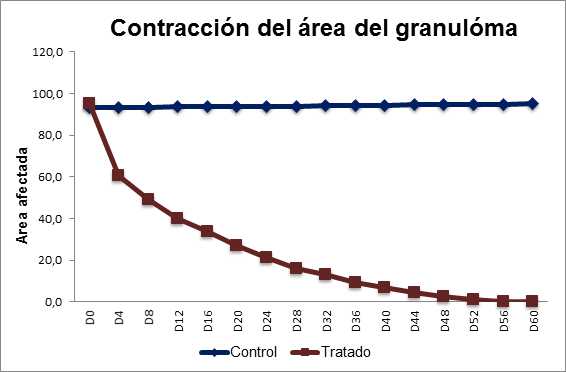
Figure 4 Mean contraction of the area of the granulomatous lesion in horses with pythiosis subject or not to treatment with triamcinolone acetonide according to the time of experiment.
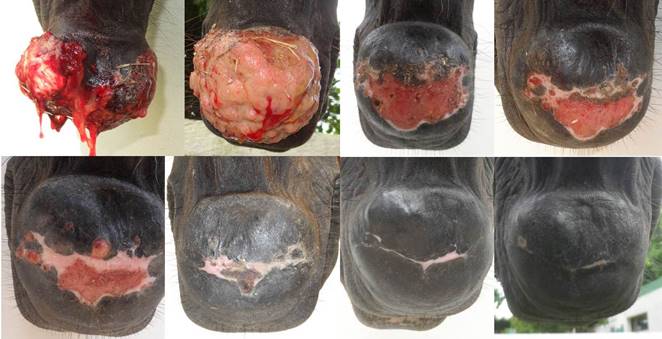
Figure 5 Contraction of the granuloma area in the TG at the chin. Note the evolution of wound contraction over a period of eight days until completely closed at 60±3.4 days after application of treatment.
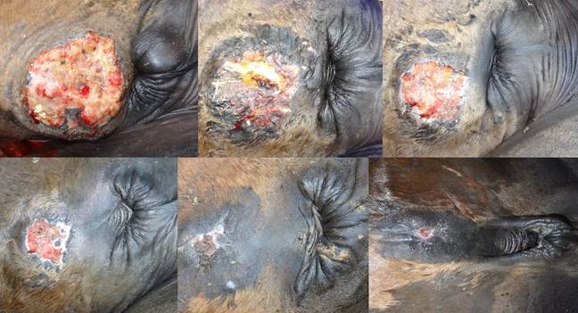
Figure 6 Contraction of the granuloma area in the TG at the abdomen. Note the evolution of wound contraction in a 10-day interval until completely closed at 60±3.4 days after application of treatment.
Regarding the radiological follow-up of chronic cutaneous lesions located in the distal parts of 3 animals in the study, the dorsum-palm and lateral-medial projections showed the development or invasion of the underlying tissues compromising bone structures, which were characterized by tissue fibrosis, moderate exostosis, as well as by osteolysis and osteomyelitis in some areas, in agreement with Zaro 20, who reported irregularities in the bone surface with severe erosions and periosteal reactions in chronic pythiosis cases in horses.
After treatment with triamcinolone acetonide, moderate radioactivity and presence of enthesophytes was observed at day 16 in the cortex of the bone. At day 32 the decrease in radio-opacity areas was more marked, and at day 64 radio-opacity was absent in the tissue, although small focal areas of radio-opacity were evident in the cortex of the bone, indicating a decrease in tissue fibrosis and the presence of exostoses as sequelae of the chronicity (Figure 7).
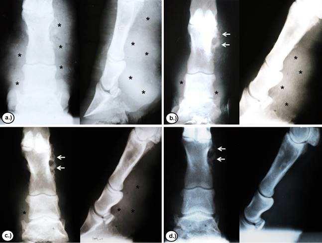
Figure 7 Radiological evaluation of the evolution of chronic skin lesions located in the distal parts of a TG animal after treatment with triamcinolone acetonide. a.) 0 days. b.) 16 days. c.) 32 days. d.) 64 days. Note the reduction in radiopaque areas in the tissue (asterisks) and the preservation of small radiopaque areas in the cortex of the bone (arrows) according to the time of experiment.
DISCUSSION
The use of triamcinolone acetonide in horses has not been reported in the treatment of dermatological diseases in horses and even less in the specific case of equine cutaneous pythiosis. For this reason this study constitutes the first report in the treatment of pythiosis with triamcinolone acetonide.
Pythiosis is undoubtedly a disease that will not be repaired by first intention, by the infectious process and the granulomatous nature of the lesion, so it will always be more aggressive and proliferative, in which case second intention healing is required due to excessive loss or severe tissue involvement, presence of kunkers, fistulous trajectories and excessive granulation tissue, requiring the administration of a medical treatment to aid in the control of the oomycete infection and to promote tissue repair 3.
Much has been studied on the treatment of equine cutaneous pythiosis, from traditional antifungals, state-of-the-art antimycotics, antibiotic therapy, surgery and immunotherapy; however, none of the treatments studied have given satisfactory results in terms of producing a complete solution for the disease 5,17,21,22.
The results obtained in this study were satisfactory with 100% clinical recovery at 60±3.4 days after treatment with triamcinolone acetonide in TG, mainly in areas of anatomical compromise where surgery was impossible. The decrease in clinical manifestations is due to the elimination of the injurious agent, which decreases the antigenic stimulus and, hence the large amount of eosinophils, which is used by the Oomycete as a defense mechanism and are the cause of the granulomatous process, pruritus and exudation; kunkers also disappear allowing for the progressive recovery of the injured tissue.
Frey et al 23 reported a 50% recovery using the Pitium-Vac vaccine associated with the curettage of the necrotic material of the lesions or surgery. Similarly, Pereira et al 21 tested in vitro carposfungin with an action of 63%; however, when tested in vivo, it showed a reduction in lesions, but when the treatment ceased the growth of the lesions restarted. On the other hand, Cavalheiro et al 22 and Argenta 24 studied state-of-the-art in vitro antifungals in combination with voriconazole with terbinafine and itraconazole with terbinafine, showing synergism against 17% of the samples from that study. Biava et al 25 reported unsatisfactory results and a recovery of less than 25% in studies where amphotericin B, 10% sodium and potassium iodide, flurocytosine, ketoconazole, and non-surgery were tested, as well as systemic amphotericin B combined with gauze pads embedded in solutions of amphotericin B and dimethyl sulfoxide (DMSO).
The results obtained with antifungal drugs are controversial, showing unsatisfactory results due to the inability of P. insidiosum to produce steroids in the plasma membrane, such as ergosterol, which is the albo-active component of most antifungal drugs 7. State-of-the-art antimycotics such as carposfungin (an inhibitor of β-glucan synthesis) showed a reduction in lesions, but when the treatment was stopped the growth of the lesions restarted 21; combinations of itraconazole and terbinafine or voriconazole and terbinafine showed some degree of success in comparison with isolates of P. insidiosum22,24. Likewise, Loreto et al 26 reported the in vitro susceptibility of P. insidiosum to macrolide antibiotics and tetracyclines by reducing the incorporation of amino acids into proteins.
Gaastra et al 27 recommend surgery as a treatment for the removal of the entire affected area and a safety margin to avoid relapses. However, in many cases it is difficult because of the degree of involvement of the anatomical structures affected, mainly in the limbs, so they also suggest to use the combination with immunotherapy, potassium iodide, state-of-the-art antimycotics and antimicrobials as the most suitable treatment for the cure of clinical pythiosis in horses.
The action mechanism of triamcinolone acetonide in the case of equine cutaneous pythiosis is still uncertain, although corticosteroids have a variety of metabolic, endocrine and immunological functions. Clares 28 and Morato 29 stated that the mechanism of the anti-inflammatory and antiallergic activity of glucocorticoid-type corticosteroids is the induction of phospholipase A2 inhibitory proteins, collectively known as lipocortins. These proteins are postulated to control potent mediators of inflammation, such as prostaglandins, thromboxanes, prostacyclins, leukotrienes, and hydroxycyatriatrienoic acids, all synthesized from arachidonic acid, which discharge is catalyzed by the enzyme phospholipase A2. As a result of this action, they will have anti-inflammatory effects, such as delayed migration of nuclear polymorphous leukocytes, decreased fibrinogenesis, decreased C-reactive protein production, suppression of mast cell degranulation and immunosuppressive effects, including the suppression of phagocytosis processes and the reduction in the number of eosinophils and lymphocytes.
A possible pharmacological explanation for the recovery of horses with pythiosis treated with triamcinolone acetonide is the mechanism of the glucocorticoid immuno-modulating activity, since it inhibits the synthesis, release and/or action of cytokines and other mediators that promote the inflammatory or immune response. These molecules include proinflammatory cytokines, such as IL-1, IL-6 and alpha tumor necrosis factor (TNF-α), clonal expansion and IL-2, subset of auxiliary T lymphocytes (Th1), IL-12 and gamma interferon (IFN-γ) and, to a lesser extent, the Th2, IL-4 and IL-5 subset, colony-stimulating factors such as granulocyte and macrophage (GM-CSF), chemokines such as RANTES and MIP-1α, adhesion molecules such as ICAM1, ELAM-1 and E-selectin, inflammatory mediators such as bradykinin, histamine, eicosanoids and nitric oxide, as well as molecules involved in the presentation of antigen and class II major histocompatibility complex (MHC). The entire spectrum of molecular albs explains pleiotropy and the anti-inflammatory and immunomodulatory force. This is why blocking the synthesis of IL-5 cytokine and granulocyte-macrophage colony-stimulating factors (GM-CSF) triggers the programmed death of these cells (apoptosis), which decreases the half-life of eosinophil, since cytokine (IL-5) is responsible for eosinophilopoiesis, increasing the function of mature eosinophils as well as degranulation, adhesion and cytotoxicity, thus prolonging the survival of that cell 30.
According to the results obtained in this study, it can be concluded that triamcinolone acetonide was effective in the treatment of granulomatous wounds by pythiosis, showing clinical recovery in all animals treated. Therefore, the use of triamcinolone acetonide can be recommended as a suitable therapeutic alternative in the treatment of equine cutaneous pythiosis. Although there are many factors that affect the action of drugs in diseased animals, such as pH, vascularization, fibrosis, idiosyncrasy, species variations, tolerance, sensitivity, nutritional status and body condition of the animal, it is necessary to perform further field trials in naturally infected animals considering triamcinolone acetonide alone or in combination with other therapeutic options.

