


 English (pdf)
 Article in xml format
 Article references
 Send this article by e-mail
 Cited by SciELO
  Cited by Google
  Similars in
SciELO
Similars in
SciELO  Similars in Google
 Permalink
INTRODUCTION
Lymphoma is the most common lymphoproliferative disorder in cats, representing 50 to 90% of all haematopoietic neoplasias. However, cutaneous lymphoma is rare, occurring in 0.2 to 1.7% of cats and corresponding to only 4.5% of all extranodal lymphomas 1.
Clinically, the lesions can be solitary, multifocal or diffuse and are often described as alopecic plaques with the presence of erythema, crust or papules 2,3. Feline cutaneous tarsal lymphoma (FCTL) corresponds to an uncommon manifestation presented as a subcutaneous mass located in the tarsal region 4.
Feline cutaneous lymphoma has been described as non-epitheliotropic and predominantly of T-cell origin; however, descriptions of FCTL suggest that this form is non-epitheliotropic, high-grade and of B-cell origin in the majority of cases 2-4. Burr et al 4 reported the average survival of FCTL to be 6.3 months and only 4% to be above 1 year, highlighting the aggressiveness of this disorder.
The aim of this study was to report the first case of FCTL in Brazil and to describe the clinical signs, diagnostic procedure and therapeutic effect using lomustine and prednisolone of this rare and aggressive form of cutaneous lymphoma in cats.
PATIENT EXAMINATION
Anamnesis. A 13-year-old, sexually intact, male, Domestic Longhair cat was admitted with a subcutaneous mass of 30 days progression at the tarsal region of its right hind limb. Pain was evident at palpation and the owner reported claudication, sporadic vomiting and weight loss. Previous cytology analysis suggested round cell neoplasia.
Clinical findings. The mass was adhered, soft, alopecic and ulcerated, measuring approx. 5 x 4 cm (Figure 1). An enlargement of the right popliteal lymph node and low body condition score (3/9) were also observed. Physiological values were within normal parameters for the species.
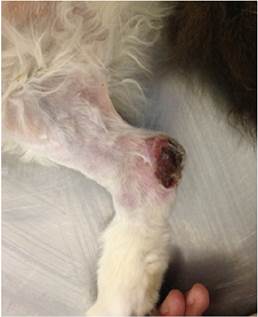
Figure 1 Medial aspect of right hind limb of a 13-years-old, male, Domestic Longhair cat with FCTL. A subcutaneous and ulcerated mass is located in the tarsus.
Diagnostics aids used. For systemic evaluation and tumor staging we performed haemogram, serum biochemical profile (creatinine, alanine aminotransferase [ALT], alkaline phosphatase), urinalysis and imaging (thoracic radiography in three views, laterolateral and craniocaudal radiography of the affected member and abdominal ultrasonography). No abnormalities were observed in the analyses. However, abdominal ultrasound revealed increased medial iliac lymph nodes, loss of typical morphology and rounded shape. Furthermore, mixed echogenicity, increase in vascularization and irregular blood flow distribution in the parenchyma were detected on Doppler ultrasound scan.
Cytology of the tarsal mass and popliteal lymph node were performed. The results indicated a population of undifferentiated round cells with malignant characteristics. Mast cell tumor, cutaneous lymphoma and cutaneous plasmacytoma were suggested as differential diagnosis. Incisional biopsy of the mass was then performed. Histopathology report was compatible with malignant neoplasia of round cells, morphologically suggestive of large cell lymphoma. Antibodies for CD3, CD79a, MUM1, PAX5 and lysozyme were used for immunohistochemistry. A positive expression of CD79A and MUM1 and a negative expression of PAX5, CD3 and lysozyme concluded the diagnosis of B-immunoblastic lymphoma.
Treatment focus. Antineoplastic treatment was instituted using lomustine (50 mg/m² PO) every 3 weeks with prednisolone at initial dose of 2 mg/kg SID with weekly reduction until 0.5 mg/kg per dose. We added tramadol (2 mg/kg PO BID) during the whole treatment. By the second round of chemotherapy, the disease was stable; however, the patient was apathetic, pallid, with persistent vomiting episodes and progressive weight loss. On physical exam, popliteal and inguinal lymph nodes were in the same size and abdominal ultrasound showed that medial iliac lymph nodes remained the same size as on the previous scan.
The haemogram showed signs of anaemia, with haematocrit of 13%, haemoglobin 4.5 g/dL, red cell count 2810 x10³/μL, white cell count 6500/μL (neutrophils 4500/μL) and thrombocytopenia (127 x10³/μL). The percentage of reticulocytes was 9% and anisocytosis was evident in the blood smear, thus classifying it as regenerative anaemia. Serum creatinine and ALT were within the normal range for the species.
Due to these results, lomustine treatment was delayed and anaemia was suspected to be associated with Mycoplasma haemofelis infection. Although PCR for mycoplasma was suggested to confirm diagnosis, the owner deny doing it. Doxycycline at 10mg/kg SID was given with prednisolone at 2 mg/kg PO SID.
After seven days, the haemogram showed improvement of the anaemia, with haemoglobin levels at 6.7 g/dL, haematocrit 20.5% and platelet count 147 x10³/uL. Clinically the patient was apathetic, lethargic and physical examination revealed an increased size of the mass and popliteal lymph node by 57% and 33%, respectively (Figure 2). The disease was classified as progressive and lomustine was re-established. However, the animal died at home six days later. Survival time was 2,1 months (63 days) Necropsy was not performed because of owner preference.
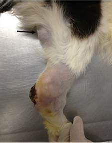
Figure 2 Lateral aspect of the right hind limb of a 13-years-old, male, Domestic Longhair cat with FCTL. Note the larger size of the subcutaneous tarsal mass compared with figure 1 and a prominent right popliteal lymph node (arrow).
DISCUSSION
The patient from this report presented an ulcerated subcutaneous mass at the tarsal region, which after histopathology and immunohistochemistry analysis was diagnosed as B-immunoblastic lymphoma. These findings are in agreement with the study by Burr et al 4, in which the majority of FCTL was classified as high-grade and of B-cell origin. The clinical characteristics observed in this study such as breed, age, evolution time and staging are similar to those described by these authors. The high-grade immunoblastic lymphoma was classified according to the National Cancer Institute working formulation (NCI WF) and, according to Valli et al 5; it is the histopathological type most frequently found in proliferative disorders in cats.
Immunohistochemistry was positive for CD79A and MUM1, and negative for PAX5, CD3 and lysozyme. CD79a and MUM1 are normally expressed by B-lymphocytes, while CD3 is typically expressed by T-lymphocytes 6,7. MUM1 expression has been evident in lymphoid differentiation, thus being mainly immunoreactive in plasma cells as well as B-lymphocytes 8. Therefore, CD3- associated with CD79a+ and MUM1+ confirmed the B-cell immunophenotype. PAX5 is also expressed by B-cells but negative immunolabeling in feline lymphomas has yet to be studied. In humans, PAX5 expression in Hodgkin lymphoma is used to differentiate the classic from the atypical form of this disease, which lacks PAX5 expression and has a less favourable prognosis than the classic form 9,10. Although it can be hypothesised that the lack of PAX5 expression could be a prognostic factor in feline lymphoma, further studies are necessary to support this assumption.
Data on cutaneous lymphoma immunophenotype in cats are scarce. The few studies available suggest the T immunophenotype to be the most common 3. However, the clinical presentation of feline T-cell cutaneous lymphoma is mainly as erythematous plaques, alopecia, ulcerative plaques and desquamation, 3 in contrast to the B-cell cutaneous lymphoma of this study, which it was presented as a subcutaneous ulcerated mass at the tarsal region.
In humans, three main subtypes of primary cutaneous B-cell lymphomas (PCBCL) have been described based on their cellular morphology. The primary cutaneous marginal zone lymphoma and the primary cutaneous follicular centre lymphoma are considered indolent. However, primary cutaneous large B-cell lymphoma, leg type (PCLBCL-LT) is a less common, but aggressive form that occurs in the distal parts of the pelvic limbs 11. The cellular characteristics of PCLBCL-LT and the areas affected are similar to those described in this report. Furthermore, in both humans and cats, this type of lymphoma is aggressive and has a poor prognosis 12.
Patient was classified as stage II based on clinical staging system for lymphoma in cats 7. In ultrasound images, the medial iliac lymph nodes showed characteristics of malignant metastatic lymph nodes such as enlargement, round shape, mixed echogenicity, increased vascularization and irregular blood flow distribution 13. Because of the patient staging, treatment was based on systemic chemotherapy.
During treatment, the coexistence of Mycoplasma haemofelis was suspected. Although the disease was not confirmed due to the owner’s objection, the severe and strongly regenerative anaemia (9% reticulocytes and anisocytosis) characterizes cats infected by this agent 14. These facts, associated with the prevalence of this disease in the region 15 and the favourable response observed after treatment with doxycycline and prednisolone, was suggestive of haemoparasitic infection by Mycoplasma haemofelis. Serologic tests for FeLV (Feline Leukemia Virus) and FIV (Feline Immunodeficiency Virus) status were not performed in this patient. Previous results highlighted the lack of association between FeLV status and cutaneous lymphoma in cats 3. Apparently, younger cats are more affected by FeLV associated lymphoma than adult cats 1.
Little information on the treatment of cutaneous lymphoma in cats has been reported 7 and no standard treatment protocol exists for this disease. Local control of the disease by surgical excision or radiotherapy could be more beneficial in selected patients when the disease remains locally confined, however most of the patients in one study, showed recurrence or metastatic disease that finally needed chemotherapy 4. It should be highlighted that in the same study, a small population of cats (n=23) were studied, so further studies need to be done before having any treatment conclusion.
The chemotherapeutic protocol prescribed to the patient in this study was based on the combination of lomustine and prednisolone. Lomustine is recommended in cases of cutaneous lymphoma in cats 3 and Komori et al 2 reported a complete remission in a cat with T-cell cutaneous lymphoma after treatment. However, Dutelle et al 16 found variable results when used as a rescue agent for lymphoma in cats, independently of the anatomic affected region, authors observed better responses for small rather than large cell lymphoma. This report are in agreement with the lack of response observed in the large cell lymphoma in this study.
The mean survival time (MST) reported for cutaneous lymphoma varies significantly, with T-cell cutaneous lymphoma patients surviving longer than those classified as B-cell. Fontaine et al 3 reported MST of 10.25 months for T-cell cutaneous lymphoma; however, it is important to note that that lymphoma consisted mainly of small or intermediary cells. On the other hand, Burr et al 4 reported the MST for large B-cell cutaneous lymphoma to be 4.5 months in patients that exclusively received chemotherapy. These results differ from this report, in which the survival time observed was 2.1 months using only chemotherapy. It is possible that the poor survival time in this patient was associated with the advanced clinical stage of the disease at the time of diagnosis. Burr et al 4 explained this by reporting a significantly lower MST in patients with B-cell cutaneous lymphoma that were treated exclusively with chemotherapy (4.5 months) when compared to surgery ± chemotherapy (13.6 months). This highlights the importance of early diagnosis at stage I, in which case, surgical removal associated with chemotherapy have longer survival time.
We concluded that antineoplastic chemotherapy with lomustine was not effective in this case of FCTL due to disease progression. This reinforces the need for larger studies to understand better the course of this disease, the role of immunohistochemistry as a prognostic factor and to develop different chemotherapy protocols aimed at increasing the survival time as well as improving the quality of life of affected cats.

