


 Inglés (pdf)
 Articulo en XML
 Referencias del artículo
 Enviar articulo por email
 Citado por SciELO
  Citado por Google
  Similares en
SciELO
Similares en
SciELO  Similares en Google
 Permalink
INTRODUCTION
EM is an economical problem for the poultry industry. In the early twentieth century, when flocks were smaller, the EM average was not above 10%. However, in the 1950s when primitive methods were replaced by modern practices of intensive production, which included confinement reproduction and artificial hatching, EM would frequently rised up to 25% or even more 1. Currently, the development of specialized breeders, its selection to ideal parameters and the technology applied into incubators have led to the establishment of limits for each strain, under which the capacity of hatching has been tested by ideal incubation conditions.
EM in chickens is not evenly distributed during the 21 days of incubation and it has been established, through mathematical models, that mortality curves present two peaks during the different phases of development, which occur between the first and third week, and are mainly related to changes in embryo metabolism 2. For this reason, EM was divided into early, intermediate and late periods.
The early EM (EEM) happens during the first week of incubation 3. It is especially high between day 3 and 5. Presumably, it is due to CO2, accumulation or to hydronephrosis 1; the latter, as a result of mechanical obstruction when the mesonephros starts working 3. Romanoff, in 1949, mentioned some other possible causes for EEM such as respiratory maladjustments before the establishment of special respiratory surfaces (area vasculosa and chorioallantois) that appear during the first three or four days of incubation. On the other hand, carbohidrates are the main source of energy for the embryo and probably enough CO2 is accumulated up to fatal levels. Also, there is a maximum concentration of lactic acid at a time when the enzyme decomposing this molecule, is also present in small quantities. Additionally, it was reported that nitrogen in the early stage of embryonic growth is excreted as ammonium, which is highly toxic for the embryo 1.
During the second week of incubation, intermediate EM (IEM) occurs and it is usually low. In quails raised in captivity, it has been related to a deficient diet in animal protein, minerals (especially calcium) or vitamins. One study showed that IEM increased when the breeder only depends on a diet based on vegetables, or during the winter when animals could not obtain enough sunlight or vitamin D. Also, vitamin E and riboflavin deficiencies may rise mortality rate during this period 1.
Late EM (LEM) occurs during the third week of incubation and raises toward the 19th day 1, when the embryonic oxygen requirements are highest 3. It may be caused by a failure in the transition from allantoic to pulmonary respiration. By the end of the developmental period, the cumulative effects from adverse conditions of incubation may result in the embryo poor viability and abnormal changes in the embryo fluids become crucial 1.
One aspect that contributes to EM variability is the flock´s age 4. Chickens, particularly from Cobb and Ross strains, get into the egg production phase around the 25th week and it is expected that by the 64th week they reach the productive levels under ideal conditions established in industrialized countries. However, it is clear that production characteristics change due to the flock age, for as time goes by, the expected EM values change 5.
For example, it is known that eggs from old flocks (compared to eggs from younger ones) are more sensible to heat increment in the nest, to long storage, to any inadecuate preincubation treatment and even, because of the lack of enough egg´s turning during incubation, possibly because of differences in the quality of albumen, yolk or shell 6.
In Colombia, the egg producing industry is highly important and it is believed that evaluation of given especially from Santander, to characterize the EM behaviour from commercial strains under the conditions of this region will provide indications which until now had only been studied in other places in the world. Present findings might aid in future plans as to increase productivity in industrial operations.
The present work was aimed to evaluate EM under controlled conditions in commercial industries.
METHODS AND MATERIALS
Population and study design. Animals were received being 1 day-old and remained in the same farm until slaughtering. The breeding of all flocks was according to the reccomendations provided by the company Ross breeders. Nutrition and vaccination plan is standarized for all farms following the requirements by the zone. Besides, a common biosecurity plan is executed by all the farms.
Every egg is picked up, classified, disinfected and transported the same day into the incubator. Later, it is stored at a temperature which ranges from 19 to 20°C and remains there according to incubation schedule and incubators availability. Eggs are removed from the cold room twelve hours before entering an incubator and they are left at room temperature; then they are incubated into the machines of multiple stages and automatic turning each hour. The temperature for the incubators is 37.5°C and 50% of relative humidity. Eggs remain in the incubators until the 18th day. From there, they are transferred to the hatcheries, to a temperature of 36.9°C.
Research was designed as a study of prospective cohort. It was performed following four groups of hatching eggs from broiler breeder (Ross 308) at 28, 40, 47 and 64 weeks of age. For each group a size of sample was calculated according to the mortality incidence established for every age in the guide of the strain. Taking these parameters into account, a sample was given for each group, in Epidat 4.1, in the sample for a proportion module, with a precision of 3%, an alpha error of 0.05% and a design effect of 2, so that the total EM proportion may be estimated. To this number as result the percentage of infertility correspondent was added to every age value, and the correspondent result, divided by the amount of eggs in the trays, the number of trays to be evaluated was obtained, this number was approximated to the upper integer as to obtain the total of eggs for the follow up.
The follow up was done by setting eggs from selected trays from days 8 and 13 through candling. The light eggs were removed from the tray and opened to determine the EM state, according to the parameters of Hamburger and Hamilton for every day of incubation 7. The day of hatching, embryo diagnosis was performed to evaluate the condition of those eggs which did not hatch. The day of death was established and the information consolidated to obtain the total number of dead embryos in every group of eggs. Furthermore, findings regarding contamination, malformations, malpositions and shell cracks were also registered.
Statistical analysis. The Kaplan Meir method was used to calculate EM incidence and the curves of incidence for every age were compared through the log-rank test. Additionally, the proportion was calculated with its respective confidence interval for infertility, contamination, malposition, malformation and cracked eggs and the variables for each age groups were compared using the Chi-square test or Fisher’s exact test. For all calculations, the statistical software STATA® 12 (Statacorp LLC, Texas, USA) was used.
Ethical considerations. This investigation fulfilled the agreements from Law 84 (National Congress of Colombia, 1989) and was approved by the Ethics committee on Scientific Investigation from Universidad Industrial de Santander (CIENCI-UIS).
RESULTS
A total of 3240 eggs were examined, 2675 chickens were born and showed a hatchability of 82.56% IC95% (81.25; 83.36). 2.90% of the eggs were infertile IC95% (2.32; 3.47). From the 3146 remaining, 471 died during the 21 day period of incubation as shown in table 1, with an EEM of 8.92%, IEM of 0.66% and a LEM of 6.5%; this is that the greater amount of deads occurred during the first week, that is 57.53% IC 95% (53.05; 62.01), followed by the third week with 38.42% IC 95% (34.01; 42.83), whereas through days 8 to 14 of incubation only 19 embryos died, which represented 4.03% of deads IC95% (2.25; 5.81).
Table 1 Number of dead embryos in every day of incubation, according to every breeding age.
| Age | n | Day of death | Total | ||||||||||||||||||||
|---|---|---|---|---|---|---|---|---|---|---|---|---|---|---|---|---|---|---|---|---|---|---|---|
| 1 | 2 | 3 | 4 | 5 | 6 | 7 | 8 | 9 | 10 | 11 | 12 | 13 | 14 | 15 | 16 | 17 | 18 | 19 | 20 | 21 | |||
| 28 | 972 | 26 | 21 | 21 | 5 | 5 | 1 | 1 | 4 | 0 | 0 | 3 | 0 | 0 | 1 | 6 | 3 | 0 | 1 | 7 | 4 | 20 | 129 |
| 40 | 648 | 9 | 10 | 10 | 5 | 6 | 1 | 3 | 1 | 0 | 0 | 0 | 0 | 0 | 0 | 1 | 0 | 1 | 3 | 5 | 4 | 32 | 91 |
| 47 | 810 | 8 | 8 | 12 | 2 | 2 | 2 | 0 | 0 | 0 | 0 | 0 | 0 | 0 | 0 | 5 | 5 | 2 | 5 | 6 | 4 | 7 | 68 |
| 64 | 810 | 39 | 19 | 28 | 11 | 10 | 2 | 4 | 0 | 3 | 1 | 2 | 4 | 0 | 0 | 6 | 1 | 4 | 9 | 19 | 9 | 12 | 183 |
| Total | 3240 | 82 | 58 | 71 | 23 | 23 | 6 | 8 | 5 | 3 | 1 | 5 | 4 | 0 | 1 | 18 | 9 | 7 | 18 | 37 | 21 | 71 | 471 |
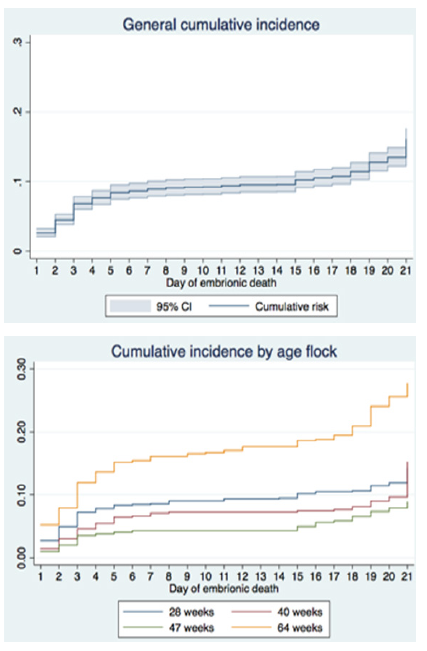
| Cumulative incidence | 2.61 | 4.50 | 6.86 | 7.65 | 8.43 | 8.64 | 8.92 | 9.09 | 9.20 | 9.23 | 9.41 | 9.55 | 9.55 | 9.58 | 10.21 | 10.53 | 10.78 | 11.42 | 12.73 | 13.49 | 16.08 | ||
The cumulative incidence of EM was 16.08% IC95% (14.69; 17.60). EM was higher in breeder flock at 64 weeks with a total of 27.66& IC95% (23.92; 31.99), followed by the 40 week old with 15.23% IC95% (12.40; 18.70), and finally the 28 weeks old with 14.27% IC95% (12.00; 16.96), whereas the 47 weeks old showed the best performance with 8.84% IC95% (6.97; 11.22). The log-rank test confirmed that there were significant differences between the cumulative incidence by every age (p<0.001). In figure 1 the incidence curve may be observed, a general one and one for every breeding age.
Other relevant findings represented 2.61% IC 95% (2.04; 3.16) of the embryos in abnormal positions inside the shell. The most frequent malposition was the peak displacement over the right wing (type VI), with 48.78%, followed by the upside down embryonic position resulting in head away from air cell (type IV), with 19.51%.
The frequency of malformations was of 0.54% IC 95% (0.28; 0.79), especially represented by deformities in two or more parts of the body such as beak, limbs or head, which reached the 58.82%, followed by visible malformations in one area of the body, such as absence of one or both eyes (23.52%) or presence of brain herniation (17.64%). Additional to this, 0.18% of the eggs showed cracked shells IC95% (0.03; 0.33) and a bacterial contamination of 0.37% IC95% (0.16; 0.57).
As shown in Table 2, several differences were seen in the percentage of infertility, malposition and PNN. It was not the case for malformations, contamination and the percentage of cracked eggs.
Table 2 Other findings observed through lighting process and embryodiagnosis
| Age | n | Egg weight (g) | Infertile (%) | Cracks (%) | Malposition (%) | Malformation (%) | Culls (%) |
|---|---|---|---|---|---|---|---|
| 28 | 972 | 57.61 | 1.02 | 0.10 | 1.45 | 0.73 | 0.51 |
| 40 | 648 | 64.61 | 1.85 | 0 | 2.67 | 0.31 | 0.15 |
| 47 | 810 | 65.10 | 1.23 | 0.12 | 2.62 | 0.25 | 0.12 |
| 64 | 810 | 70.78 | 7.65 | 0.49 | 4.01 | 0.80 | 0.37 |
| General | 3240 | 2.90 | 0.18 | 2.61 | 0.54 | 0.37 | |
| P value | <0.001 | 0.116 | 0.012 | 0.328 | 0.565 |
DISCUSSION
Ever since the early reports from Payne in 1919, it was observed that embryos did not die uniformly during the first 21 days of incubation, and that on the contrary, there were two critical periods: the first one between days 4 and 6, and the second one between the days 18 and 20, where a percentage of mortality next to 65% was present 8. Because of this, several mathematical multiphasic models were developed to characterize the EM distribution and they ratified this pattern.
The first model was developed in 1996 by Jassim et al. 2, who made predictions on infertility, total EM and on the accumulated distribution of the two phases for time until the EM starting from the adding of two logistics distributions. These authors found an EM of 11% 2, which is closer to the one found in the present study, which was of 16.08%. In another approximation, Kuurman et al. 9, perfectioned the predictive model using a distribution of Weibull, starting from a lower EM, of 10% 9.
Jassim et al 2, determined that the first phase had its peak of EM on day 2, with a lasting of 4.6 days, and that the second phase had its peak on the day 18, with a duration of 4.8 days 2. The previous information contrasts with the present study, where the EM peak for the first phase was day 1 and lasted around 3 days, and the peak in the second phase was day 21 and lasted three days as well. This allows to establish that even if the biphasic pattern remains, there are conditions in the eggs, like handling during incubation processes that could affect the embryo in different states, speed up or delay the death peak in every stage, although it was always associated to vital changes in the beginning as well as in the end of incubation.
Payne also showed that the third week of incubation was more critical than the first one (with a 60% of deads after the 14th day of incubation 8, whereas in the present study it was during the first week where the greater amount of deads were reported (57.24%). Something similar happened in the Jassim et al. study, where mortality was reported between 63 and 73%, but including the second week of incubation 2.
In this study the EEM was 8.92%, however, in Canada the 8% was considered as a value unusually high just like in this follow up excedes the expected limits for the first week of incubation 10. Besides, this percentage also overpasses the maximum values allowed by the Ross 308 strain guide, in which the highest value allowed for an EEM is 5.5% when eggs come from flocks between the ages of 25 and 30 weeks old. Nevertheless, it is important to take into account that these guides are formulated by the matrix house of the strain under ideal conditions for poultry and incubation, none of which can be compared to the own mode of breeding and production in Santander, Colombia.
When detailing the moment of death of embryos during the first week, it is observed that the pick of death was day 1, followed closely by day 3. In contrast, Scott and Mackenzie 10 specifically evaluated death according to the stages provided by Hamburger and Hamilton 7 and found a peak of death between the stages HH14 to HH18 and a lower one in the stages HH24 and HH28.
The first one matches with the present study giving that stages HH14 to HH18 occur during day 3 of development and is related to changes that include the amnion, heart and circulatory system formation; the second peak matches the days 4 and 5 of incubation and is related to possible alterations in the formation of the visceral arches 10, the corion and allantois fusion and the beginning of the functioning of the mesonephros 3. This agrees with the characterization given by the multiphasic models previously mentioned, but it differs from the present study given that it was on day 1 where the highest peak of mortality was seen.
As to IEM, the present research found an incidence of 0.66%, which remains within the limits of the Ross 308 guiding, where the maximum value allowed is 1%. During the second week of incubation the mineralization of bones begins, the secreted hormonal activity is established in thyroid, pituitary and gonads. Also, nutrients actively are taken from albumin and progressively from the yolk 11. For these reasons, to supply a balanced diet to the breeder flocks is vital in order to transfer into the egg the necessary elements for embryo development and, for the same reason, excess or deficiency of some nutrients will influence EM during the second week of incubation.
The LEM in this study was 6.5%, which excedes the one reported by Elibol et al 12 and Lourens 13 10, 12, or 14 d of incubation at standard conditions to determine if an increased turning frequency would facilitate an early cessation of turning. Turning was discontinued after the respective days were completed. Eggs remained in setter trays until combined at 18 d to complete hatching in a single machine. The young flocks exhibited significantly better fertile hatchability, as expected, but there was no overall effect due to differences in cessation of turning from 8 to 14 d of incubation (range 88.9 to 89.2%, and by the incubation guide Ross 308, in which the maximum allowed is 4.5% (included those embryos who pip the shell) for the breeder flocks between 25 to 30 weeks of age. However, it matches to the mortality found in eggs that were storaged for long periods of time 14 or in incubated eggs with cracked shell 15.
The last week of incubation showed a peak in the mortality on the day 19, which agrees with the findings in the multiphasic models 2,9. At this time, the allantoic blood vessels are responsible for respiration and excretion, but when the embryo breaks the shell membranes using the beak in order to access to the air cell, lung breathing is completed 8, and any failure in this transition may interfere with successful birth of the embryo 1. Also, it may be associated to other changes present after day 16 and may contribute to less vigorous embryos weakening. The first change is that nutrients for the embryo are obtained primarly from the albumen and after day 16, it starts relying mainly on the yolk. Besides, during day 19, the yolk has not been absorbed yet, but it is in the process of going into the abdominal cavity 8, and this can alter the viability of the embryo.
This study allowed to describe the EM according to the ages of the breeder flocks, determining aspect in the evaluation of the EM and that has been widely discussed in multiple experimental studies 5,12,16. The EM was higher in eggs from the 64 weeks old breeders, followed by the 40, 28 and 47 weeks. This behaviour is attributed, among other issues, to differences in the egg weight and the quality of the shell measured through variables such as specific gravity, thickness, conductance or porosity 17.
It was reported that the shell generally gets thiner with age 17 and that young hens tend to produce eggs with a thicker shell compared to the older ones 18. Although the quality of the shell was not measured in this study, it was evident that those eggs from the 64 weeks old broke or fractured easier, they had the higher percentage of cracked shells although with no differences between the other ages, and also the shell was thiner and with imperfections regarding color and texture.
A strong association between the LEM and specific gravity was reported, particulary in old hens. According to this, when gravities are under 1.080, more embryos died, which was attributed to a greater water loss due to thinning of the shell 19. Although the specific gravity was not evaluated, the physical characteristics could give an idea of its decrease, which could have impacted LEM in the 64-old weeks flock. This LEM turned out to be the highest among all evaluated ages.
Hatchability is usually maximum in the middle part of the hens productive period, when shell thickness values decrease and the porosity becomes higher 17. These findings coincide with the present ones, since the best results found were obtained in the 47 weeks old beeders group. On the other hand, a study done on ducks shows that thickening of the shell may increase EEM because there is less porosity and besides the distribution of the pores tends to be on the blunt and sharp end of the egg, in constrast to a higher porosity in the equator of the eggs of embryos who manage to hatch 20. This could be related to the finding in this study which is that embryos of breeder flock at 28 weeks had the second highest incidence of EEM given that the eggs with the previous characteristics are typical from breeders who iniciate the production.
A positive correlation has also been found between the flock age and the egg size 18, as a result of an increase in yolk deposition caused by changes in the ovarian function influenced by age, translated into follicle weight gaining, and therefore, a heavier egg 5. These findings agree with present ones, given that the biggest and smallest eggs were from the breeder flocks at 64 and 28 weeks, respectively.
Besides, keeping in mind that yolk provides the nutrients for embryonic development, it is to be expected that eggs from younger breeders have a lower availability of nutrients and, therefore, this has a negative effect in embryonic development 5, which could explain in part the EM in eggs from the breeder flock at 28 weeks.
Moreover, egg size is correlated to embryo size 21) and in consequence, it is expected to obtain big chickens from big eggs. Expected causes for the abovementioned relation include a wider surface for O2 difusion, higher circulation and extraction of nutrients from the tolk, and overhydration by fewer water loss 22. The latter was presently encountered as shown by residual albumen observed in some embryos who died toward the end of incubation and, although the abovementioned incidence was not recorded, it is an important characteristic in embryodiagnosis, since this may be related to adverse conditions during incubation. On the other hand, the EM increase associated to large eggs, especially in the LEM period, was postulated as a consequence of the difficulty for the embryo to lose metabolic heat during the last periods of incubation 23.
Shell´s weight and thickness are negativily associated to the relative rate of water loss. Regarding that, it is believed that the main osmoregulatory mechanism of the embryo is the chorioallantoic membrane (CAM), which acts as a selective barrer, allowing the absorption of electrolites and water from allantoic fluid and functions as an effective defense against intraluminal toxic contents 24.
The CAM degenerates toward day 19 when pulmonary ventilation begins. This change in oxygenation may affect the blood flow due to local hipoxia and vasoconstriction 24 and under adverse conditions it may increase LEM.
Another aspect related to age and EM augmentation is the difference in embryo development associated to egg laying sequence, which makes reference to the production pattern determined by days where hens lay eggs consecutively, followed by a “pause” due to ovulation delay of the F1 follicle 25.
The follicle destined to be the first egg of the new sequence remains in the ovary around 16 hours more than the remaining follicles, and consequently, the first hatched embryo tends to be more developed than the rest of the embryos. Even, low incubability has been observed, which may be caused by the preovulatory aging of the oocyte or by changes in the yolk composition, which affect embryonic growth 25.
If it is taken into account that the laying sequence of the eggs normally decreases as the flock production period finishes, low hatchability may be related to the incidence increase from first eggs of laying sequences in flocks growing older 25. The above could explain the fact that the highest EM mortality and infertility rate was provided by breeder flock at 64 weeks. However, other studies attribute the increase of these parameters directly to the effect of age, and not to the laying sequence 26.
Regarding other findings, malpositions near the end of incubation are a cause of LEM. From those embryos who died from day 18 on, almost 50% manifested some malposition that made extremely difficult or impossible to hatch because the chicken could not reach the air chamber, was unable to pip the shell due to lack of movement, or for the combination of both reasons. This matches with other previous reports where between 50 and 85% of embryos that did not hatch, showed some malposition 27.
The increase of malpositions is related to fails in the egg turning, abnormal temperature during incubation, genetic influences, and even the presence of environmental contaminants 27. The above leads to think that malposition is not always the primary cause of death, but it may be the result of unfavorable environmental conditions or lethal factors 1.
The present malformations observed could be due to genetic alterations of low lethality, which allow development of the embryo up to advanced incubation states. However, all embryos with any malformation are not necessarily related to genetic anomalies given that, in a greater proportion, they are due to adverse incubation conditions such as abnormal temperature, O2 excess or restriction and excessive quantities of CO21. The percentage of embryos with structural abnormalities (such as anophthalmia, excencephaly or limbs alteration) was 0.54%, value considered too low as to be attributed to abnormal handling conditions. If that was the case, a larger number of affected embryos could be expected, fact which does not match to present data, and allows to consider that there is a relation between malformations and genetics.
On the other side, malformations are intimately linked to EEM. Heart, head, eyes, thorax or other vital organs defects from embryos who died in the “blood ring” state of development with visible embryo. It was evident that embryos from broiler strains showed a higher amount of structural abnormalities compared to the laying strains 10. This, however, was not presently found, since only visual inspection was performed and on early development states is not always feasible to watch in detail these anomalies.
In conclusion, to define the EM behavior, allows to have a base to measure any variation of a key parameter regarding incubation and to perform control before the evidence of abnormal changes and adverse conditions that may alter each and every one of the developmet stages of the embryo, and that are subject to change facing corrective implementation measures.
Although this study was performed under industrial incubation conditions, the EM biphasic behaviour was evidenced as described in the literature, but minor variations were found regarding the specific day of EM ocurrence from the two peaks of mortality. Hence, it is clear that particular incubation conditions of any industrial company, may alter EM occurrence, but the biological mechanisms tendency inherent to the chick embryo are conserved.

