


 English (pdf)
 Article in xml format
 Article references
 Send this article by e-mail
 Cited by SciELO
  Cited by Google
  Similars in
SciELO
Similars in
SciELO  Similars in Google
 Permalink
INTRODUCTION
Reproductive infectious diseases represent a serious limitation of bovine production. The etiology of these disease is diverse and comprises a variety of viral, bacterial, protozoan, chlamydial and fungal agents, some of which are zoonotic (1). Brucellosis, neosporosis, leptospirosis, bovine viral diarrhea and infectious bovine rhinotracheitis are the most well-known diseases and cause the greatest economic losses for the livestock industry (2). The main alterations caused by these agents are abortions and infertility. These alterations make diagnosis difficult, as more than 50% of abortions have an indeterminate etiology (3). Regardless of the individual losses caused by each agent, the association between them generates different co-infections that can be subclinical and can increase economic losses (4,5).
In contrast to the prevalence of these agents in some European countries, where they are being eradicated (6,7,8), their prevalence in Colombia continues to be high in some regions, and no short-term strategies are available for effective control measures.
This situation may affect the competitiveness of the livestock sector in the future, since disease-free countries can impose health and commercial barriers to meat and other products originating from countries that are not disease-free.
Colombia has a cattle population of 22.689.420, of which 1.412.313 are located in the department of Santander, which makes this department the sixth largest cattle-producing department (9). Although isolated serological studies have been performed in other regions of cattle importance (10,11,12), no studies have been conducted for the department of Santander.
The objective of this study was to determine the serological status of bovine females against IBR, BVD, leucosis, Leptospira and Neospora caninum in the department of Santander, Colombia.
MATERIALS AND METHODS
Location. The work was conducted in 460 cattle farms of 23 municipalities of Santander. Santander is located in the northeastern region of the country and is composed of 87 municipalities grouped into six provinces: Comunera, García Rovira, Guanentá, Mares, Soto and Vélez.
Location. The work was conducted in 460 cattle farms of 23 municipalities of Santander. Santander is located in the northeastern region of the country and is composed of 87 municipalities grouped into six provinces: Comunera, García Rovira, Guanentá, Mares, Soto and Vélez.
A total of 440 blood samples from 460 farms with no vaccination history were collected during the second half of 2015 and assessed by ELISA. The municipalities included in the study were Piedecuesta, Mesa de los Santos, San Gil, Betulia, Guadalupe, San Vicente de Chucuri, Charalá, Puerto Wilches, Barbosa, Cerrito, Málaga, Suaita, Guapota, Barrancabermeja, Puerto Parra, Simacota, Sabana de Torres, Matanza, Miranda, Mogotes, Rio Negro, Vélez and Cimitarra.
Obtaining blood samples. A total of 5.0 ml of blood was obtained from the coccygeal vein of each animal using an 18-gauge needle and collected in Vacutainer ® tubes without anticoagulant. The samples were duly identified, transported at 4°C to the Clinical Veterinary Laboratory of the Cooperative University of Colombia (UCC) and centrifuged at 2,500 revolutions per minute (rpm) for 10 minutes. The resulting sera was fractioned into 2.0 ml vials and stored at -20°C prior to analysis.
ELISA test. Samples were processed using commercial indirect ELISA kits for the detection of antibodies against bovine viral diarrhea (BVD), infectious bovine rhinotracheitis (IBR), enzootic bovine leukosis and Neospora caninum (Svanova Biotech ® ). Indirect ELISAs use immobilized antigens on microplates and detect antibodies in bovine sera.
For the diagnosis of Leptospira, a double antibody sandwich assay (DAS-ELISA) (Linnodee bovine Leptospira Elisa kit) was used to detect antibodies in response to lipopolysaccharides (LPSs) of Leptospira borgpetersenii serovar Hardjo (hardjp bovis) and Leptospira interrogans serovar Hardjo (lPS) hardjo pratjino. In all tests, the procedures used in the laboratory followed the manufacturer’s instructions.
Ethical aspects. The authors state that respectful treatment was given to the animals during the execution of the project proper and that the blood collections were always performed by professionals in veterinary medicine and animal husbandry with the consent of the owners.
Statistical analysis. The information obtained was recorded in a database in Excel and analyzed using descriptive statistics.
RESULTS
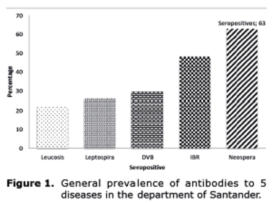
Animals exhibited seroreactivity to all diseases studied, with general prevalence rates as follows: leucosis, 21.8%; Leptospira, 26.1%; BVD, 29.7%; IBR, 48.2%; and Neospora, 63% (Figure 1).
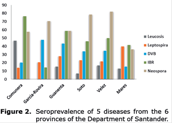
Province-wide, Neospora caninum had the highest seroprevalence, with the highest values in the provinces of Vélez (82.2%), Soto (78.6%), García-Rovira (70.8%), Guanenta (59%), Comunera (57.8%) and Mares %). Regarding IBR, the highest prevalence values corresponded to Comunera (76.6%), Guanenta (59%) and Velez (50%). The highest prevalence of BVD was observed in Garcia-Rovira (47.9%), and the lowest was found in Mares (15.6%), whereas for bovine leucosis, the highest prevalence was detected in Comunera (46.9%), and no reactors were present in Garcia-Rovira (0%). Finally, the highest values for Leptospira were found in Mares (40%), and the lowest values were observed in Comunera (14.1%) (Figure 2).
Regarding the municipalities, the largest numbers of reactors to Neospora caninum were found in Guapota and Simacota and to IBR in Guapota, Mesa de los Santos and Betualia; no reactors were found in Miranda. The distribution of BVD seropositive sera was homogeneous among the municipalities, whereas in some municipalities the prevalence of leucosis was medium to low, and no reactors were present in Cerrito, Malaga, Miranda, Mogotes, Piede Cuesta and Puerto Wilchesin. The lowest values for Leptospira were observed in Sabana de Torres, Suaita, Guapotá, Matanzas and Mesa de los Santos, and no reactors were found in Miranda (Table 1).
Table 1 Table 1. Seropositivity to the five diseases in the six provinces of the department of Santander according to municipality.
| Province | Municipality | Seropositivity | ||||
| Leucosis | Leptospira | BVD | IBR | Neospora | ||
| Comunera (64) | Suaita | 2/9 | 1/9 | 1/9 | 3/9 | 4/9 |
| Guadalupe | 9/14 | 5/14 | 4/14 | 12/14 | 1/14 | |
| Simacota | 9/21 | 2/21 | 4/21 | 15/21 | 16/21 | |
| Guapotá | 10/20 | 1/20 | 4/20 | 19/20 | 16/20 | |
| García-Rovira (48) | Cerrito | 0/20 | 5/20 | 12/20 | 5/20 | 14/20 |
| Málaga | 0/13 | 5/13 | 7/13 | 2/13 | 10/13 | |
| Miranda | 0/15 | 0/15 | 4/15 | 0/15 | 10/15 | |
| Guanenta (39) | San Gil | 1/8 | 2/8 | 3/8 | 6/8 | 2/8 |
| Charalá | 5/14 | 3/14 | 4/14 | 8/14 | 5/14 | |
| Mogotes | 0/17 | 6/17 | 10/17 | 9/17 | 16/17 | |
| Soto (56) | Rio negro | 2/19 | 10/19 | 1/19 | 3/19 | 13/19 |
| Piedecuesta | 0/8 | 1/8 | 6/8 | 8/8 | 7/8 | |
| Matanzas | 1/17 | 1/17 | 4/17 | 4/17 | 12/17 | |
| Mesa de los Santos | 1/12 | 1/12 | 8/12 | 11/12 | 12/12 | |
| Vélez (118) | Cimitarra | 6/16 | 8/16 | 3/16 | 8/16 | 4/16 |
| Vélez | 1/17 | 4/17 | 9/17 | 2/17 | 11/17 | |
| Barbosa | 5/40 | 10/40 | 12/40 | 17/40 | 29/40 | |
| Puerto Parra | 8/45 | 4/45 | 17/45 | 32/45 | 43/45 | |
| Mares (115) | Betulia | 8/13 | 4/13 | 1/13 | 13/13 | 0/13 |
| San Vicente | 1/20 | 9/20 | 4/20 | 3/20 | 2/20 | |
| Barrancabermeja | 22/52 | 22/52 | 9/52 | 25/52 | 25/52 | |
| Sabana de Torres | 5/15 | 2/15 | 1/15 | 5/15 | 5/15 | |
| Puerto Wilches | 0/15 | 9/15 | 3/15 | 2/15 | 10/15 | |
DISCUSSION
The study of reproductive diseases in the main cattle regions of Colombia must be prioritized to establish control plans and enable zonal eradication programs for the improvement of the health status of the country’s herd.
The results of this study show a high presence of antibodies against Neospora caninum (63%) that coincides with studies in several countries (13), indicating that this disease is the major cause of economic losses in dairy and beef cattle worldwide (14). A similar prevalence (57.5%) has been reported in the Sugamuxi Valley (Boyacá) (15), and a higher prevalence has been reported in bulls of Middle Magdalena Valley (79.3%) (16), which is in contrast with the low seropositivity (10.2%) in animals from Montería with reproductive problems (17). The high prevalence in bovine females in this study should be a cause for concern, since this infection has been associated with epidemic abortion in dairy cattle (18), and vertical transmission of the infection can infect the entire herd.
Concomitantly, seropositive animals are more likely (12-19 times) to abort than seronegative animals (19), but this outcome should be contrasted with clinical findings in the field. If this relationship exists, increasing the region’s livestock population in response to the increased demand for meat and milk by domestic and foreign markets will be difficult.
However, this information should be carefully analyzed, since cross-reactions have been found with other cyst-forming parasites of the family Sarcocystidae, including Toxoplasma gondii, Neospora spp., Sarcocystis spp., Hammondia spp. and Besnoitia besnoiti (20), which have not been studied in the country and should be investigated.
The prevalence of IBR (48.2%), which had the second highest prevalence in this study, was similar to that reported in different countries (21). However, within the country, IBR is much more prevalent in other regions, including Antioquia (85.5%), Valle (69.8%) (10), Montería, Córdoba (74.7%) (22), Colombian Altillanura (84%) (23) and Caquetá (90%) (11). In another study, the prevalence in bulls from Middle Magdalena was 92.5% (24). However, bovine herpesvirus 5 (HVB-5), which is a recently detected agent in the country (23), presents a cross-reaction with bovine herpes virus 1 (HVB-1), which is the etiological agent of IBR, and may interfere with these results; this possibility warrants further research. In addition, the latent nature of both herpesviruses suggests that they will continue to circulate indefinitely on farms with seropositive animals with subsequent new infections and reinfections.
Regarding BVD, studies in the country have found a high prevalence in the Bogota savannah (89%) (25) but a progressively lower prevalence in other regions, such as Caquetá (58%) (11), Valle del Cesar (46%) (26) and Montería, Córdoba (29.4%) (12). Notably, this antibody response possibly corresponded to genotype I of the BVD virus, since at the time of the study, genotype 2 did not exist in Colombia (27). The persistence of this virus on the farms studied will depend on the presence of persistently infected (PI) animals, which can affect the entire herd. Thus, further investigation is warranted to detect these individuals.
The prevalence of Leptospira hardjo (26.1%) was lower than the prevalence reported in Central America (31-83%) (28) and other countries in Latin America (16.4-100%) (29). Although establishing comparisons with international and national studies that use microscopic agglutination tests (MAT), which are considered the standard and are based on the use of live antigens, is difficult, the use of ELISA has the advantage of detecting LPS antibodies to the antigenically similar serovar hardjo type hardjo-bovis virus (30).
With this clarification, the results obtained are similar to those found in Montería, Córdoba (20.8%) (31) and Caquetá (28%) (32) but are in contrast to those obtained in the coffee-growing region (45.7%) (33).
With respect to the bovine leukemia virus, which causes enzootic bovine leukosis, the prevalence was lower than the prevalence reported in Peru (42.3%), Paraguay (50%), Bolivia (30%), Argentina (77.4%) and Chile (29.1%) (34) and was similar to that found in the department of Córdoba (21.5%) by ELISA (35). However, the prevalence was high (83% and 60%) in some breeds (Harton del Valle and Chino Santanderano cattle, respectively), which contrasted with the absence of reactors in Blanco Orejinegro, Sanmartinero and Romosinuano cattle and suggested some degree of resistance to the virus (36).
Another study in the Mesa de los Santos (Santander) showed a high prevalence (73%) in dairy cattle (37). In contrast, a lower prevalence (44%) detected by PCR was reported in dairy cattle in the department of Antioquia (38). The use of molecular techniques will likely increase the detection of seropositive animals compared to the use of traditional immunoenzymatic and agglutination techniques, resulting in an increased prevalence in future studies. Although the prevalence found for this virus is not high and economic losses have not been directly associated with reproductive problems, the impact of this virus should not be ignored, since the infected animals have compromised immune systems, which makes them susceptible to suffering from other secondary diseases (39).
The absence of serological reactors to leucosis in the three municipalities of the province of García-Rovira is noteworthy and can be explained by geographic isolation and the absence of roads, which prevent animals from circulating throughout the province. An exception is Cimitarra (Vélez province), where the cattle population is larger and the mobilization of cattle is greater due to a cattle auction that allows greater commercialization of cattle to neighboring farms.
In conclusion, the prevalence of the diseases analyzed in the department of Santander among the bovine population fluctuates from medium to high and thus requires official control measures.

