


 Inglês (pdf)
 Artigo em XML
 Referências do artigo
 Enviar este artigo por email
 Citado por SciELO
  Citado por Google
  Similares em
SciELO
Similares em
SciELO  Similares em Google
 Permalink
INTRODUCTION
The understanding immunity and pathogenesis of giardiasis has been notably developing, due to disparity in several investigations persist, remaining lot to be learned (1,2). Different parasites have long been recognized to affect various aspects of their host’s pro-inflammatory responses (3,4,5). Furthermore available evidence (i.e. findings from selected human researches and to those of experimental proof) suggested that Giardia sp., are able to modulate host pro-inflammatory response (6, 7). The latter knowledge has to be taken into consideration as giardiasis is frequently detected in relation with a variety of pro-inflammatory gastrointestinal (GI) pathogens. Human patients with chronic assemblage B infection were detected to present duodenal inflammation (6). “In vivo” studies showed that experimental infections with G. duodenalis assemblage B were related to post-infectious neutrophil infiltration in ileum or intestinal inflammatory response (8,9). Investigation polarated from human (10,11), and ruminants (12), suggested that some of the Giardia infections might be related to eosinophilia; as was also described previously by “in vivo” infections with Giardia assemblage B H3 infections (13) or parasite excretory/secretory products (14). Duodenal inflammation (6) as detected by microscopy was detected in human patients post-giardiasis (15). Results from various studies indicated roles of specific pro-inflammatory cytokines in giardiasis (16). Proinflammatory conditions in critically ill hospitalized human caused increased D-dimer levels via cytokine activation of the coagulation cascade and subsequent inhibition of fibrinolysis (17). Contrarily no such relation has been made among D-dimer levels, proinflammatory cytokines and giardiasis.
The elevated incidence of thromboembolic events in Inflammatory Bowel Disease (IBD) seems like having multifactorial etiology, where as the inflammatory process might itself participate (18, 19). Furthermore, a defective or impaired intestinal mucosa barrier function may as well contribute to the procoagulative state, especifically in humans with IBD (20, 21). The pathogenic mechanisms proposed for Giardia sp. infections also composed of induction of IBD (22, 23). To this context it may not be unwise to speculate that probable IBD in canine giardiasis might be promoting to a procaogulative state. The alteration of intestinal barrier function in IBD (21), and giardiasis (22,23,24,25,26,27,28) may be a cofactor promoting a procoagulative state.
Relevant data strongly suggested the persence of multifactorial post-infectious complications in giardiasis. One area of developing interest in giardiasis is that initiation of delayed immune-mediated pathophysiology during the acute stage of the infection. Furthermore a better knowledge of the conditions responsible for post-infectious manifestations of giardiasis might help explore common pathways leading to this disease to this context, a biochemical parameter, such as D-dimer, would have helped better understanding the relationship between probable inflammation and thrombosis. This may be briefly explained by some selected hypoythesis. Given the relationship between D-dimer and C-reactive protein, indicating a connection between inflammation and thrombosis (29), each quartile in which C-reactive protein increased, there was a correlational elevation in D-dimer. The latter correlation suggested that as inflammation progresses, the likelihood of thromboembolism rises (29). Previous studies have investigated the responsible mechanisms in which inflammation might potentiate blood clotting. Inflammatory mediators might cause the expression of tissue factor on monocytes and, probably, endothelium, there by stimulating the coagulation cascade. Inflammation-induced thrombosis has long been recognized, indeed its pathogenesis remains complicated. Complex interactions between inflammation and hemostasis, composed of proinflammatory cytokines, and other relevant mediators might occur. Inflammation elevates procoagulant factors, additionally inhibits natural anticoagulant pathways and fibrinolytic activity, resulting with a thrombotic tendency.
To the present authors’ knowledge no such correlation regarding probable thrombosis and inflammation in dogs with giardiasis were detected previously. To verify whether dogs might display a prothrombotic condition, plasma D -dimer was assessed for its value in inflammatory disease activity in dogs with giardiasis. A furthermore another purpose was to analyze correlation between cyst excretion and D-dimer levels to those of dogs naturally infected with Giardia sp.
MATERIALS AND METHODS
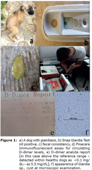
Demographic data. All clinical records from November 2014 to January 2017 were searched to identify dogs with giardiasis for which Snap Giardia test kit analysis (Figure 1b), microscopical detection and a D-dimer test had been performed. A total of 51 cases were found that solely 25 met the criteria for involvement in the present study. These cases were then classified into groups I, II and III.
Classification of the dogs. Three groups of dogs were studied: group I consisted of 15 dogs naturally infected with giardiasis treated with antigiardial protocole (secnidazole at a dose of 10 mg/kg at a single oral dose). Group II consisted of 10 dogs naturally infected with giardiasis without any treatment application, left as untreated control. Finally group III (n=17) were composed of those of healthy dogs, without giardiasis, presented for routine health screen and physical examination, without any apparent clinical signs.
Study duration. The study was performed in 10 days duration. Day 0 (D0) was designated as the initial day, prior to a single dose treatment, and Day 10 (D10) was designed as after treatment analysis day.
Laboratory analysis
Fecal Examination. During the allocation period all dogs (Figure 1a showed one of them) were screened twice with a 10 days interval to confirm the presence/absence of Giardia sp. cysts (Figure 1f). For an initial diagnosis two native smear was prepared to those of dogs with altered fecal consistency (Figure 1c). This was followed by fecal material mixed with 33% ZnSO4 solution, added on to centrifuge tubes, which then was consequently spunned in centrifuge at 880 x g for 300 seconds, similary to a prior study (30). Following centrifugation, some of the fecal mixture was separated, then were added on a microscope slide containing Lugol iodine, which was covered by a slip. The slide was microscopically examined under 40x power for detecting Giardia cysts by a specialized parasitologist. To those of solely mono infected dogs with giardiasis were proven only by microscopical examination was included. Furthermore Giardia antigens in canine feces were tested by use of a rapid enzyme immunoassay for detection of (SNAP ® Giardia Test, IDEXX, USA), (Figure 1b).
Methodology for point of care D-dimer analysis. Two mililiters of sera samples were withdrawn from the vena cephalica of each dog into plain tubes without anticoagulant. Circulating D-dimer concentration was analyzed by use of the Point-of-Care fluorescent immunoassay (Figure 1d). The presentauthor’s private analytic room was specialized and utilized for the Wondfo Finecare FIA meter (FIAm) Fluorescence Immunoassay Rapid Quantitative Test, manufactured by; Guangzhou Wondfo Biotech Co. Ltd.; imported by RDA Grup, Turkey) which is automated. The use of FIAm D-dimer analyzer (Figure 1d, e) has been validated in veterinary practice in the present authors experience since 2014.
Briefly, the FIAm utilizes a Fluorescence Immunoassay methodology, in which 10 μl of undiluted serum is required. A human anti-D-dimer monoclonal antibody was implanted into the well on a supplied test cartridge via the manufacturer. Obtained sera sample were then added; along with buffer solution, which were then shaked for half a minute. Out of those mixtures, 75 μL was obtained, put on to the reading stribes and finally forwarded to the analyzer. The required duration for reading was 180 seconds, also composed of incubation. FIAm D-dimer analyzer is currently able to read ranges between 0.1-10 mg/L. Methodology was adapted with in the manufacturer directions. Elevated D-dimer levels was set as (>0.1mg/L).
Statistical Analysis. Descriptive statistics of the patient and control groups were obtained as follows Unpaired Two-way analysis of variance was performed to determine the differences between groups. ROC (reciever operator characteristics curve) analysis method was used for predicting cutt-off values among D-dimer levels. Results were deemed statistically significant at p<0.05 with 95% confidence interval. Regarding cyst counts on the basis of the number of 0 versus, 0 plus in dogs with giardiasis defining measurement of youden ROC analysis index >0.09 cuttoff value was determined. The average data for performing two-way analysis of variance between groups, and the number of cysts were subjected to log transformation of the time domain. Charts were presented as the median number of cysts.
RESULTS
D-dimer levels in dogs with giardiasis. Median D-dimer levels to those of dogs with giardiasis before (day 0) and after (day 10) treatment. Secnidazol treatment and untreated control groups were involved. Boxplots of median D-dimer levels in dogs with giardiasis before (day 0) and after (day 10) treatment were shown in Figure 2 (Table 1).
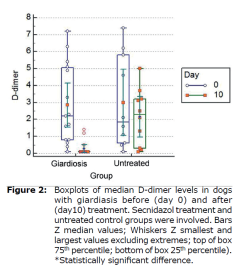
Table 1. Table 1. The interactions between log cyst count and circulating D-dimer levels before (day 0) and after (day 10) treatment. Log cyst count per gram of feces (mean±SE, min-max at 95%CI) were compared to those D-dimer levels at 95% confidence interval. The effects of group, time and group x time were analyzed. Effects (mean±SE, min-max at 95%CI).
| Effect | Factor | Log Cyst (/g) Mean± SE (95% CI) | d-dimer (ng/L) Mean± SE (95% CI) | |
| Group x Time Interaction | Untreated | 0 | 5.35±0.27 (4.81-5.88) | 2.99±0.61 (1.76-4.22) |
| 10 | 5.18±0.26 (4.64-5.71) | 2.14±0.61 (0.91-3.37) | ||
| Treated | 0 | 5.28±0.21 (4.85-5.72) | 2.84±0.50 (1.84-3.85) | |
| 10 | 0.85±0.21 (0.42-1.30) | 0.27±0.50 (0.72-1.28) | ||
| p | <0.001 | 0.13 | ||
ROC curves analyses (Figure 3) were used to select D-dimer cutoff points of below 0.1 mg/L. The FIA meter assay had high specificity (90.9 %) at the 0.09 mg/L cutoff and relatively high sensitivity (87.2%) at the 0.09 mg/L cutoff, as shown in Figure 3 (Table 2).
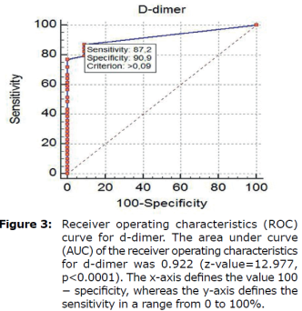
Table 2. Table 2. The sensitivity and specificity of d-dimer determined by fluorescent immunoassay for cases with giardiasis.
| Threshold Value1 (ng/l) | Sensitivity2 (95% CI) | Specificity3 (95% CI) | Likelihoodratio (95% CI) | Predictivevalue (95% CI) | P-value | ||
| Positive4 | Negative5 | Positive6 | Negative7 | ||||
| >0.09 | 87.18 (72.6-95.7) | 90.91 (58.7-99.8) | 9.59 (8.7-9.9) | 0.14 (0.1-0.2) | 97.16 (85.1-99.9) | 66.38 (38.2-87.9) | <0.01 |
| 1 Threshold value for d-dimer determined by the receiver operating characteristic (ROC) analysis. 2 Sensitivity: Probability that a test result will be positive when 0thedisease is present (truepositive rate). 3 Specificity: Probability that a test result will be negativewhenthedisease is not present (truenegative rate). 4 Positivelikelihoodratio: Ratio between the probability of a positive test result given the presence of the disease and the probability of a positive test result given the absence of the disease. 5 Negative likelihood ratio: Ratio between the probability of a negative test result given the presence of the diseaseand the probability of a negative test result given the absence of the disease. 6 Positive predictive value: Probability that the disease is present when the test is positive. 7 Negative predictive value: Probability that the disease is not present when the test is negative | |||||||
NOTE: * Area Under Curve (AUC), the following is interpreted:
90-1.00 = excellent
80-.90 = good
70-.80 = medium
60-.70 = weak
50-.60 = failed
Therefore, the AUC value of 0.922 obtained in this test can be considered excellent.
*The greater the positive portion of Likelihoodratio, the smaller the negative portion, the higher the reliability. Therefore, 9.59 + LR and 0.14-LR are quite good results.
*Treshold value means that animals with values greater than 0.09 ng/L are likely to be sick.
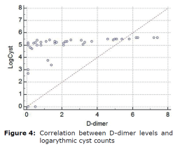
Correlation between D-dimer levels and logarythmic cyst counts were shown in figure 4.
DISCUSSION
Giardia infections cause acute or chronic diarrhea, dehydration, abdominal disturbance and weight loss. Despite suffering from Giardia’s disease, the underlying pathophysiological characteristics of the intestine and the disturbance in Giardia’s disease are not fully understood.
Giardiasis does not spread to other parts of the gastrointestinal tract, such as blood, but is limited to the lumen of the small intestine.
Interestingly, epithelial abnormalities responsible for intestinal malabsorption and responsible for diarrhea in giardiasis share similarities with those observed in other enteric disorders such as bacterial enteritis, small intestine bacterial over growth and chronic food allergies. It is understood that the pathology of the Giardia has emerged. These include disruption of the epithelial barrier, defects at the epithelial brush border, secretion of chloride ions, and hypermotility of the intestinal smooth muscles. The role of parasite virulence factors and host immune responses in these mechanisms has begun to be clarified, but much remains to be understood. The understanding immunity and pathogenesis of giardiasis has been notably developing, due to disparity in several investigations persist, remaining lot to be learned (1, 2). Different parasites have long been recognized to affect various aspects of their host’s pro-inflammatory responses (3,4,5). Furthermore available evidence (i.e. findings from selected human researches and to those of experimental proof) suggested that Giardia sp., are able to modulate host pro-inflammatory response (6, 7). The latter knowledge has to be taken into consideration as giardiasis is frequently detected in relation with a variety of pro-inflammatory gastrointestinal (GI) pathogens. Proinflammatory cytokines are generally released by activated macrophages and are important in the inflammatory response. It has been suggested that D-dimer activation, which is induced by the activation of clotting factors due to the activation of inflammatory processes in this disease, might briefly support our findings as a preliminary marker. In this study, we demonstrated by statistical analysis that d-dimer concentrations, which are considered as proinflammatory markers, are important and specific in this disease by stimulating inflammatory processes after arithmetic and geometric averages of cyst counts before and after treatment. The interactions between log cyst count and circulating D-dimer levels before (day 0) and after (day 10) treatment was interesting. Log cyst count per gram of feces (mean±SE, min-max at 95%CI) was compared to those of D-dimer levels at 95% confidence interval. The D-dimer range in healthy dogs was <0.1 mg/L. In dogs with giardiasis, the D-dimer concentrations were greater than those of healthy dogs (p<0.05) and (p<0.01), respectively. The mean initial plasma D-dimer level was 2.84±0.50 and 2.99±0.61 ng/L in treated and untreated control groups. At the final follow-up evaluation on day 10 was 0.27±0.50 and 2.14±0.61 ng/L, in treated and untreated control groups, respectively, which was significantly lower in treated group (p<0.001). The area under curve (AUC) of the receiver operating characteristics for d-dimer was 0.922 (z-value =12.977, p<0.0001). (95%CI: 0.780–0.885). At a cut-off value of 0.1 ng/L, the D-dimer measurement had a sensitivity of 87.2%, a specificity of 90.9%. There was a correlation between D-dimer levels and logarythmic cyst counts.
Biochemical markers are now being closely scrutinized for detecting disease activity. D-dimer, as a valuable and non-invasive thrombotic predictor, has currently been investigated for supporting a connection between thrombosis and inflammation along with other parameters. As a result, these D-dimer concentrations measured in this disease were concluded to be a pre-biomarker in the diagnosis of these and similar diseases. It should not be unwise to draw preliminary conclusions that D-dimer levels support the link between probable thrombosis and inflammation, namely low grade.

