


Servicios Personalizados
Revista
Articulo
 Inglés (pdf)
 Articulo en XML
 Referencias del artículo
 Enviar articulo por email
Indicadores
-
 Citado por SciELO
 -
 Accesos
Links relacionados
-
 Citado por Google
 -
 Similares en
SciELO
Similares en
SciELO -
 Similares en Google
Compartir

 Permalink
CT&F - Ciencia, Tecnología y Futuro
versión impresa ISSN 0122-5383
C.T.F Cienc. Tecnol. Futuro vol.6 no.2 Bucaramanga jul./dic. 2015
3rd IMRMPT+
SYNTHESIS AND CHARACTERIZATION OF NANOSTRUCTURED MATERIALS BASED ON Fe50Co50 AND Fe75Co25
SÍNTESIS Y CARACTERIZACIÓN DE MATERIALES NANOESTRUCTURADOS BASADOS EN Fe50Co50 Y Fe75Co25
SÍNTESE E CARACTERIZAÇÃO DE MATERIAIS NANO-ESTRUTURADOS BASEADOS EM Fe50Co50 E Fe75Co25
Zulia-Isabel Caamaño-De Ávila1*, María-Angélica Consuegra-Peña1, María-Dolores Baró2 and Santiago Suriñach2
1Departamento de Física, Universidad del Atlántico, Barranquilla, Atlántico, Colombia
2Departamento de Física, Universidad Autónoma de Barcelona, Bellaterra, España
e-mail: zuliacaamano@mail.uniatlantico.edu.co
How to cite: Caamaño-De Ávila, Z. I., Consuegra-Peña, M. A., Baró, M. D. & Suriñach, S. (2015). Synthesis and characterization of nanostructured materials based on Fe50Co50 and Fe75Co25. CT&F - Ciencia, Tecnología y Futuro, 6(2), 33-44.
*To whom correspondence should be addressed
+3rd International Meeting for Researchers in Materials & Plasma Technology - 1st Symposium on Nanoscience and Nanotechnology, Universidad Industrial de Santander and Universidad Pontificia Bolivariana, Colombia, May 4-9, 2015.
(Received: Sep. 14, 2015; Accepted: Nov. 20, 2015)
ABSTRACT
Nowadays, nanostructured magnetic materials are of great interest both in basic research on magnetism and in technological applications. The interest lies in the nanometric particle size, which affects their magnetic properties. Nanostructured magnetic materials based on FeCo have relevant soft magnetic properties, useful in potential applications in information storage equipment and devices. In this paper, nanostructured materials based on Fe50Co50 and Fe75Co25 were synthesized using the mechanical alloying method. The influence that the composition and milling time have on the morphology, structure and magnetic properties of nanostructured powders was studied by means of X-ray diffraction, Scanning Electron Microscopy (SEM) and Vibrating Sample Magnetometer (VSM). After 30 h milling, X-ray diffraction results confirmed a body-centred cubic nanostructured Fe phase with a crystallite size of about 13.6 nm for Fe50Co50 and 22 nm for Fe75Co25, and SEM analysis showed the morphology evolution of the powder particles and the lamellar structure. A maximum saturation magnetization (Ms) of 275 emu/g was obtained for Fe75Co25 which had been milled for 30 h.
Keywords: Mechanical alloying, Magnetic alloy, Nanotechnology, X-ray diffraction, Scanning electron microscopy, Magnetic properties.
RESUMEN
Actualmente, los materiales magnéticos nanoestructurados son de gran interés en la investigación básica del magnetismo y en aplicaciones tecnológicas. El interés radica en el tamaño nanométrico de las partículas, lo cual repercute en sus propiedades magnéticas. Los materiales magnéticos nanoestructurados a base de FeCo tienen las mejores propiedades magnéticas blandas, útiles en potenciales aplicaciones en equipos y dispositivos de almacenamiento de información. En este trabajo, se sintetizaron materiales magnéticos nanoestructurados a base de Fe50Co50 y Fe75Co25 por el método de aleado mecánico. Se estudió la influencia de la composición y del tiempo de molienda en la morfología, estructura y propiedades magnéticas de los polvos nanoestructurados por medio de la difracción de rayos X, Microscopía Electrónica de Barrido (SEM) y Magnetometría de Muestra Vibrante (VSM). Los resultados de difracción de rayos X confirmaron la fase nanoestructurada de Fe cúbica centrada en el cuerpo con un tamaño de cristalito cerca de 13.6 nm para el Fe50Co50 y de 22 nm para el Fe75Co25 después de 30 h de molienda, y el análisis por SEM mostró la evolución de la morfología de las partículas de polvo y la estructura laminar. Se obtuvo una magnetización de saturación (Ms) máxima de 275 emu/g para el Fe75Co25 molido 30 h.
Palabras clave: Aleamiento mecánico, Aleación magnética, Nanotecnología, Difracción de rayos X, Microscopía electrónica de barrido, Propiedades magnéticas.
RESUMO
Atualmente, os materiais magnéticos nano-estruturados são de grande interesse para a pesquisa básica do magnetismo e em aplicações tecnológicas. O interesse radica no tamanho nanométrico das partículas, sendo que o tamanho repercute em suas propriedades magnéticas. Os materiais magnéticos nano-estruturados a base de FeCo contam com as melhores propriedades magnéticas brandas, úteis em potenciais aplicações em equipamentos e dispositivos de armazenagem de informação. Neste trabalho, sintetizamos materiais magnéticos nano-estruturados a base de Fe50Co50 y Fe75Co25 pelo método de ligação mecânica. Estudou-se a influência da composição e do tempo de moagem na morfologia, estrutura e propriedades magnéticas dos pós nano-estruturados por meio da difração dos raios X, Microscopia Eletrônica de Barrido (SEM) e Magnetometria de Amostra Vibrante. Os resultados da difração de raios X confirmaram a fase nano-estruturada de Fe cúbica centrada no corpo com um tamanho de cristalito de aproximadamente 13.6 nm para o Fe50Co50 e de 22 nm para o Fe75Co25, depois de 30 h de moagem, e a análise por SEM mostrou a evolução da morfologia das partículas de pó e a estrutura laminar. Obteve-se uma magnetização de saturação (Ms) máxima de 275 emu/g para o Fe75Co25 moído 30 h.
Palavras-chave: Ligação mecânica, Ligação magnética, Nanotecnologia, Difração de raios X, Microscopia eletrônica de barrido, Propriedades magnéticas.
1. INTRODUCTION
Over the last decades, nanostructured materials have been widely studied as they have shown new physical, mechanical and chemical properties when compared to the coarse-grained polycrystalline materials (Akkouche et al., 2011; Bergheul, Otmane & Azzaz, 2012).
In particular, nanostructured Fe-Co alloys display interesting soft magnetic properties, such as high permeability, high Curie temperature and the highest saturation magnetization of all known magnetic materials (Khajepour & Sharafi, 2011; Chermahini, Rahimipour & Pakseresht, 2014). Although Fe-Co alloys exhibit soft magnetic properties in the vicinity of ~ 30 at. % Co concentration (Sharifati & Sharafi, 2012) and equiatomic composition (Khajepour & Sharafi, 2011), they can be further enhanced by microstructure refinement (Sharifati & Sharafi, 2012). All these notable soft magnetic properties make these nanostructured materials useful in a wide variety of technological and industrial applications (Khajepour & Sharafi, 2011; Bergheul et al., 2012; Yousefi & Sharafi, 2012; Chermahini et al., 2014).
In the search for synthesis methods of nanostructured magnetic alloys, Mechanical Alloying (MA) is one of the most commonly used methods, because it is relatively inexpensive and easy to handle and scale up for future technological applications. The obtained nanostructured powders can then be compacted in a great variety of desired shapes and sizes, which would not be possible with other kinds of conventional synthesis methods (Akkouche et al., 2011; Khajepour & Sharafi, 2011; Bergheul et al., 2012; Sharifati & Sharafi, 2012; Yousefi & Sharafi, 2012).
In this study, the synthesis of nanostructured Fe50Co50 and Fe75Co25 alloys has been achieved by mechanical alloying, presenting the evolution of microstructure and magnetic properties over different milling times (7.5, 15 and 30 h).
2. EXPERIMENTAL PROCEDURE
In the present study binary Fe-Co powder alloys with nominal compositions Fe50Co50 and Fe75Co25 were mechanically alloyed in high energy ball milling Spex 8000 mix/mill. The elemental powders of Fe (99.5 %, 6-9 μm) and Co (99.8 %, 2 μm) were accurately weighted to obtain the desired compositions. The mixture of the powders with one stainless steel ball was charged into a stainless steel container under Ar atmosphere inside a glove box. The mechanical alloying was performed at room temperature using a milling intensity of 825 rpm, a ball to powder mass ratio of 1:1 and milling times of 7.5, 15, and 30 h. The milling was stopped every 100 minutes to avoid overheating.
Morphological, microstructural and compositional studies were also conducted on slightly polished, embedded alloyed powder particles by means of scanning electron microscopes (FESEM Merlin Zeiss microscopes) coupled with Energy Dispersive X-ray analysis (EDX).
Structural characterization was performed on a Phillips X'Pert diffractometer. XRD analysis was carried out at room temperature, using Cu radiation over 30-120° 2θ and 0.02°/8s step. XRD patterns for milled samples at 7.5, 15 and 30 h were obtained. The mean crystallite size, phase crystalline percentage, lattice parameter and micro strains were estimated by diffraction peaks refinement software MAUD (Material Analysis Using Diffraction) (Lutteroti & Scardi, 1990).
Magnetic hysteresis curves were recorded on an Oxford Instruments VSM, in order to study the magnetic properties of saturation magnetization and coercive field of milled powders. All measurements were performed at room temperature with an applied magnetic field of one Tesla.
3. RESULTS
Morphological Characterization by SEM
Figure 1 shows the morphological evolution of Fe50Co50 alloy as a function of milling time (0, 7.5, 15 and 30 h). In Figure 1a, the different shapes of the alloying elements particles for the un-milled powder can be seen. The Co particles have spherical shape while the Fe particles have cylindrical shape. In Figure 1b, the formation of Fe and Co particles agglomerates with flattened shape can be observed for 7.5 h milling time compositions, due to the collisions that occur during the milling process.
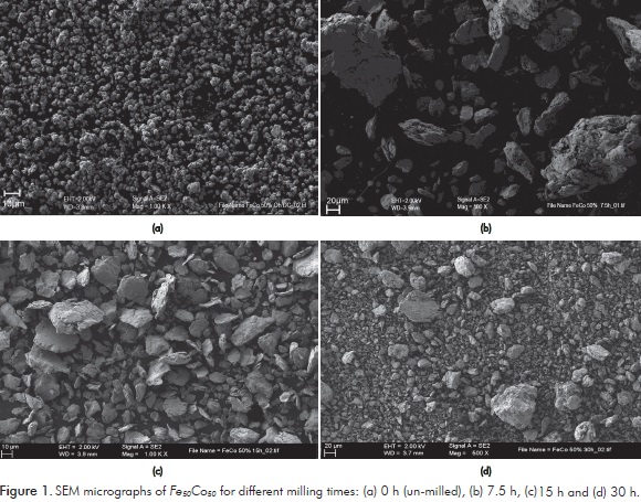
SEM micrographs for the powders milled 15 h are shown in Figure 1c. Aggregates of powder particles can be seen, as a result of the flattening, welding, fracture and rewelding of powders, which is a typical feature of the mechanical alloying process for ductile-ductile systems, as reported in the literature (Zeng et al., 2007; Chermahini et al., 2009a; Chermahini et al., 2009b; Shokrollahi, 2009; Akkouche et al., 2011; Chitsazan et al., 2011; Yepes et al., 2014).
A narrower distribution of particle size can be observed as compared to the samples milled for 7.5 h, as previously reported by Chermahini et al. (2009a) for powders of similar compositions.
The continuous cold welding and fracture events evidenced by the refinement of powders particles can be seen in SEM microphotographs of the alloy milled for 30 h, as shown in Figure 1d. The decrease in size of aggregates particles as the milling time increases is characteristic of the milling process. All the observations for Fe50Co50 agree with those obtained for the same composition milled for 8, 20 and 35 h (Yepes et al., 2014).
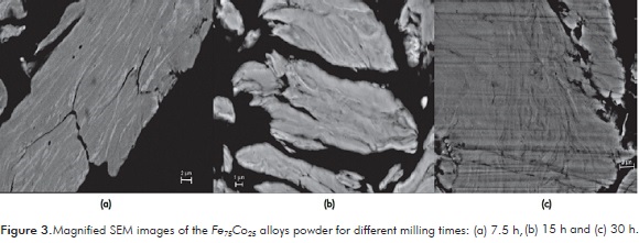
Figures 2 and 3 show SEM micrographs for the Fe50Co50 and Fe75Co25 7.5, 15 and 30 h milled alloys. The lamellar structure of the aggregates of Fe and Co particles for both compositions can be seen, which is characteristic of the milling process of ductile-ductile elements, as reported in precedent studies (Suryanarayana, 2001; Chermahini et al., 2009a).
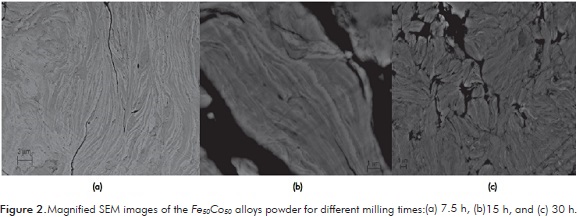
Magnified SEM in Figure 2a at 7.5 h of milling shows clear and dark layers nearly in equal proportion, which evidences the concentration of Fe and Co. At 15 h of milling, a lower proportion of clear, mostly linear layers is observed, as shown in the magnified SEM of Figure 2b. At 30 h of milling, the separations between layers cannot be observed and the elements of the alloy cannot be identified. Also, Figure 2c shows the random welding of the powder particles sizes, resulting in a lamellar structure, which indicates the segregation of Co atoms and the formation of the alloy. Analogue results were reported by Bergheul et al. (2012) for powders of composition Fe60Co40 milled for 12 and 36 h.
For 7.5 h milling times, Figure 3a, clear and dark layers can be distinguished, corresponding to the phases of the alloying elements. In the figure, the darker layer predominates as opposed to the alloy of composition Fe50Co50, due to the higher concentration of Fe (75 %) in this alloy (darker layer). Magnified SEM in Figure 3b for the alloy Fe75Co25 milled 15 h, clear layers cannot be observed as they were observed for the sample Fe50Co50. This indicates that Co diffusion for Fe75Co25 is higher than for Fe50Co50 alloy, because Co concentration is lower and it can be faster diffused, occupying Fe atoms places easily. At 30 h of milling, the distance between layers decreases, causing a higher diffusion of Co, so it can be said that a solid solution is formed (Figure 3c). Similar results were reported in literature by Bergheul et al. (2012) and Chermahini et al. (2014).
Figures 4 and 5 show the maps of Fe and Co distributions for the selected areas of samples Fe50Co50 and Fe75Co25 milled 7.5 h and 15 h (FeKα1 and CoKα1 images), respectively.
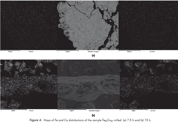
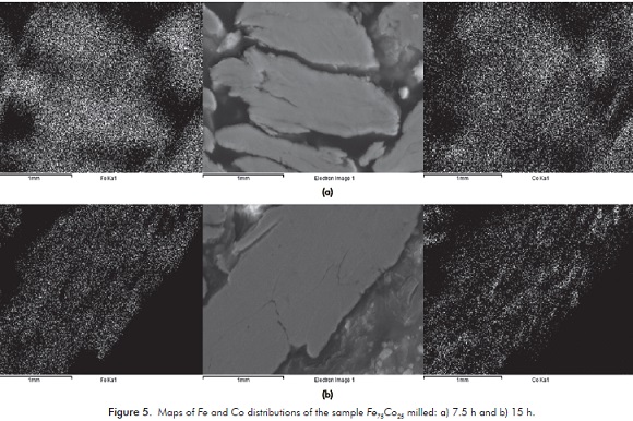
In both Figures, white and black zones corresponding to Fe and Co can be seen. For 7.5 h milling time, as shown in Figures 4a and 5a, it is difficult to distinguish between the two types of particles (Fe and Co); this indicates that the mixture of Fe and Co has begun. Nevertheless, at 15 h of milling (Figures 4b and 5b), a narrower distribution between Fe and Co for Fe75Co25 is observed, which reveals the formation of a FeCo solid solution, in comparison with the alloy Fe50Co50 milled at the same time, where a solid solution is just beginning. A similar behavior was reported by Akkouche et al. (2011) for the Fe50Co50 milled 100 h and 200 h.
Characterization by XRD
Figure 6 shows the evolution of the XRD patterns for Fe50Co50 and Fe75Co25 alloys as a function of the milling time. In Figure 6a, for the unmilled powders of Fe50Co50 (0 h), the presence of Fe (bcc), Co (fcc) and Co (hcp) is confirmed. From 7.5 h of milling, Co phases disappear, as the Fe phase remains, and an observable broadening of the bcc Fe-Co diffraction peaks occurs due to the deformation processes (at macroscopic level) and internal stresses during the milling process. This behavior is attributed to the diffusion of Co atoms into the Fe bcc lattice, as it was reported by Chermahini et al. (2009a; 2009b).
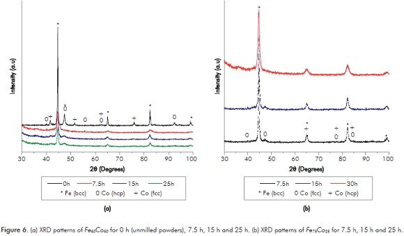
Figure 6b presents the XRD patterns for Fe75Co25 milled at different times. The same behavior of Figure 6a is observed here, as the milling time increases, Co phases disappear. However, for the Fe50Co50 alloy, Co diffraction peaks are more broadened than for Fe75Co25 alloy, indicating that the equiatomic composition has a lower crystallite size than that of Fe75Co25. Figure 7 shows the variation of the crystallite size, lattice parameter and phase percentage of Fe and Co for Fe50Co50 and Fe75Co25 alloys, respectively.
The values for the crystallite size, lattice parameter and phase percentages of Fe and Co for both alloys were calculated from the diffraction peaks refinement Maud software.
From Figure 7a, it could be observed that the crystallite size decreased from a value of 173.7 nm at 0 h down to a value of 13.6 nm at 30 h of milling for the Fe phase. These results differ from the values reported by Yepes et al. (2014) who found a crystallite size of 7.5 nm at 35 h of milling. The difference between these values could result from the experimental conditions used for the mechanical alloying process of the powders. Nevertheless, these values are similar to those reported in the literature for the same composition (Zeng et al., 2007; Chermahini et al., 2009a; Chermahini et al., 2009b; Shokrollahi, 2009; Akkouche et al., 2011; Chitsazan et al., 2011; Khajepour & Sharafi, 2011; Bergheul et al., 2012; Yousefi & Sharifati, 2012; Chermahini et al., 2014).
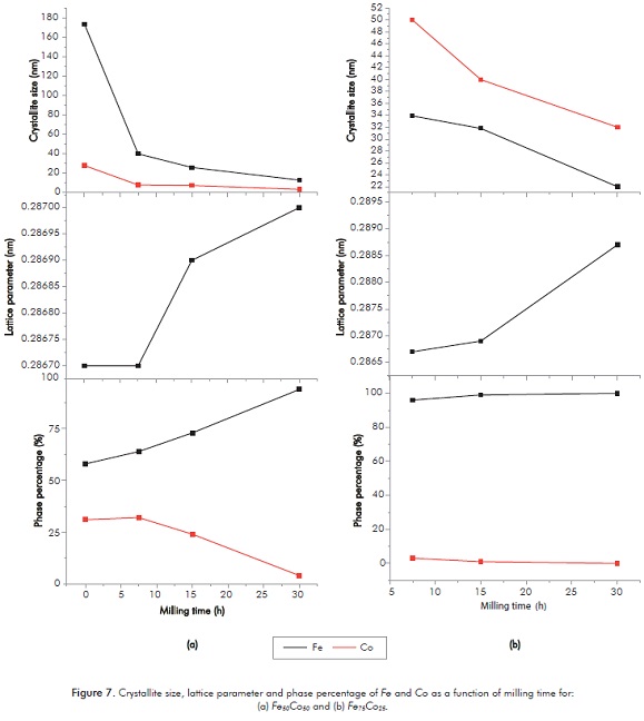
For the hcp Co phase, crystallite size values of 28.5 nm at 0 h until 3.4 nm at 30 h of milling were obtained, which corroborates the reduction of the crystallite size of Co with increased milling time.
From Figure 7b, corresponding to the crystallite size for the sample Fe75Co25 milled at various times, it can be noticed that the crystallite size decreases from a value of 33.9 nm at 7.5 h to a value of 22 nm at 30 h of milling for the Fe; and for the Co, crystallite size values from 50.4 nm at 7.5 h up to 32 nm at 30 h of milling are observed.
In Figures 7a and 7b it is observed that the lattice parameter tends to increase with the milling time for the two studied compositions. This behavior differs from that reported in the literature (Zeng et al., 2007; Chermahini et al., 2009a; Chermahini et al., 2009b; Shokrollahi, 2009; Akkouche et al., 2011; Chitsazan et al., 2011; Khajepour & Sharafi, 2011; Bergheul et al., 2012; Sharifati & Sharafi, 2012; Yousefi & Sharafi, 2012; Chermahini et al., 2014), in which the lattice parameter decreases as milling time increases. The increase of the lattice parameter observed in this work could be due to:
-
The substantial amount of Co atoms segregated into grain boundaries, which contain high amount of vacancies, dislocations, impurities and other defects that become, suitable places for Co atoms segregation (Chermahini et al., 2014).
-
As the volume fraction of grain boundaries increases, some compressive and tensile force on the interfaces and lattice may appear, which can increase lattice parameter (Chermahini et al., 2014).
From Figure 7a and 7b it is also seen that, as milling time increases, the Co phase decreases while the Fe phase increases, which confirms the formation of Fe (Co) solid solution for both compositions. Nevertheless, for the sample Fe75Co25, the Fe phase percentage slightly changes (from 96.1 to 100 %), unlike the Fe phase percentage for the sample Fe50Co50, which changes from 64.5 % to 94.5 %. These results indicate that for the Fe75Co25 alloy the solid solution at 30 h of milling is completely formed, while for Fe50Co50 alloy the solid solution is not yet completed. XRD results are consistent with the SEM results for the alloys milled at different times.
Characterization by VSM
Figure 8 shows the magnetic hysteresis curves of the Fe50Co50 and Fe75Co25 milled at different times. For the Fe50Co50 (Figure 8a), an increase of saturation magnetization is observed with at milling time of 7.5 and 15 h; however, saturation magnetization decreases at 30 h of milling. In the meantime, for the Fe75Co25 alloy (Figure 8b), the highest saturation magnetization (275 emu/g) was obtained for the sample milled for 30 h. This value is higher than those obtained by Yousefi and Sharafi (2012) and Poudyal et al. (2012) for a similar composition milled for 40 h (200 emu/g) and 20 h (240 emu/g), respectively.
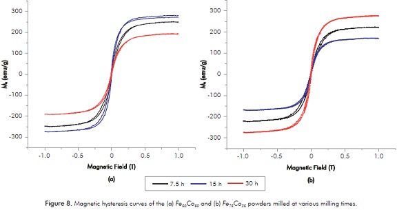
The magnetization increase is related to the decrease of the crystallite size at higher milling times and with the diffusion of Co atoms in the crystalline lattice of Fe, allowing the homogeneity of the alloy.
4. CONCLUSIONS
-
Nanostructured alloys of Fe75Co25 and Fe50Co50 were synthesized by the high energy mechanical alloying method for milling times of 7.5, 15 and 30 h.
-
The evolution of the crystallite size and the lattice parameter as a function of the milling time was evidenced by XRD. After 30 h of milling a coherent domain diffraction of 13 and 22 nm was obtained for the Fe50Co50 and Fe75Co25 alloys respectively. As the crystallite size decreases, the lattice parameter increases, probably due to the substitution of Co atoms into the Fe crystalline lattice.
-
Morphological study by SEM showed fracture and welding of the initials particles with the milling time for both compositions. Also, the lamellar structure, which is characteristic of the ductile-ductile systems, was evidenced by magnification SEM images.
-
The evolution of powders morphology, as well as the crystallite size and lattice parameter, are attributed to the deformation processes that powders undergo when they are mechanically alloyed within a high energy ball milling.
-
SEM, EDX and XRD results confirm the formation of the Fe (Co) solid solution in both compositions of study. For the alloy Fe50Co50, the solid solution was formed at 30 h of milling, while for the Fe75Co25 alloy, the solid solution was formed at 15 h of milling, indicating that the concentration of Co in the alloy promote the formation of the solid solution of Co in Fe.
-
A maximum saturation magnetization value of 275 emu/g was obtained for the Fe75Co25 sample at 30 h of milling from the magnetic hysteresis curves by VSM; until now, this is the highest value of saturation magnetization of Fe-Co alloys.
ACKNOWLEDGMENTS
To Universidad del Atlántico (Barranquilla, Colombia) and COLCIENCIAS (research seeds project - 2013) for financial support for the internship of M. A. Consuegra Peña at the Universitat Autònoma de Barcelona, (Bellaterra, Spain).
The first author highly appreciates Dr. P. Solsona for their support. The technical assistance of the Servei de Microscòpia and the Servei de Difracció de Raigs X of the UAB are greatly appreciated.
To Claudia Rondón for initial grammatical corrections and Juan David Nieto for the edition of the SEM micrographs.
REFERENCES
Akkouche, K., Guittoum, A., Boukherroub, N. & Souami, N. (2011). Evolution of structure, microstructure and hyperfine properties of nanocrystalline Fe50Co50 powders prepared by mechanical alloying. J. Magn. Magn. Mater., 323(21), 2542-2548. [ Links ]
Bergheul, S., Otmane, F. & Azzaz, M. (2012). Structural and microwave absorption properties of nanostructured Fe-Co alloys. Adv. Powder Technol., 23(5), 580-582. [ Links ]
Chermahini, D., Zandrahimi, M., Shokrollahi, H. & Sharafi, S. (2009a). The effect of milling time and composition on microstructural and magnetic properties of nanostructured Fe-Co alloys. J. Alloys Compd., 477(1-2), 45-50. [ Links ]
Chermahini, M. D., Sharafi, S., Shokrollahi, H., Zandrahimi, M. & Shafyei, A. (2009b). The evolution of heating rate on the microstructural and magnetic properties of milled nanostructured Fe1-xCox (x = 0.2, 0.3, 0.4, 0.5 and 0.7) powders. J. Alloys Compd., 484(1-2), 54-58. [ Links ]
Chermahini, M. D., Rahimipour, M. R. & Pakseresht, A. H. (2014). Microstructure and magnetic properties of nanostructured Fe-Co powders prepared by series of milling and annealing treatments. Adv. Powder Technol., 25(1), 462-466. [ Links ]
Chitsazan, B., Shokrollahi, H., Behvandi, A. & Mirzaee, O. (2011). Characterization and magnetic coercivity of nanostructured (Fe50Co50)100-xVx=0,2,4 powders containing a small amount of Co3V intermetallic obtained by mechanical alloying. Powder Technol., 214(1), 105-110. [ Links ]
Khajepour, M. & Sharafi, S. (2011). Structural and magnetic properties of nanostructured Fe50(Co50)-6.5 wt % Si powder prepared by high energy ball milling. J. Alloys Compd., 509(29), 7729-7737. [ Links ]
Lutteroti, L. & Scardi, P. (1990). Simultaneous structure and size-strain refinement by the Rietveld method. J. Appl. Cryst., 23: 246-252. [ Links ]
Poudyal, N., Rong, C., Zhang, Y., Wang, D., Kramer, M. J., Hebert, R. J. & Liu, J. (2012). Self-nanoscaling in FeCo alloys prepared via severe plastic deformation. J. Alloys Compd., 521: 55-59. [ Links ]
Sharifati, A. & Sharafi, S. (2012). Structural and magnetic properties of nanostructured (Fe70Co30)100-xCux alloy prepared by high-energy ball milling. Mater. Design, 41: 8-15. [ Links ]
Shokrollahi, H. (2009). The magnetic and structural properties of the most important alloys of iron produced by mechanical alloying. Mater. Design, 30(9), 3374-3387. [ Links ]
Suryanarayana, C. (2001). Mechanical alloying and milling. Prog. Mater. Sci., 46(1-2), 181-184. [ Links ]
Yepes, N., Orozco, J., Caamaño, Z., Mass, J. & Pérez, G. (2014). Preparation and characterization of Fe50Co50 nanostructured alloy. J. Phys.: Conf. Ser., 480(1), 1-4. [ Links ]
Yousefi, M. & Sharafi, S. (2012). The effect of simultaneous addition of Si and Co on microstructure and magnetic properties of nanostructured iron prepared by mechanical alloying. Mater. Design, 37: 325-333. [ Links ]
Zeng, Q., Baker, I., McCreary, V. & Yan Z. (2007). Soft ferromagnetism in nanostructured mechanical alloying FeCo-based powders. J. Magn. Magn. Mater., 318(1-2), 28-38. [ Links ]
AUTHORS
Zulia-Isabel Caamaño De Ávila
Affiliation: Universidad del Atlántico
Bachelor in Education Sciences: Mathematics and Physics, Universidad del Atlántico
Master in Materials Science, Universitat Autònoma de Barcelona
Doctorate in Sciences Physics, Universitat Autònoma de Barcelona
e-mail: zuliacaamano@mail.uniatlantico.edu.co
María-Angélica Consuegra-Peña
Affiliation: Universidad del Atlántico
BSc. in Physics, Universidad del Atlántico
e-mail: mangie0121@hotmail.com
María-Dolores Baró
Affiliation: Universitat Autònoma de Barcelona
BSc in Physics, Universitat de Barcelona
Doctorate in Sciences (Physics), Universitat Autònoma de Barcelona
e-mail: dolors.baro@uab.es
Santiago Suriñach
Affiliation: Universitat Autònoma de Barcelona
BSc in Physics, Universidad Autónoma de Barcelona
Doctorate in Sciences (Physics), Universitat Autònoma de Barcelona
e-mail: santiago.surinyach@uab.es
