


Services on Demand
Journal
Article
 English (pdf)
 Article in xml format
 Article references
 Send this article by e-mail
Indicators
-
 Cited by SciELO
 -
 Access statistics
Related links
-
 Cited by Google
 -
 Similars in
SciELO
Similars in
SciELO -
 Similars in Google
Share

 Permalink
Boletín de Investigaciones Marinas y Costeras - INVEMAR
Print version ISSN 0122-9761
Bol. Invest. Mar. Cost. vol.45 no.1 Santa Marta Jan./June 2016
NOTA:
TWO CASES OF TWO-HEAD SHARK EMBRYOS, SMALLEYE SMOOTH-HOUND MUSTELUS HIGMANI AND THE BLUE SHARK PRIONACE GLAUCA
DOS CASOS DE DOS CABEZAS EN EMBRIONES DE TIBURÓN, VIUDA AMARILLA MUSTELUS HIGMANI Y EL TIBURÓN AZUL PRIONACE GLAUCA
Nicolás Ehemann1,2,5, Javier Marín-Sanz1,2,3 and Michelle Barany-González2,4
1 Universidad de Oriente Núcleo Nueva Esparta (UDONE), Escuela de Ciencias Aplicadas del Mar (ECAM), Boca de Río, Nueva Esparta, 6304, Venezuela. nehemann@yahoo.com
2 Proyecto de Iniciativa Batoideos (PROVITA), Boca de Río, Nueva Esparta, 6304, Venezuela.
3 Museo Marino de Margarita, Departamento de Investigación y Educación, Nueva Esparta, 6304 Venezuela. javiermarin308@gmail.com
4 Instituto Venezolano de Investigaciones Científicas (IVIC), Altos del Pipe, Miranda, 1201 Venezuela. michellebarany@yahoo.com
5 Centro Interdisciplinario de Ciencias Marinas, (CICIMAR), IPN, La Paz, Baja California Sur, 23000, México.
ABSTRACT
This study reports the first dicephalic shark's embryo of smalleye smooth-hound Mustelus higmani and the eighth case in blue shark Prionace glauca. Each embryo was removed from the womb of a pregnant female, caught in coastal waters of the Caribbean Sea, near to Nueva Esparta state. The dicephalic embryos are exhibited in the Marine Museum of Margarita.
KEY WORDS: Elasmobranchii, Caribbean Sea, Venezuela, Abnormality, Bicephaly.
RESUMEN
Este estudio reporta el primer embrión dicefálico de la viuda amarilla Mustelus higmani, y el octavo caso en el tiburón azul Prionace glauca. Cada embrión fue removido del vientre de una hembra preñada, capturada en aguas costeras del mar Caribe en las cercanías del estado Nueva Esparta. Los embriones dicefálicos son exhibidos en el Museo Marino de Margarita.
PALABRAS CLAVES: Elasmobanchii, Mar Caribe, Venezuela, Anormalidad, Dicefalia.
The blue shark, Prionace glauca (Linnaeus), has a circumglobal distribution in temperate and tropical waters, probably the widest ranging chondrichthyian. It is a highly migratory species that has been included in the Annex I of the 1982 Convention on the Law of the Sea. This species can be found on a depth range from 1 to 1000 m, usually between 80 and 220 m. It has been reported that P. glauca has a total length of 400 cm, a maximum weight of 205.9 kg, and an oldest estimated age of 20 years (Froese and Pauly, 2013).
On the other hand, the smalleye smooth-hound, Mustelus higmani Springer and Lowe, is a western Atlantic exclusive species (northern coast from Venezuela to the southward to Brazil). It can be found on the continental shelf muddy, sandy and calcareous bottoms, also occurs in shallow brackish water. It is a bycatch species utilized for human consumption and for the preparation of traditional dishes. Mustelus higmani has a maximal total length of 70 cm (Cervigón and Alcalá, 1999; Froese and Pauly, 2013).
Cases of dicephalia in sharks have been documented previously in many species: Bosinceano (1934) for Squalus acanthias, Lozano-Cabo (1945) for S. blainvillei, Gopalan (1971) for Rhizoprionodon acutus, Delpiani et al. (2011) for Galeorhinus galeus, Goto et al. (1981), Hevia-Hormazábal et al. (2011), Galván-Magaña et al. (2011) for Prionace glauca, and more recently Muñoz-Osorio et al. (2013) and Wagner et al. (2013) for Carcharhinus porosus and Carcharhinus leucas, respectively. This article reports the eighth case of dicephalia in the blue shark Prionace glauca and the first case in the smalleye smooth-hound shark Mustelus higmani.
In the nineties, pregnant females of Mustelus higmani and Prionace glauca were captured by artisanal fishermen in the Southeastern Caribbean (Margarita Island, Venezuela, and close to Guyana, respectively), but unfortunately they did not record the pregnant shark measurement or other important data; however, both embryos were donated to the Museo Marino de Margarita and initially preserved in formaline 10% and later in ethylic alcohol 70%. The specimens were identified in fresh, by Cervigón F. and Rodriguez P. (com. pers.). All the measurements were made with a calibrated calliper.
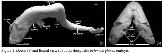
The dicephalic embryo of P. glauca was a female with 236 mm total length (TL) and each head had a length of 46.5 mm from the union of both heads, representing 19.7% TL. The embryo had two developed heads with almost the same size and shape, (Figure 1) with two pair of ocular pockets, two mouths and also ten gill openings at each head. Goto et al. (1981) reported four dicephalus specimens of the blue shark with a mean total length from both heads of 175, 236, 107, 117 mm (one male and three female respectively). Three new dicephalic specimens of P. glauca were reported in 2011, all of them in the Pacific. The first specimen was an unborn female embryo of 160 mm TL reported in the coast of Chile by Hevia-Hormazábal et al. (2011). The other specimens were two females of 227 and 239 mm of TL, respectively reported in the coast of Mexico (Galván-Magaña et al., 2011). The embryo described by us is the second largest ever recorded amongst the eight P. glauca individuals reported with this abnormality.
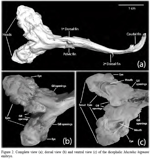
The male embryo of M. higmani was 74.8 mm TL, with a head length of 16.7 mm representing 22.3% TL. (Figure 2) This embryo had a single body with two dorsal and pectoral fins, one anal fin and one caudal fin. The two heads originated posterior to the gills, each head with one mouth, two pairs of eyes and no cephalic organ shared with the other head. Delpiani et al. (2011) reported a dicephalic male embryo of a Triakidae species, Galeorhinus galeus, a 162 mm TL specimen caught in Argentina. Likewise, the Smithsonian National Museum of Natural History possesses under the catalog number (USNM 39499) a dicephalic female embryo of Mustelus sp. which has a total length of 140 mm and was captured in the coast of Peru.
Due to the delicate conditions of both embryos, an X-ray plate was impossible to do without causing an irreparable damage of these specimens that already had suffered excessive dehydration and previous bad manipulation. Nevertheless, there is no doubt that we did not encounter a diprosopus case as Hevia-Hormazabal et al. (2011), because the embryos did not share their cephalic organs. Furthermore, our observations suggested that the bodies of both embryos were well formed and the head malformation would have affected their survival. Both embryos here described constitute the first report of dicephalia in sharks caught in the Venezuelan coastal waters and the Caribbean Sea area.
There are diverse hypotheses for the origin of these two heads monsters, twins or aberration development. Some suggest incomplete division of the embryonic disk and secondary fusing adjacent embryos, poor nutrition, parasitic infections, arthritis, tumors, genetic abnormalities, environmental degradation such as pollution, the number of embryos and uterine size and genetic abnormality, indicative of inbreeding within a small gene pool (Heupel et al., 1999; Kaufman, 2004; Thorburn and Morgan, 2004; Mancini et al., 2006; Saidi et al., 2006; Molina et al., 2008; Delpiani et al., 2011; Galván-Magaña et al., 2011; Muñoz-Osorio et al., 2013).
ACKNOWLEDGMENTS
To the artisanal fishermen for donating both embryos, the Museo Marino de Margarita for letting us photographing and measuring the embryos and finally to the editors and anonymous referees for their comments and suggestions that improved this manuscript.
LITERATURE CITED
Bosinceano, A. 1934. Sur un cas de monstre double incomplet chez Squalus acanthias. Ann. Scientif. l'Univ. Jassy, 19: 339-344. [ Links ]
Cervigón, F. and A. Alcalá. 1999. Los peces marinos de Venezuela vol. V. Fundación Museo del Mar, Caracas. 230 p. [ Links ]
Delpiani, S., Y. Deli Antoni, S. Barbini and D. Figueroa. 2011. First record of a dicephalic specimen of tope Galeorhinus galeus (Elasmobranchii: Triakidae). J. Fish Biol., 78(3): 941-944. [ Links ]
Froese, R. and D. Pauly. 2013. Fishbase. www.fishbase.org. 09/2013. [ Links ]
Galván-Magaña, F., O. Escobar-Sánchez and M. Carrera-Fernández. 2011. Embryonic bicephaly in the blue shark, Prionace glauca, from the Mexican Pacific Ocean. Mar. Biodiv. Rec., 4: 1-4. [ Links ]
Gopalan, U. 1971. On two abnormal sharks from Gujarat. J. Bombay Nat. Hist. Soc., 68: 465-466. [ Links ]
Goto, M., T. Taninuchi, N. Kuga and M. Iwata. 1981. Four dicephalous specimens of blue shark, Prionace glauca, from Japan. Japan. J. Ichthyol., 28: 157-165. [ Links ]
Heupel, M., C. Simpfendorfer and M. Bennett. 1999. Skeletal deformities in elasmobranchs from Australian waters. J. Fish Biol., 54: 1111-1115. [ Links ]
Hevia-Hormazábal, V., V. Pastén-Marambio and A. Vega. 2011. Registro de un mounstruo diprósopo tiburon azul (Prionace glauca) en Chile. Internat. J. Morphol., 29(2): 509-513. [ Links ]
Kaufman, M. 2004. The embryology of conjoined twins. Childs Nerv. Syst., 20(8-9): 508-525. [ Links ]
Lozano-Cabo, D. 1945. Nota sobre un caso de bicefalismo en el Squalus blainvillei. Bol. Real Soc. Esp. Hist. Nat., Sec. Biológ., 43: 147-148. [ Links ]
Mancini, P., A. Casas and A. Amorim. 2006. Morphological abnormalities in a blue shark Prionace glauca (Chondrichthyes: Carcharhinidae) foetus from southern Brazil. J. Fish Biol., 69: 1881-1884. [ Links ]
Molina, S., J. Duque, A. Motta, C. Alvarado and C. Torres. 2008. Duplicación facial Diprosopus: Reporte de un caso y revisión de la literatura. Latin Am. J. Dysmorphol., 1: 10-4. [ Links ]
Muñoz-Osorio, L., P. Mejía-Falla and A. Navia. 2013. First record of a bicephalic embryo of smalltail shark Carcharhinus porosus. J. Fish Biol., 82(5): 1-5. [ Links ]
Saidi, B., A. Bradaï, S. Marouani, O. Guélorget and C. Capapé. 2006. Atypical characteristics of an albino embryo of Carcharhinus plumbeus (Chondrichthyes: Carcharhinidae) from the Gulf of Gabés (southern Tunisia, central Mediterranean). Acta Adriatica, 47: 167-174. [ Links ]
Thorburn, D. and D. Morgan. 2004. The northern river shark Glyphis sp. C (Carcharhinidae) discovered in western Australia. Zootaxa, 685: 1-8. [ Links ]
Wagner, C., P. Rice and A. Pease. 2013. First record of dicephalia in a bull shark Carcharhinus leucas (Chondrichthyes: Carcharhinidae) foetus from the Gulf of Mexico, U.S.A. Journal of Fish Biology, 82(4): 1419-1422. [ Links ]
RECEIVED: 07/04/2014 ACCEPTED: 11/12/2015

