


 English (pdf)
 Article in xml format
 Article references
 Send this article by e-mail
 Cited by SciELO
  Cited by Google
  Similars in
SciELO
Similars in
SciELO  Similars in Google
 Permalink
1. Introduction
The need to adapt the agricultural sector to the demands and requirements (present and future) of the population has caused the second green revolution or agriculture 4.0. Its basis is collecting and analyzing data on the field to improve crop quality, take data in real-time, generate corrective actions, reduce environmental pollution, and deal with climate change. This improvement is possible with the use of technologies such as drones, sensors, software, mobile applications, among others that allow the development of monitoring systems, traceability, geolocation, soil mapping, guidance systems, crop data management technification of the countryside 1.
Agriculture remains an important sector for most countries. Because it represents the main source of food for the world's population, however, it faces a significant challenge: producing more and better while increasing sustainability with reasonable use of natural resources, reducing environmental degradation and adapting to climate change, reducing the environmental impact of plant protection products, and preventing the introduction and spread of quarantine diseases. Therefore, there is a great interest in developing legislative, scientific, and technological tools to detect diseases and pests in crops 2. Modern technologies such as expert systems, artificial intelligence, and computer vision have made it possible to solve problems in different applications such as rice, corn, grape, palm, banana fields, and the detection of different types of diseases 3,4.
On the other hand, 5 used artificial intelligence techniques to estimate the visual characteristics of different plant leaves through image processing. The results showed that the artificial vision techniques models could be of great use in agriculture if good training and validation of the virtual tools is carried out. Similar studies have been reported by 6-10.
Currently, pests monitoring, and crop diseases are also done by remote sensing through the leaves level, canopy, and field. In this way, hyperspectral and multispectral aerial data have been used to monitor the disease in tomatoes and rice, for example, according to the study carried out by Phone et al.11. Likewise, authors such as 12 and 13 have carried out review studies establishing different variables that intervene in plant diseases in agriculture and how they have been treated with artificial intelligence techniques, determining that the type of soil and the climate (temperature and humidity) is one of the most important variables In the agricultural sector.
Huang et al. 14 analyzed the spectral characteristics of yellow rust (pest type) and its influences on winter wheat based on multitemporal push-broom hyperspectral imaging (PHI). Besides, they used a regression model to predict the severity of yellow rust successfully. On the other hand, Barman et al. 15 classified citrus from oranges with 60% accuracy based on hyperspectral images in the air. However, information on the control of crop pests and diseases is still scarce because large-scale detection due to the area's magnitude is difficult to study and process.
The application of image processing techniques and machine learning algorithms in the detection and recognition of diseases and pests in crops is an active area of research that shows great potential for study. Currently, several methods are used for the detection of pests and diseases, among which are the direct chain 16, fuzzy system 17, K-Means clustering 18, decision tree 19,20, computer vision, and artificial intelligence 21-23, deep convolutional neural network 24,25, Markov algorithms 26, fuzzy inference system 27,28, convolutional neural network (CNN) 29,30, and Bayesian networks 31, as the important methods currently studied.
Considering the above, digital image processing is an important study area due to the need to innovate and improve agricultural processes, with which computer technologies can be applied together with innovative methods for agriculture 4.0. such as sensors based on the analysis of host responses, e.g., differential mobility spectrometer and lateral flow devices, provide instant results and can effectively detect early infections directly in the field, phage display-based biosensors and biophotonics can also detect infections instantly although can be integrated with other systems. Therefore, this review study aims to identify approaches, methods, and technologies of a plant pest and disease detection system through articles collected in current databases.
2. Methodology
The information collection was carried out in different academic sources, as following IEEE, Scopus, and Science Direct due to being the most prominent. This with the aim to identify works/articles in the databases related to the subject of detecting pests and diseases in crops using digital processing techniques of images. Figure 1 shows the process carried out during the review.
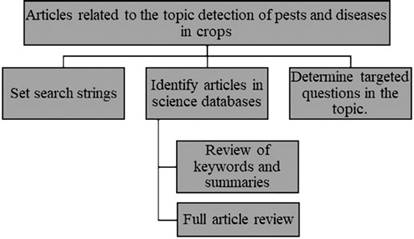
The following questions were presented to establish the search for articles: 1) What digital image processing techniques are used to detect pests and diseases in crops? and 2) In what types of crops have digital image processing techniques been used to detect diseases and pests? Table 1 shows the information search conditions using defined text strings. The search equation was selected considering the following words: diseases in crops, plants, and artificial intelligence.
Table 1 Information search conditions
| Database | Search string | Number of articles |
|---|---|---|
| Science Direct | Digital crop imaging techniques, automatic crop disease detection, crop pest detection | 30 |
| IEEE | Automatic detection of diseases and pests in crops | 9 |
| Scopus | Automatic detection of diseases and pests in crops | 22 |
Source: own elaboration
3. Results
In the literature review 63 articles were gathered, where the information was collected and analyzed around the search questions that were selected, and the results are presented below.
3.1. Digital image processing techniques for detecting diseases in crops
The articles found that the extraction of artisanal characteristics (visual method) of the leaves of the crops is the most common technique used for the recognition of crop diseases, as shown in Figure 2.
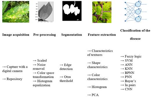
Image acquisition begins with transferring electronic signals from a sensor to a numerical representation by a device such as a camera. Mainly, two types of cameras can be used that are distinguished by way of scanning: area or line. Conventional or area scan cameras generate an image with each exposure cycle. In contrast, line scan cameras capture only one line of pixels at a time. To acquire two-dimensional images, it is necessary to move the object to be captured employing a protractor or move the camera along a stationary object. The quality of the image acquired by a computer vision system is directly affected by the lighting used during the acquisition phase.
In this way, all the effort invested in the use of adequate lighting will increase the performance and reliability of the system, reducing the complexity of the software used in the pre-processing stage 32. Examples of procedures carried out at this stage are image resizing, filtering, color space conversion, and histogram equalization. In-plant disease recognition applications, the segmentation is twofold. First, segmentation is carried out to isolate the leaf, fruit, or flower from the background, and then a second segmentation is carried out to isolate healthy tissue from diseased tissue 33, taking into account the image colors previously programmed.
Characteristics extraction involves extracting information from the segmented image that could facilitate the precise classification of the anomaly (the pest or disease identified by agricultural crop). The characteristics that can be extracted are texture (energy, contrast, homogeneity, and correlation), shape, size, and color 34. Texture features can be extracted using statistical measures such as local binary patterns (LBP), gray level co-occurrence matrix (GLCM), color co-occurrence matrix (CCM), and spatial gray level dependency matrix (SGLDM). Texture features can also be extracted using model-based methods, such as Auto-Regressive (AR) and Markov Random Field (MRF) models 35.
3.2. Methods for classifying diseases in crops
3.2.1. Support Vector Machines “SVM”
SVM is a non-probabilistic linear classifier that constructs a decision threshold with the greatest possible distance between the examples. An SVM attempts to find the hyperplane by dividing the data into two subspaces with the maximum distance between the data closest to the hyperplane (called a separate hyperplane). The distance between the hyperplane and the data is called the margin. The data closest to the hyperplane are called support vectors. Because data is divided into two subsets of data, there are two types of support vectors: those of a subset of data and those of a different subset of data. They are often called positive and negative support vectors, respectively. In this way, it is possible to create linear separators without the original space of the inputs, it is impossible to define them linearly. Finally, SVM is a non-parametric method, so it has the flexibility to represent complex functions and at the same time is resistant to overfitting 36.
3.2.2. Neural network
A neural network is composed of nodes connected by directed links with an associated weight that determines the strength and signal of the connection. Weights are the main form of long-term storage in neural networks and learning algorithms can generally update the weights. The input and output nodes are those with connections to the external world. Each node is independent of the others and therefore can perform your calculations only from their input values and their respective weights. Each unit receives the values of the incoming links, calculates the activation level, and when activated, sends the signal to all its outgoing links. The four main architectures used are unsupervised pre-trained networks, convolutional neural networks, recurrent neural networks, and recursive neural networks. These networks are already used to solve various problems such as text-to-speech conversion, cite fan2014 synthesis, language identification, translation, signal processing, automatic character recognition, and automatic generation of subtitles. In the classification of images, convolutional neural networks are the most used due to their nature. The main purpose of a convolutional neural network (CNN) is to learn the data characteristics of convolution operations. Therefore, this type of network is more suitable for the recognition of patterns in images 37.
3.2.3. K-Means grouping technique
The K-means clustering method is an unsupervised machine learning algorithm used to segment the interest region from plant leaf images. Also, divide the data objects into K-groups based on the similarity between the objects. K-means is a technique used to segmentation wheat diseases such as leaf rust, powdery mildew, and striped rust from images using automatic data grouping (clustering), which is achieved by considering the absolute difference between each pixel and the clustering center in the color space.
3.2.4. Edge detection techniques
The edge is defined as the boundary with different properties between two regions. It is the transition point from one entity to another. This detection technique is used to describe the physical scale of objects because there is often a significant change in intensity at the area’s boundaries. Edge detection is a critical step in understanding image function and is also used to separate images into specific target regions according to needs 17.
3.2.5. Convolutional neural network
A convolutional neural network (CNN) is a type of data processing breakthrough neural network with a grid-like topology. CNN is one of the best algorithms in deep learning methodologies because it can easily understand the image’s content and, therefore, aid in different classifications. CNN can be classified into seven different types based on architectural modifications, including ResNet, VGG, GoogLeNet, AlexNet, DAG Net, and AlexNet. Transfer learning methods are developed to increase the learning capacity of the methodology 20.
Transfer learning is one of the machine learning methods in which the model developed for a particular task can be reused for the second model. In transfer learning, the training of the base network is carried out first on the data set. Then it is passed to the next layer, where the target data set is trained again. The whole process would work if the characteristics extracted in the target and base tasks are general and not specified for particular tasks.
3.3. Classification of pests in crops
In the articles consulted, it was found that the classification of pests in crops utilizing digital image processing techniques is carried out with the methodology shown in Figure 3.
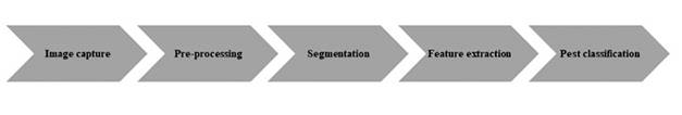
3.3.1. Image acquisition and pre-processing
Processing began with the acquisition of the cropped image. Then, pre-processing operations are performed to improve the image by eliminating unwanted distortion and enhancing the desired characteristics for further processing 38. In image pre-processing, image quality enhancement techniques are applied to reduce noise and sharpen the image for greater precision. This process is carried out to improve the image’s quality to obtain a better detection and classification of insects, pests, and diseases.
3.3.2. Segmentation
The next step in the discovery process is segmentation. Segmentation is performed on the image converted to color to separate the object of interest from the sheet. Generally, Gaussian mixture models and segmentation algorithms are employed, as described in the reference 25. These algorithms involve many arithmetic calculations in the form of divisions, multiplications, and calculation average, which in turn leads to an exponential increase in execution time 39.
3.3.3. Feature extraction
Feature extraction plays an important role in image processing. Feature extraction techniques are applied to extract features useful for image recognition and classification. A feature set contains information to distinguish one object from another. Features can be classified into two, shape feature extraction and texture feature extraction. Some image properties are considered in the extraction of characteristics, such as region properties, gray covariance, among others 40.
Shape feature extraction: Shape characteristics are the essential characteristics that are not affected due to scale, rotation, and translation and are applied in computer vision and automatic object recognition systems. The classification of insects is carried out based on the finite shape characteristics extracted from the images of insects. RGB-shaped insect images were converted to grayscale images for further feature extraction. Image processing techniques are applied to extract shape features using Sobel's edge detection algorithm and morphological operations. The nine shape features include area, perimeter, major axis length, minor axis length, eccentricity, circularity, solidity, shape factor, and compactness stored in feature vectors and then applied to classifier models 41.
Texture feature extraction: Color is a property of pixels, while texture can be measured from a group of pixels. Examples of texture characteristics are energy entropy, contrast, among others. According to the domain from which the texture feature is extracted, they can be classified into spatial texture feature extraction and spectral texture feature extraction 42,43.
3.3.4. Pest classification
This final classification categorizes all the pixels in the image into different classes according to their similarities or differences. Two types of classification methods are supervised classification and unsupervised classification.
On the other hand, in unsupervised pest classification, pixel clusters are based on an image’s software analysis without the user providing a training data set. The computer uses techniques to determine which pixels are related and groups them into classes. Supervised classification is based on the idea that a user can select sample pixels in an image representing tutorials. Then the image processing software uses these training data sets as references for the classification process of all the pixels of the image. The training data sets are selected based on the user's knowledge. Table 2 shows some works related to the technique of extraction of characteristics for diseases in crops. On the other hand, Table 3 shows some works related to the pests and diseases classification in crops.
Table 2 Summary of recent works on the recognition of diseases through the extraction of artisanal characteristics
| Reference | Processing techniques | Precision |
|---|---|---|
| 3 | Crop: Wheat | 78% |
| Techniques: Color constancy normalization, automatic SLIC segmentation, and manual clipping of the region of interest (RoI) using the touch screen of the mobile device and the Chan-Vese algorithm | ||
| 44 | Crop: Rice | Probability ratio of brown spot and leaf scald diseases was 25 and 50%, respectively. |
| Techniques: Histogram intersection, grouping of K-means based on thresholds. | ||
| 45 | Crops: Apple, Corn, Grape, Potato, Tomato, Tobacco | Apple: 83.3% Corn: 87.2% Grape: 86.8% Potato: 81.9% Tomato: 87.3% Tobacco: 82.5% |
| Techniques: Automatic image segmentation algorithm and deep learning with an expanded data set. | ||
| 46 | Crops: Beans and Potatoes | LIRA, an 88% recognition rate. |
| Techniques: Neural classifiers RSC (Sub-space Random Classifier) and LIRA (Limited Receptive Area) | With CSR, or 89%. | |
| 24 | Crops: Corn | 88.4% |
| Technique: convolutional neural networks | ||
| 47 | Crop: Banana | 98.6%. |
| Technique: Correlation coefficient | ||
| 48 | Crop: Onion | 87.2% |
| Technique: Neural Networks | ||
| 49 | Crop: Apple, Banana, Cabbage, Yucca, Celery, Corn Cucumber, Grape | 99.5% |
| Technique: Convolutional Neural Networks |
Source: Own elaboration.
Table 3 Summary of recent works on classification and detection of pests in crops
| Article | Processing techniques | Precision |
|---|---|---|
| 50 | Convolutional neural networks | 92.4% |
| 51 | Sparse multi-task coding, multi-core learning technique | Colour: 70.2% Texture: 63.5% Form: 80.2% |
| 52 | Deep residual learning method | Classification of 98.6% |
| 53 | Multilayer classifier based on the random threshold classifier | Recognition rate 85%. |
| 54 | Multiple Instance Learning (MIL) | 59.8% |
| 55 | Deep learning with UAV imaging | 91.2% |
| 56 | SVM classification | With an error less than 2.5%. |
Source: Own elaboration.
3.4. Image-based techniques for the detection of diseases in crops
Optical imaging technology, also called "image-based technique", has gained great interest in detecting diseases and pests in crops, which has become a valuable tool in all significant areas of application due to different technological advances and developments that help process improvement 57. Known emerging imaging-based techniques for food safety and quality include traditional imaging (TI), hyperspectral imaging (HSI), laser backscatter imaging (LBI), ultrasound imaging (UI), magnetic resonance imaging (MRI), X-ray imaging (XRI), thermal imaging (TI), fluorescence imaging (FI), Raman imaging (RI), microwave imaging (MI), and 3D imaging. These emerging imaging techniques have shown promising potential in the non-destructive quality control of various food products58.
3.4.1. 3D image
In 2D imaging, the data are taken from two different dimensions that differ given some plant names, such as development, total height, and yield estimation. Therefore, the need for 3D images appears as an essential element for the automatic detection of plant diseases. There are two main 3D representations: one related to the surface and the other to the volume presentations. The first involves the details of depth, the surface element, and the different points given by their dimension coordinates. The volume is also given by defining the volumetric component and a frequency component of the model coordinates.
Nowadays, different types of affordable sensors are available besides exhibit highly advanced technology for different domains like nutrient content, growth level, crop presence, biomass estimation, height, and health status. Plant leaf diseases are further analyzed using these sensors because the collected data can be used to quantify the various previously identified production traits.
3.4.2. Fluorescence images
The fluorescence imaging system is one of the practical ways to distinguish the state of metabolism in plants. The fluorescence method that emits light focuses on the chlorophyll complex of the plant. The light irradiates the chloroplast with blue light resulting in re-emission of the light that was absorbed by the chlorophyll. As a result, fluorescent light reflects light to assimilate blue light. The combination of the blue light source with the saturation of the blue pulse could be used to estimate the efficiency and other physical properties of plants.
The UV light source for fluorescence excitation is obtained from a xenon or halogen lamp. A charge-coupled device (CCD) camera is used as fluorescence detection to record the excitation of fluorescence at specific wavelengths. CCD cameras respond with sensitivity to visible and ultraviolet light radiated by the fluorescence signal along with other light sources, including pulsed lasers, pulsed flash lamps, or light-emitting diodes (LEDs) 59.
3.4.3. Tomography
Tomography is one technique that involves obtaining images of a single plane or an object that a tomography generates. There are different tomography types such as linear, polytomography, zonography, computerized or computerized axial type, and positron emission tomography. For the detection of diseases in plants, tomography is used to evaluate the plant state, being a non-invasive technique, capable of offering information on the entire cross-sections of the fruit and the plant with only one measurement, the tomography is based on the transformation of the data, which are given as ranges of speeds, in colors. It is done through software that interpolates the data obtained. The interpretation of the result depends directly on the meaning of each color.
3.4.4. Multispectral images
Multispectral imaging techniques use different types of wavebands such as green, red, or near-infrared wavebands to capture all kinds of images rather than invisible or visible fruits or crops, or vegetation. To detect plant diseases, multispectral images are integrated with classification algorithms and machine learning that give the information to meaningful data 60.
3.4.5. Hyperspectral images
Hyperspectral Imaging (HSI) technology combines the advantages of traditional computer vision and spectroscopy. This technology enables the spatial and spectral variation in a sample to be measured simultaneously.
This technique aims to find the spectrum of the involved pixels that contribute to the image under consideration. The hyperspectral imaging device is implemented for wavelength dispersion, and a transport stage is present. Besides different from the traditional computer vision system 61.
The spectral variation is the product of the interaction of the matter's radiation for hundreds of 10 wavelengths and sampling points simultaneously. It can be quantified by reflectance, which is defined as the percentage of light emitted by a sample concerning the total light that falls on it. This technique aims to obtain information through a group of spectral bands, thus detecting the affected leaves and fruits, classification. From a hyperspectral image, the spectral signature (Spectrum) of a pixel can be obtained. It is possible to try to detect damage caused more efficiently than with conventional RGB systems.
3.4.6. Magnetic resonance images
Nuclear magnetic resonance (NMR) is a unique technology that measures the magnetic properties of spins related to subjects' physical or chemical properties. NMR is the physical process in which the nucleus, whose magnetic moment is different from zero, resonantly absorbs radiation of a certain frequency under an external magnetic field. The detectors detect and receive the emitted NMR signals as electromagnetic radiation. These signals can be sent to the computer and converted into an image through data processing 62.
In the detection of diseases in crops, magnetic resonance imaging is used to identify in advance, in a non-destructive way, the composition and internal structure of the quality of fruits and vegetables. Magnetic resonance images represent different parameters of the analyzed object: the density of atoms, the time it takes for the atoms to return energy to the medium, and the time it takes for the atoms to lose coherence with each other.
3.4.7. Thermal images
Thermography is a non-destructive temperature monitoring technique based on infrared radiation emitted by an object. Thermal imaging evaluation provides useful key data on the dimension, heat distribution, and structural analysis. Thermal imaging can aid in the early detection of plant diseases and therefore allow the design of timely control treatments, allowing for presymptomatic monitoring of plant diseases and pests. Thermal imaging can efficiently monitor temperature irregularities at an early stage of development using thermal imaging analysis.
3.5. Trends and challenges in digital image processing techniques for the detection of pests and diseases in crops
After reviewing different articles mentioned in this work, some challenges to be solved in the agricultural sector are presented below:
The data set should be considered in large quantity due to the cultivation areas in the agricultural industry.
Acquired images are affected by background data and noise (i.e., image quality).
Segment the exact place on an insignificant leaf disease-preparation of training and test samples from the input image.
Classification plays a role in recognizing a segmented point in disease in a pest.
The color, size, and texture of the crop leaf vary when the weather changes depending on the year's month.
Regular observation is needed for particular crops to identify important characteristics.
In Fuzzy C-Means grouping techniques, the calculation can be concentrated, and the determination of the membership operation is a difficult task for detecting diseases in crops.
Identifying diseases and pests for different leaves, stems, and fruits of crops is a challenge through visual inspection.
The reviews suggest that image processing and machine learning techniques have great potential to find diseases and pests in crops, so you should continue to subject research. In this way, it contributes to the economic development of the agricultural sector that is affected widely due to the problem of damage to crops due to pests and diseases generated during the growth of plants that are part of the production process.
4. Conclusions
This article presents a review of image processing techniques for detecting diseases and pests in crops. Also, it presents different algorithms used in the identification of diseases and pests in the agro-industrial sector.
This article reviews the non-destructive techniques that have been used to detect plant diseases and crops. Two non-destructive techniques have been discussed for detecting plant diseases, image-based approaches, and image-processing approaches. Image-based approaches include fluorescence and hyperspectral imaging, while image-processing approaches consist of image acquisition, image pre-processing, image segmentation, feature extraction, and classification. Image processing techniques require a combination of camera and software for further image analysis. Based on this method, the classifier selections are chosen to produce reliable results.
Conventional methods to identified diseases and pests in crops it is focused on the recognition of a single disease or a small number of diseases and generally worked well only under controlled conditions in the laboratory rather than in wild settings containing many Influence factors, such as uniform illumination, scale or constant position of the disease in the image. Although progress has been made regarding disease identification, most traditional methods are only capable of identifying some specific types of diseases in nature.

