SciELO - Scientific Electronic Library Online

 
vol.23 issue2Population-based cancer registriesUpdating the indications for the use of Intensity-Modulated Radiation Therapy in the Instituto Nacional de Cancerología - Colombia. Clinical protocol reported in the evidence. author indexsubject indexarticles search
Home Pagealphabetic serial listing  

Services on Demand

Journal

Article

Indicators

Related links

  • On index processCited by Google
  • Have no similar articlesSimilars in SciELO
  • On index processSimilars in Google

Share


Revista Colombiana de Cancerología

Print version ISSN 0123-9015

rev.colomb.cancerol. vol.23 no.2 Bogotá Apr./June 2019

https://doi.org/10.35509/01239015.58 

Artículos originales

Immunophenotypic characteristics of diffuse large b-cell lymphoma and expression of C-MYC protein: single Ecuadorian center experience

Características inmunofenotípicas del linfoma difuso de células grandes B y expresión de la proteína C-MYC: experiencia en un centro de Ecuador

Johanna Cevallosa  * 

Nelson Montalvoa 

a Laboratorio de Patología, Hospital Metropolitano, Quito, Ecuador


Abstract

Background:

Diffuse large B-cell lymphoma (DLBCL) makes up from 25% to 40% of all non-Hodgkin lymphomas (NHL) and is the most common histological subtype worldwide. In Ecuador, DLBCL makes up 49% of all NHL cases, but there have been no studies on the immunophenotypic classificationof DLBCL in germinal center (GC) and non-germinal center (NGC)subtypes.This study was conducted to ascertain the immunophenotypic profile of DLBCL in an Ecuadorian hospital.

Methods:

A total of 38 DLBCL cases from 2006 to 2015 were compiled from the Pathology Service at Metropolitan Hospital (HM) in Quito, Ecuador. Eleven of these cases failed to meet the inclusion criteria; thus, the final sample consisted of 27 cases. Manual tissue microarrays were constructed, and three immunohistochemical markers (CD10, BCL6, and MUM1) were applied according to the Hans algorithm; in addition, the expression of the c-myc protein expression was also investigated.

Results:

The results showed that 77.8% of cases were of the GC subtype, 11.1% were NGC, and 11.1% were unclassifiable according to the Hans algorithm.

Conclusions:

The most frequent DLBCL subtype was GC, with 21 cases; and 40.7% of these cases overexpressed c-myc.

Keywords: Lymphoma, Large-Cell, Diffuse; Germinal Center; Proto-Oncogene Proteins c-myc; Ecuador

Resumen

Antecedentes:

El linfoma difuso de células grandes B (LDCGB) constituye el 25 al 40% del total de los linfomas no Hodgkin (LNH) y es el subtipo histológico más frecuente en el mundo. En Ecuador el LDCGB corresponde al 49% del total de los casos de LNH, sin embargo no hay estudios de clasificación inmunofenotípica del LDCGB en centro germinal (CG) y no centro germinal (NCG). Este estudio se realizó para conocer el perfil inmunofenotípico del LDCGB en un hospital de Ecuador.

Métodos:

Se recopiló del Servicio de Patología del Hospital Metropolitano de Quito, Ecuador, un total de 38 casos de LDCGB desde el 2006 al 2015, de los cuales 11 no cumplieron con los criterios de inclusión. La muestra final fue de 27 casos. Se realizaron microarreglos tisulares manuales para la aplicación de tres marcadores de inmunohistoquímica según el algoritmo de Hans (CD10, BCL6 y MUM1) y luego se correlacionó con la sobreexpresión de la proteína c-MYC.

Resultados:

El 77,8% de casos fue tipo CG, 11,1% fue NCG y 11,1% fueron inclasificables según Hans.

Conclusiones:

El subtipo de LDCGB más frecuente fue CG con 21 casos y de estos 40,7% sobreexpresaron c-MYC.

Palabras clave: Linfoma de Células B Grandes Difuso; Centro germinal; Proteínas Proto-Oncogénicas c-myc; Ecuador

Introduction

Diffuse large B-cell lymphoma (DLBCL) is a heterogeneous group of neoplasms that have in common certain morphological, immunophenotypic, and genetic characteristics. DLBCL makes up from 25% to 40% of all non-Hodgkin lymphomas (NHL), and is the most common histological subtype worldwide. In Ecuador, it accounted for 49% of all NHL cases from 2006 to 20101-3.

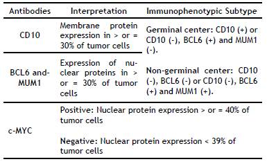
In 2008, the World Health Organization (WHO) put forward a new DLBCL classification that includes morphological and molecular variants and immunophenotypic subgroups, resulting in two subtypes: germinal center (GC) and non-germinal center (NGC). This division is based on the Hans immunohistochemical algorithm (CD10, BCL6, and MUM1), which has a high reproducibility and concordance (80-96%) with gene expression profiling using DNA microarrays4-8.

The results of scientific studies on the immunophenotypic division of DLBCL have been published in Europe, Asia, Latin America (Colombia, Brazil, and Guatemala), and the United States. Despite its recognized prognostic and therapeutic value, the immunophenotypic profile of DLBCL in Ecuador is unknown. This study is the first immunophenotypic classification of DLBCL and its correlation with the overexpression of c-MYC protein in the Ecuadorian population8-14.

Materials and Methods

Patients: We compiled all the DLBCL cases between January 2006 and December 2015 (n = 38) at the Pathology Service of the Metropolitan Hospital in Quito, Ecuador. We excluded 11 cases because they contained insufficient histopathological material to perform the required analyses, ending up with a final sample of 27 cases.

Histopathology: The selected paraffin blocks were surgical specimens and biopsies processed according to the Service's protocols. DLBCL cases were selected if the paraffin blocks contained adequate tissue for manual tissue microarray construction. The DLBCL diagnoses were based on the current 2008 WHO criteria for classification of tumors of hematopoietic and lymphoid tissues.

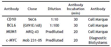
Manual Tissue Microarrays and Immunohistochemistry: The best-preserved area with appropriate morphology was selected to construct manual tissue microarrays with a diameter of 0.4 cm. Sections with a thickness of 4(jm were cut and then stained with hematoxylin-eosin (H&E); then immunohistochemical techniques for CD10, BCL6, MUM1, and c-MYC (Table 1 and 2).

Table 1 Antibodies Used 

Table 2 Interpretation of Immunohistochemistry Results 

Statistical Analysis: The epidemiological design was descriptive as a whole regarding all the samples from DLBCL-diagnosed patients in whom the frequency of the immunophenotypic subtypes GC and NGC was determined. The qualitative variables were expressed in simple frequencies and percentages, and the quantitative variables were expressed in averages and standard deviation. Moreover, the expression of the c-myc protein expression was also investigated for each immunophenotypic subtype of DLBCL.

Results

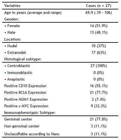
Table 3 summarizes the patients' clinical-pathological characteristics. The average age of the 27 patients was 69.9 ± 16.9 years (range: 39 to 106 years); 51.9% of the subjects (n = 14) were female. The average age of the studied women was 70.5 ± 16.8 years; it was 69.2 ± 17.6 years (p > 0.05) for men. The most frequent locations were extra-nodal (n = 10), predominantly the colon and stomach. H&E staining showed that all the cases had a centroblastic morphology. The immunophenotypic subtype was established according to Hans algorithm. The GC subtype accounted for 77.8% of cases; 11.1% of cases were NGC and 11.1% were unclassifiable. The GC subtype predominantly overexpressed c-MYC (in 40.7% of cases).

Table 3 Clinical-pathological features of the DLBCL Cases 

Discussion

Classification of DLBCL in GC and NGC subtypes has been carried out in a number of studies worldwide. In Latin America (Guatemala, Brazil, and Colombia), very little has been published on this subject 7,8,9,10. In Ecuador, our results show GC to be the most frequent immunophenotypic subtype of DLBCL (42.9% versus 33.3% NGC). These results are similar to those obtained by Perry in Guatemala, where the GC subtype predominated. In Colombia, Ospina found GC to be the most frequent subtype (n = 61), whereas Castellanos reported an NGC predominance (n = 40)1, 2,9,10. It is worth noting that two studies in Latin American countries with very different population figures -Brazil, with 210,616,910 inhabitants, and Ecuador, with 16,507,400- were based on the same number of cases (n = 27), but they show a different immunophenotype, with predominance of the NGC subtype in Brazil and the GC in Ecuador8,11-14.

Studies published in Asian countries such as Korea (n = 381), Taiwan (n = 153) and China (n = 114) used Hans algorithm; they concluded that the most common subtype was NGC9,10.

The international multi-institutional study "Confirmation of the molecular classification of diffuse large B-cell lymphoma by immunohistochemistry using a tissue microarray" compiled data from 152 patients at different universities and cancer centers [Nebraska Lymphoma Study Group Registry (n = 49); British Columbia Cancer Center (n = 30); University of Würzburg (n = 29); Norwegian Radium Hospital (n = 29); University of Barcelona (n = 8); and the Southwest Oncology Group (n = 7)]. This study concluded that NGC was the most frequent immunophenotype (n = 88). Ten cases could not be classified according to Hans algorithm, requiring the use of other algorithms, such as Choi et al.8,15

The WHO recommends Hans algorithm for DLBCL classification because of its high reproducibility and concordance (80-96%) with gene expression profiling through DNA microarrays. Nevertheless, some cases cannot be classified through this algorithm, and organizations such as the College of American Pathologists recommend other algorithms, such as Choi's (MUM-1, CD10, BCL6, and FOXP1) or Tally's (MUM-1, CD10, FOXP1, and LMO2) in such cases3. In our study (n = 27), three cases were unclassifiable according to Hans algorithm. Castellanos (n = 103) reported six such cases in Colombia, and, as previously mentioned, the international multi-institutional study of 152 patients reported ten of such cases. These figures indicate that the majority of DLBCL cases are properly classified by the Hans algorithm, and a minimal number of cases require the application of other algorithms8,12.

Our study identified c-MYC overexpression in 40.7% (95% CI: 22.1 to 59.2%) of cases with GC subtype predominance (42.9% of cases). These results are analogous to those of Gupta et al. (n = 40), in which 50% of cases overexpressed c-MYC by IHC and 65% were of the GC subtype. They are also similar to the results of the Perry study (n = 106), in which 46% of patients who overexpressed c-MYC were of the GC subtype. Johnson et al. (n = 72) and Kluk M et al. (n = 77) reported overexpression of c-MYC by IHC in 33°% and 11.5°% of their DLBCL cases, respectively14,16-18.

This study is the first immunophenotypic classification of DLBCL with the overexpression of c-MYC protein in the Ecuadorian population. The GC subtype accounted for 77.8% of cases; 11.1% of cases were NGC and 11.1% were unclassifiable. The GC subtype predominantly overexpressed c-MYC (in 40.7% of cases).

Currently, overexpression of the c-MYC protein is considered an IPI-independent prognostic factor for both disease-free and overall survival. Patients who overexpress the c-MYC protein have larger tumors, higher chemo-resistance, a worse prognosis, and shorter survival19-25.

It is required to complement the amplification study of the myc gene by fluorescent in situ hybridization (FISH).

References

1. American Cancer Society. Linfoma no Hodgkin. Cancer Facts & Figures. Atlanta: American Society 2017; 21. [ Links ]

2. Ioachim H, Medeiros J. Nomenclature and classification of lymphomas. In: Ioachim's Lymph Node Pathology. 4th edition. Philadelphia: Wolters Kluwer; 2009. p. 296-305. [ Links ]

3. Cueva P, Yépez J. Sociedad de Lucha Contra el Cáncer/ Registro Nacional de Tumores. Epidemiología del cáncer en Quito 2006-2010. Quito; 2014. p. 152-62. [ Links ]

4. Stein H, Warnke RA, Chan WC, Jaffe ES, Chan JKC, Gatter KC et al. Diffuse large B-cell lymphoma, not otherwise specified. In: Swerdlow SH, Campo E, Harris NL, Jaffe ES, Pileri SA, Stein H, et al (eds). WHO Classification of Tumours of Haematopoietic and Lymphoid Tissues. Lyon: IACR Press; 2008. p. 233-7. [ Links ]

5. O'Malley D, Grimm K, Banks P. Inmunohistology of Non- Hodgkin Lymphoma. In: Dabbs D. Diagnostic Inmunohistochemistry. Theranostic and Genomic applications. 4th edition. Elsevier Saunders; 2014. p. 148-16. [ Links ]

6. Young K, Medeiros LJ, Chan W. Diffuse Large B-cell Lymphoma. In: Orazi A, Weiss L, Foucar K, Knowles D. Knowles' Neoplastic Hematopathology. 3th edition. Philadelphia: Wolters Kluwer ; 2014. p. 502-56. [ Links ]

7. Grande C. Actualización en el tratamiento del linfoma difuso de células grandes B. En: Fernández- Rañada J, Alegre A (Eds). Cuadernos de Hematología. 2011; 1-15. [ Links ]

8. Hans CP, Weisenberger DD, Greiner TC, Gascoyne RD, Delabie J, Ott G, et al. Confirmation of the molecular classification of diffuse large B-cell lymphoma by inmunohistochemistry using a tissue microarray. Blood. 2004; 103(1):275-82. [ Links ]

9. Chen Y, Xiao L, Zhu X, Lu C, Yu B, Fan D, et al. Immunohistochemical classification and prognosis of diffuse large B-cell lymphoma in China. Zhonghua Bing Li Xue Za Zhi. 2014; 43(6):383-8. [ Links ]

10. Chang ST, Chen SW, Ho CH, Kuo CC, Sakata S, Takeuchi K, et al. Immunophenotypic and genetic characteristics of diffuse large B-cell lymphoma in Taiwan. J Formos Med Assoc. 2016; 115(11):961-7. [ Links ]

11. Ospina JA. Caracterización de los pacientes con diagnóstico de Linfoma B Difuso de células grande en el Instituto de Cancerología [tesis de investigación]. Bogotá: Universidad Nacional de Colombia; 2015. [ Links ]

12. Castellanos OW. Acuerdo intraobservador en estudios de inmunohistoquímica para Linfoma B difuso de célula grande según protocolo de Hans [tesis de investigación]. Bogotá: Universidad Nacional de Colombia; 2012. [ Links ]

13. Silva FC. Valor pronóstico do perfil inmunotípico em linfoma difuso de grandes células B [tese de Mestrado]. Porto (Portugal): Instituto de Ciências Biomédicas Abel Salazar da Universidade do Porto; 2010. p. 1-18. [ Links ]

14. Perry AM, Molina-Kirsch H, Nathwani BN, Diebold J, Maclennan KA, Müller-Hermelink HK, et al. Classification of non-Hodgkin lymphomas in Guatemala according to the World Health Organization system. Leuk Lymphoma. 2011; 52(9):1681-8. [ Links ]

15. Choi WW, Weisenburger DD, Greiner TC, Piris MA, Banham AH, Delabie J, et al. A new immunostain algorithm classifies diffuse large B-cell lymphoma into molecular subtypes with high accuracy. Clin Cancer Res. 2009; 15(17):5494-502. [ Links ]

16. Gupta M, Maurer MJ, Wellik LE, Law ME, Han JJ, Ozsan N, et al. Expression of Myc, but not pSTAT3, is an adverse prognostic factor for diffuse large B-cell lymphoma treated w'th epratuzumab/R-CHOP. Blood. 2012; 120(22):4400-6. [ Links ]

17. Johnson NA, Slack GW, Savage KJ, Connors JM, Ben-Neriah S, Rogic S, et al. Concurrent expression of MYC and BCL2 in diffuse large B-cell lymphoma treated with rituximab plus cyclophosphamide, doxorubicin, vincristine, and prednisone. ClinOncol. 2012; 30(28):3452-9. [ Links ]

18. Kluk MJ, Chapuy B, Sinha P, Roy A, Dal Cin P, Neuberg DS, et al. Immunohistochemical detection of MYC-driven diffuse large B-cell lymphomas. PLoS One. 2012; 7(4):e33813. [ Links ]

19. Batlle-López A, González de Villambrosía S, Francisco M, Malatxeberria S, Sáez A, Montalban C, et al. Stratifying diffuse large B-cell lymphoma patients treated with chemoimmunotherapy: GCB/non-GCB by immunohistochemistry is still a robust and feasible marker. Oncotarget. 2016; 7(14):18036-49. [ Links ]

20. Duncavage E, Advani RH, Agosti S, Foulis P, Gibson C, Kang L, et al. Template for Reporting Results of Biomarker Testing of Specimens from patients with diffuse large B-cell lymphoma, not otherwise specified. Arch Pathol Lab Med. 2016; 140(11):1225-17. [ Links ]

21. Valentino C, Kendrick S, Johnson N, Gascoyne R, Chan WC, Weisenburger D, et al. Colorimetric in situ hybridization identifies MYC gene signal clusters correlating with increased copy number, mRNA, and protein in diffuse large B-cell lymphoma. Am J Clin Pathol. 2013; 139(2):242-54. [ Links ]

22. Akyurek N, Uner A, Benekli M, Barista I. Prognostic significance of MYC, BCL2, and BCL6 rearrangements in patients with diffuse large B-cell lymphoma treated with cyclophosphamide, doxorubicin, vincristine, and prednisone plus rituximab. Cancer. 2012; 118(17):4173-83. [ Links ]

23. Horn H, Ziepert M, Becher C, Barth TF, Bernd HW, Feller AC, et al. MYC status in concert with BCL2 and BCL6 expression predicts outcome in diffuse large B-cell lymphoma. Blood. 2013; 121(12):2253-63. [ Links ]

24. Xu X, Zhang L, Wang Y, Zhang Q, Zhang L, Sun B, et al. Double-hit and triple-hit lymphomas arising from follicular lymphoma following acquisition of MYC: report of two cases and literature review. Int J Clin Exp Pathol. 2013; 6(4):788-94. [ Links ]

25. Mahmoud AZ, George TI, Czuchlewski DR, Zhang QY, Wilson CS, Sever CE, et al. Scoring of MYC protein Expression in diffuse large B-cell lymphomas: concordance rate among hematopathologists. Mod Pathol. 2015; 28(4):545-51. [ Links ]

Received: August 29, 2018; Accepted: February 26, 2019; Published: June 25, 2019

* Autor para correspondencia: Johanna Cevallos Correo electrónico: johannacevallosespinel@gmail.com; jace77_7@hotmail.com

Conflict of interest

The authors state they have no conflicts of interest

Creative Commons License This is an open-access article distributed under the terms of the Creative Commons Attribution License