


Services on Demand
Journal
Article
 text in
  English (pdf)
 Article in xml format
 Article references
 Send this article by e-mail
Indicators
-
 Cited by SciELO
 -
 Access statistics
Related links
-
 Cited by Google
 -
 Similars in
SciELO
Similars in
SciELO -
 Similars in Google
Share

 Permalink
Acta Colombiana de Psicología
Print version ISSN 0123-9155
Act.Colom.Psicol. vol.22 no.1 Bogotá Jan./June 2019
https://doi.org/10.14718/acp.2019.22.1.9
Artículos
Brain electrical activity of attention in polydrug adolescents using an equipment BCI (Brain Control Interface)
*Universidad Católica de Colombia ORCID: https://orcid.org/0000-0002-0815-4480. Avenida Caracas # 46-72, Bogotá, Colombia, (57) 3277300-50-62. Joven Investigador Colciencias 2016, aadiaz93@ucatolica.edu.co.
** Universidad Católica de Colombia ORCID: https://orcid.org/0000-0002-5983-075X. Avenida Caracas # 46-72, Bogotá, Colombia, (57) 3277300-50-62. Dic. Línea de Investigación en Procesos Psicobiológicos. smcamelo@catolica.edu.co Artículo de investigación Joven Investigador financiado por el Departamento Administrativo de Ciencia, Tecnología e Investigación (Colciencias) y la Facultad de Psicología de la Universidad Católica de Colombia, bajo la convocatoria 645.
The consumption of psychoactive substances is a public health problem that increasingly affects the adolescent population. This investigation had the objective of record the brain electrical activity (EEG) in attention tasks (sustained and selective) in a group of polyconsumers. Employment a retrospective ex post-facto design with a quasi-control group with 46 adolescents between 12-17 years old: 23 polyconsumers and 23 quasi-controls. For the recording of brain electrical activity, it was used a equipment BCI (Brain Control Interface) research grade 14 Channel Mobile EEG and applied the Brain Training Virtual Program "Brain HQ" module “focus my attention” to evaluate the attention. The results showed an increase in beta-β (1330 Hz), theta-θ (4-7 Hz) and delta-δ (3-4 Hz) brain waves in frontal and prefrontal areas in adolescent polyonsumers versus the quasi-control group in attention tasks. Likewise, identified a significant difference with respect to the response time between adolescents consuming psychoactive substances in relation to the quasi-control group in both types of attentional tasks.
Keywords: attention; BCI (Brain Control Interface); electroencephalography; emotiv EPOC; polydrug; teenagers
El consumo de sustancias psicoactivas es un problema de salud pública que afecta cada vez más a la población adolescente. La presente investigación tuvo como objetivo registrar la actividad eléctrica cerebral (EEG) en tareas de atención (sostenida y selectiva) en un grupo de adolescentes policonsumidores. Se empleó un diseño ex post-facto retrospectivo con grupo cuasi control, en 46 adolescentes con edades entre los 12 los 17 años: 23 policonsumidores y 23 cuasi-controles. Para el registro de la actividad eléctrica cerebral se utilizó un equipo de BCI (Brain Control Interface) Emotiv EPOC research grade 14 Channel Mobile EEG y se aplicó el Programa virtual de entrenamiento cerebral Brain HQ con el módulo “enfoco mi atención” para la evaluación de la atención. Los resultados mostraron un incremento de ondas cerebrales beta-β (13-30 Hz), theta-θ (4-7 Hz) y delta-δ (3-4 Hz) en áreas frontales y prefrontales en los adolescentes policonsumidores en tareas de atención en comparación con el grupo cuasi-control. Se identificó una diferencia significativa con respecto al tiempo de respuesta entre los adolescentes consumidores de sustancias psicoactivas frente al grupo cuasi-control en ambos tipos de tareas atencionales.
Palabras clave: adolescentes; atención; BCI (Brain Control Interface); electroencefalografía; Emotiv EPOC; policonsumo
O consumo de substâncias psicoativas é um problema de saúde pública que afeta cada vez mais a população adolescente. Esta pesquisa teve como objetivo registrar a atividade elétrica cerebral (EEG) em tarefas de atenção (sustentada e alternada) num grupo de adolescentes policonsumidores. Foi empregado um desenho ex post-facto retrospectivo com grupo quasecontrole, em en 46 adolescentes entre 12 e 17 anos de idade: 23 policonsumidores e 23 quase-controles. Para o registro da atividade elétrica cerebral, foi utilizado um equipamento de Brain Control Interface (BCI) Emotiv EPOC research grade 14 Channel Mobile EEG e foi aplicado o Programa Virtual de Treinamento Cerebral Brain HQ, com o módulo “foco minha atenção” para a avaliar a atenção. Os resultados mostraram um aumento de ondas cerebrais beta-β (13-30 Hz), theta-θ (4-7 Hz) e delta-δ (3-4 Hz) em áreas frontais e pré-frontais nos adolescentes policonsumidores em tarefas de atenção em comparação com o grupo quase-controle. Foi identificada uma diferença significativa a respeito do tempo de resposta entre os adolescentes consumidores de substâncias psicoativas ante o grupo quase-controle em ambos os tipos de tarefas de atenção.
Palavras-chave: adolescentes; atenção; BCI (Brain Control Interface); eletroencefalografia; Emotiv EPOC; policonsumo
Introduction
According to the United Nations Children's Fund (UNICEF) (2011), adolescence is defined as a stage that goes from 10 to 19 years of age, characterized by emotional, cognitive, behavioral and physiological changes. Among the latter, multiple neuro-anatomical differences are recognized in the literature (Golarai, et al., 2007, Mota & Corral, 2011), neuro-biochemical (López, et al., 2014) and neuro-physiological (Weise, Eisenhofer & Merke, 2002). Likewise, the cognitive mechanisms of inhibition and the search for reward are also in a process of modification during this stage (Lacono, Malone & McGue, 2008) and generate a greater vulnerability in adolescents for assuming risk behaviors, such as the consumption of psychoactive substances (Corominas, Roncero, Bruguera & Casas, 2007; Garcia, Garcia, & Rivera, 2015; Lima, 2007).
The National Study of Consumption of Psychoactive Substances in Colombia carried out by UNICEF in 2013, indicated that the consumption of alcohol and tobacco is the most prevalent in the country and that the substance most consumed by young people between 12 and 17 years old is marijuana. The National Government of the Republic of Colombia (2013) with the support of the United Nations Office on Drugs and Crime (UNODC), the Inter-American Commission for the Control of Drug Abuse (CICAD, for its Spanish acronym) of the Organization of American States (OAS) and the Embassy of the United States in Colombia (INL) indicated that 13% of people in Colombia had used an illicit substance at least once. Likewise, it established that the first substance most consumed was Cannabis and that of the group of respondents, 62% correspond to adolescents aged 12-24 years and the second substance identified concerns cocaine. Similarly, in 2016 the UNODC pointed out that 250 million people aged between 15 and 64 years of age had consumed at least one illicit substance during 2014, and it was identified that 29 million consumers suffered from a disorder associated with this consumption, finding an increase in cocaine consumption from 14 million to 18.8 million in 2014 and a maintenance of cannabis consumption of 3.8% registered from 1998 to 2014, among others.
The consumption of psychoactive substances, according to DSM-IV, is understood as the dependence to various substances that an individual experiences, characterized as a consumption of at least three substances (caffeine or nicotine are not included) for a period of at least 12 months (APA, 2005). Along this same line, craving is defined as the intense desire to consume one or several psychoactive substances which occurs during the period of abstinence in drug dependent individuals. This desire is generated by learning mechanisms since it is elicited from a stimulus present in the environment that has been previously associated with drug consumption (Acosta, Cervantes, Pineda, Torre, López & Cárdenas, 2011). Addiction and craving after polydrug use are explained from neurobiology, identifying dopamine as the neurotransmitter with the greatest involvement. Redolar (2008) considers this neuro-biochemical as one of the most important catecholamines of the central nervous system involved in the reinforcement of learning and memory processes. According to Daza (2009), a decrease in the function of the mesolimbic dopaminergic system has been observed in adolescence, identifying a reduction of dopamine in the synaptic cleft, which contributes to emotional and behavioral changes since there is evidence of a decrease in the level of motivation causing significant behavioral alterations and characteristics of this stage, which revolve around boredom, dissatisfaction and anhedonia. These neuro-biochemical changes contribute to the explanation of adolescents' involvement in reinforcing behaviors, such as substance abuse.
In reviewing the research literature, several studies are identified showing the neurological changes generated by psychoactive substances which alter neurobiochemistry and brain physiology both in adolescents and adults. The following citations refer to this issue: (1) concerning alcohol (Esel, 2006; Fagundo, Martin, Abanades, Farré & Verdejo, 2008; García & Barriguete, 2012; Gilpin & Koob, 2008; León, González, León, Armas, Urquiza & Rodríguez, 2014; López, et al., 2014); (2) with respect to cocaine (Caballero, 2005; Connolly, Foxe, Nierenberg, Shpaner & Garavan, 2012; Crespo & Rodríguez, 2007; Fernández, 2006; Guardia, 2001; Madoz, Ochoa & Martínez, 2009; Moratalla, 2008; National Institute on Drug Abuse, 2010; Nestler, 2005; Urigüen & Callado, 2010); (3) regarding ecstasy (Colado, 2008, Gaviria, 2010, Partilla, Dempsey, Nagpal, Blough, Baumann & Rothman, 2006); (4) about heroin (Kosten & George, 2002; Kreek, Levran, Reed, Schlussman, Zhou & Butelman, 2012); (5) as for nicotine (D'Souza & Markou, 2011; Pérez et al., 2011); and in relation to cannabis (Mena et al., 2013; Rodríguez, Carrillo & Soto, 2005; Van Hell, Vink, Ossewaarde, Jager, Kahn & Ramsey, 2011).
Attention and electroencephalography in polydrug users
Attention is defined as a coordinated set of mechanisms that fulfill a function in the identification and selection of environmental stimuli that are important for the resolution of cognitive tasks (Ríos, Muñoz & Paúl, 2007). Within the classification of the different subtypes of attention, two classes are identified: 1) selective attention, which refers to the ability to focus on a relevant environmental stimulus, eliminating those stimuli that are not pertinent at a certain moment, and 2) sustained attention, relating to the ability to maintain focused attention in the performance of a task for long periods of time (Sánchez, Vázquez & Valiente, 2011). The presence of an attention deficit in polydrug adolescent consumers coexists with psychophysiological alterations, since the consumption of different psychoactive substances produces changes on the normal cerebral activity, causing variations with respect to the frequency and amplitude of brain waves around different cerebral areas (Acosta, Cervantes & Puentes, 2009; Colzato, Van & Hommel, 2009; Coullaut, Arbaiza, Arrúe, Coullaut & Bajo, 2011; Fernández, Pérez & Verdejo, 2011; Jacobus &Tapert, 2013; Madoz & Ochoa, 2012; Mena, et al, 2013; Mota & Corral, 2011, Pagani, 2014; Rosselli &Ardila,1996). Among the brain areas involved in attention, the participation of right frontal circuits in sustained attentional tasks is highlighted, while the right posterior parietal cortex is related to selective and sustained attention (Cuervo & Quijano, 2008). Other associated areas are the corticostriatal circuit (anterior cingulate gyrus, amygdala, nucleus accumbens), in which, in turn, there is an increase in dopamine as a result of drug use and the predisposition for the subject to fix their attention on the same (Franken, 2003). Likewise, those stimuli that are associated with drug use will help provoke and maintain the search for drugs (Belin, Belin-Rauscent, Murray & Everitt, 2013)
The recording of brain waves is carried out from the electroencephalogram (EEG), which is defined as an extracellular recording technique that measures the current density of cortical potentials (Silva, 2011) and a functional brain test that allows determining cortical electrical activity from a graphic record of it (Talamillo, 2011). The EEG is made up of a series of electrodes, whose cerebral location depends on the International System 10-20 (González, Ortiz, Gutiérrez & González, 2011). Cortical activity is represented by brain waves which have a low voltage, and therefore, an amplifier is required to observe and interpret them, being usually very small (Silva, 2011).
The brain waves associated with the attention process are theta-θ (4-7hz) and alpha-α (7-13hz) waves (Carretie, 2009; Salgado, 2003). They are present in healthy subjects: the former are related to the support attention provides to cognitive processes, including learning (Lee, 2013) and the latter are identified in relaxed alert states (Lomas, Ivtzan & Cynthia, 2015). With respect to attention deficit, the presence of theta-θ (4-7hz) and beta-β (13-30hz) waves in EEG has been documented in subjects with Attention Deficit Hyperactivity Disorder (ADHD) (Lansbergen, Arns, Van, Spronk & Buitelaar, 2011; Van et al., 2010).
Several studies have shown the presence of differences in the wave spectral power after the consumption of psychoactive substances. In the case of alcohol consumption, Courtney & Polich (2010) identified the presence of delta-δ (0-4 hz) and fast beta-β (10-35 hz) waves; Ehlers & Phillips (2007) found a reduction of alpha-α waves (713hz); Camelo, Rojas, Mejía & Castro, 2015; Quesada, Díaz, Herrera, Tamayo & Rubio (2007) detected an increase of theta-θ (4-7hz) and delta-δ (0-4hz) bands, associated with cortical atrophy, suggesting the possible existence of a frontal dysfunction and explaining the lower performance in psychological attention tests; and Bauer (2001) came across an increase in fast bands helping predict the relapse to consumption.
With regard to EEG studies in cocaine or benzoylmethyleneggonin, there have been several findings like the following: an increase in beta-β (13-30hz) in central-frontal cortical regions and an increase in alpha-α (7-13hz) in frontal-temporal regions, being betaβ greater than alpha-α (Heming, Glover, Koeppl, Phillips & London, 1994); an increase of theta-θ (4-7hz) and beta-β (13-30hz) in prefrontal cortex (FP1-FP2) (Reid, Flammino, Howard, Nilsen & Prichep, 2006); a decrease in theta-θ ( 4-7hz), beta-β 1 and beta-β 2 in posterior brain regions and a tendency of delta-δ decrease in parietal regions (Copersino, Heming, Better, Cadet & Gorelick, 2009); a decrease in alphaα (7-13hz), a tonic increase in theta-θ (4-7hz) and a tonic decrease in delta-δ (0-4hz) (Kiyatkin & Smimov, 2010); a reduction in alpha-α (713hz) after exposure to stimuli associated with cocaine use (Liu, Vaupel, Grant & London, 1998); a left hemispheric imbalance caused by the increase in delta-δ (0-4hz) in response to gratifying conditions in dependent subjects (Balconi & Finocchiaro, 2015). Moreover, the EEG in individuals who have consumed Cannabis and are in a period of abstinence is characterized by the presence of beta and gamma waves (Allsop & Copeland, 2016).
EEG research on ecstasy users or 3,4 Methylenedioxymethamphetamine, as that ofAdamaszek, Khaw, Buck, Andresen, and Thomasius (2010) showed that consumers of this substance: a) had an increase in beta-β (13-30hz) compared to controls; b) a low activity of alpha-α (7-13hz) and theta-θ (4-7hz) and c) an increase of theta-θ (4-7hz) in users of medium and high consumption.
In relation to EEG studies in nicotine-consuming subjects, there is an increase in beta-β (13-30hz) over the left and posterior frontal lobe in smokers exposed to smoking-related stimuli (Littel, Franken & Van, 2009) and a significant increase in alpha-α frequency (7-13hz) before cigarette stimuli (Cui et al., 2013).
EEG research in opioid users identify an increase in alpha-α (7-13hz) and a decrease in delta-δ (0-4hz) (Phillips, Heming & London, 1994), and also a significant increase in deltaδ (0-7hz) in heroin users (Greenwald & Roehrs, 2005). There are other studies whose objective has been examining EEG changes in consumers of different psychoactive substances (Ceballos, Tivis, Prather &Nixon, 2008; Lansbergen, Dumont, Van, Buitelaar &Verke, 2011).
Recording of brain electrical activity through BCI
Brain Control Interface (BCI) is defined as the technology that allows the individual, through the acquisition of brain waves, to interact with their environment (Santana, Ramírez & Ostrosky, 2004). Brain activity is recorded by means of an EEG and processed in order to distinguish tasks or mental states (Flórez, Azorín, Úbeda & Fernández, 2011) as a result of changes in neuronal activity (Zhao, and Zhang, 2007). Over the last years, this technology has motivated many researchers to develop an efficient and reliable brain-computer interface; this technology follows the same basic principle (recording of brain activity, signal processing and characterization, and interaction with the environment according to the user's purposes), regardless of the way the technology of the devices used in the different research groups is operated. Most studies with BCI technology have been carried out with different aims, such as the following: a) restoring the movement of neuro-prosthesis in patients with tetraplegia by means of brain electrical activity, which represents messages (Nicolas & Gómez, 2012); b) enhancing communication in tetraplegic patients from a 6x6 board, which is related to the P300 evoked potential that has been used as a device that provides communication (Duvinage, Castermans & Dutoit, 2012); c) envisaging moving images according to audiovisual signals presented by a computer and a loudspeaker (Choi, Ryu, Lee & Lee, 2011); d) evaluating the corresponding EEG artifacts with movement or blinking of the eyes (Bobrov, Frolov, Charles, Fedulova, Bakhnyan & Zhavoronkov, 2011); e) identifying which type of physiological signals registers EPOC EMOTIV (Duvinage et al., 2012); f) recording EEG in relation to mental tasks in healthy subjects (Flórez et al., 2011); and g) developing and evaluating the BCI commands that tetraplegic patients could learn to control (Kauhanen, Jylanki, Lehtonen, Rantanen, Alaranta & Sams, 2007).
In the study of cognitive processes and even more, of problems such as the consumption of psychoactive substances, there are few studies that show the application of these devices. Therefore, this research aimed to record brain electrical activity (EEG) in attentional tasks (sustained and selective) in a group of polydrug adolescent consumers versus a quasi-control group, by means of an Epoc Emotiv BCI (Brain Control Interface) instrument.
Method
Design
For this research, a retrospective ex post facto design with a quasi-control group was used (León & Montero, 2003). The first of these, the target group, was made up of a group of adolescents who had the same value in the independent variable (presence of polydrug use) which was the variable under examination; and the quasi-control group, characterized by a group of adolescents who did not present this value in the independent variable (absence of polydrug use) but who were very similar to the previous group in those variables to be controlled.
Participants
The sample consisted of a group of 46 male adolescents (23 polydrug consumers belonging to the Centro de Orientación Juvenil Luis Amigó [Luis Amigo Juvenile Orientation Center] and 23 quasi-controls of a state school in Bogotá) aged 12-17 (M = 14.60, SD = 1.67) chosen by a self-selective non-probabilistic sampling, with similar sociodemographic characteristics (basic primary educational level, 1-2 socioeconomic status), with no history of neurological problems, psychiatric disorders, organic diseases and / or diagnosis of cognitive deficit. The group of polydrug users showed a history of polydrug use of at least one year, with a period of abstinence of at least one month and a minimum consumption of three psychoactive substances.
Instruments
The Alcohol, Smoking and Substance Involvement Screening Test (ASSIST), version 3.1 (World Health Organization, 2011).
This test is aimed at people with psychoactive substance consumption, whose objective is to inform the risk for each substance that the user reports having consumed; it consists of 8 questions with a running time of 5-10 minutes.
Emotiv EPOC research grade 14 Channel Mobile EEG (Designed by the company Emotiv Inc).
It is a system that detects and amplifies the brain electrical signal from a helmet that contains 14 surface electrodes (AF3, F7, F3, FC5, T7, P7, O1, O2, P8, T8, FC6, F4, F8, AF4) and 2 reference ones (P3 and P4), based on the international system 10-20. EPOC Emotiv uses the sequential sampling method, only ADC (analog-to-digital conversion), at a speed of 128 or 256 SPS * (2048 internal Hz) operating at a resolution of 14 bits. The bandwidth is between 0.2-43 Hz, with digital notch filters at 50 Hz and 60 Hz. The filtering is built in synchronous and fifth order digital form. The dynamic range (reference input) is 8400μV peak-to-peak. The coupling mode is AC (alternative current). The connectivity is wireless 2.4GHz band.
Virtual Brain Training Program "Brain HQ" Module "I focus my attention". (Merzenich, 2003).
This module allows to measure and assess sustained and selective attention, and also to obtain the response time of each participant. It consists of the presentation of a series of pairs of geometric figures (visual stimuli) of different colors and shapes through a computer, whose exercise includes two tasks: A) selection by color: the participant must press the "YES" key each time both geometric figures have the same color, and press "NO" each time both figures have a different color; and B) selection by shape: the participant must press the "YES" key each time both geometric figures have the same shape and press "NO" each time both figures have a different form. The execution time for each task corresponds to 2 to 3 minutes, which depends on the execution of each participant. Both tasks involve processes of sustained and selective attention under a Go no Go paradigm, but they differ in the type of information (color or shape) that the subject must discriminate, and in turn, inhibit erroneous responses.
Procedure
Once the sample was selected, it was organized by age groups (12, 13, 14, 15, 16 and 17 years), assigning 8 adolescents to each one, corresponding either to the target or the quasi-control groups. However, for the group of 12 years only 6 adolescents were included. The following phases were included: 1) initially, a clinical history and the application of ASSIST test were performed. 2) Subsequently, a preparation phase took place, which consisted of providing instructions and recommendations to the participant to be applied during the recording of the brain electrical activity throughout the tasks. 3) The Cerebral Electrical Record was carried out consecutively by the Emotiv Xavier Test Bench software in a controlled environment, placing the subject in front of the computer and initiating the capture of the cerebral electrical activity corresponding to the channels AF3, AF4, F3, F4 , F7, F8, FC5, FC8 for both tasks (A and B) of the Virtual Brain Training Program "Brain HQ" Module "I focus my attention".
Data analysis
Afrequency analysis was used for the data corresponding to the ASSIST test, which allowed obtaining the number of users for each one of the psychoactive substances and the frequency of consumption for each one. To determine the type of distribution of the variable response time of the virtual brain training program "Brain HQ" Module "I focus my attention", the Kolmogorov-Smirnov test was used, yielding a non-normal distribution (p <0.05); based on this, the Mann-Whitney U-test, corresponding to a nonparametric test, was applied. The statistical analyzes were performed using the SPSS Software Version 20.
In relation to the analysis of cerebral electrical records, obtained from Emotiv Xaier Test Bench software, the reading and description of each of them was carried out, identifying the graphic elements associated with the selective and sustained attention in both groups for both tasks. Finally, the quantitative analysis of brain electrical records was performed by means of the Fourier Transform Scale, using bandpass filters that employ the MATLAB software's firpmode function, from which the frequencies of 3-4 HZ for delta -δ were taken into account; 4-7 HZ for theta-θ; 7-13 HZ for alpha-α, and 13-30 HZ for beta-β. The filtering process was started from 3 HZ since it was probable that there would be noise at frequencies between 0-2 Hz and greater than 45 HZ. EPOC EMOTIV also has a 0.2-45 Hz band pass filter in order to control impedance effects. Likewise, a segmentation process was performed for each of the records, processing only 70-90 seconds; this time was associated with the attention tasks in order to obtain the signal related to them. This allowed obtaining power spectral density graphs for each one of the participants with respect to each one of the selected channels and attentional task.
Ethical considerations
This study took into account the following considerations set forth in the Code of Ethics of the Psychologist: (1) confidentiality (Law 1090 of 2006, title II, article 2, number 5), which establishes that the psychologist cannot reveal the patient’s confidential and private information unless it is done under their consent or that of their legal representative; (2) professional secrecy (Law 1164 of 2007, article 36), which establishes that confidentiality, reliability and credibility in the fulfillment of commitments must be maintained; also, article 74 of the Political Constitution of Colombia) establishes that professional secrecy is inviolable (Colegio Colombiano de Psicólogos, 2013)
Results
Following is the frequency analysis for the data corresponding to the ASSIST, the quantitative analysis of the response times of the virtual brain training program "Brain HQ" Module "I focus my attention", and the quantitative analysis of the electrical brain records through the Fourier Transform Scale supported by some of the electroencephalographic records.
The Alcohol, Smoking and Substance Involvement Screening Test (ASSIST)
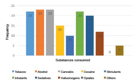
The consumer screening test allowed identifying the substances consumed with respect to the polydrug user group. Within this group, 95.7% of the participants reported having used tobacco, 100% alcohol, 100% cannabis, 95.7% inhalants and 87% sedatives. (See Figure 1).
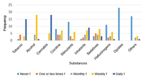
Similarly, in relation to the frequency of consumption for each of the substances by the polydrug group, most of the subjects reported having consumed tobacco (65%), cannabis (78.3%) and inhalants (39 ,1%) daily, compared with those who reported having consumed alcohol (78.3%) and sedatives (34.8%) on a weekly basis (See Figure 2).
Response times of the virtual brain training program "Brain HQ" Module "I focus my attention"
A non-parametric U-Mann-Whitney statistic was used to identify differences between the group of polydrug adolescent users and the quasi-control subjects with respect to the response times for both attention tests (A and B).
Once the Mann-Whitney U test was performed, a difference in the average range was identified for each group with respect to each attention task, whose difference was significant at 0.01. In this regard, polydrug adolescent users obtained a greater median in both tasks with respect to the quasi-control adolescents. Therefore, it can be stated that the consumers group had greater response times in each of the tasks with respect to the quasi-control group (See Table 1).

Recording of brain activity during the attention test
After the qualitative analysis allowing to identify the graphic elements, and the quantitative one, from the Fourier Transformation Scale of the EEG records allowing to obtain spectral power density graphs, a predominance of combined theta-θ (4-7hz) beta-β (13-30hz) waves (See Figure 3) and theta-θ (4-7hz) delta-δ (3-4hz) waves (See Figure 5) on channels AF3, AF4, F3, F4, F7, F8, FC5, FC8, was observed in the group of polydrug users, corresponding to prefrontal and frontal brain areas during tasks A and B for the majority of participants. In relation to the quasi-control group of adolescents, a prevalence of combined theta (47hz) alpha-α (7-13hz) waves was identified in channels AF3, AF4, F3, F4, F7, F8, FC5, FC8 during the execution of both tasks A and B for the majority of participants of the different age groups (See Figures 4 and 6).
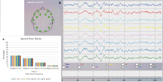
Figure 3 A) Spectral Power Density and B) EEG Record with the Bench Test: during task A of selective and sustained attention of the Virtual training program "Brain HQ" Module "I focus my attention" in a polydrug adolescent consumer. Graph and EEG Record associated with channels AF3, AF4, F3, F4, F7, F8, FC5, FC6 showed a predominance of theta-θ delta-δ waves.
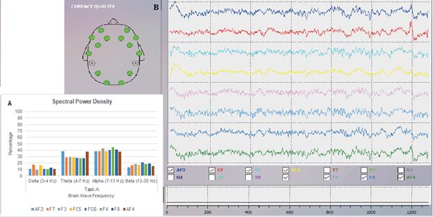
Figure 4 A) Spectral Power Density and B) EEG Record with the Bench Test: during task A of selective and sustained attention of the Virtual training program "Brain HQ" Module "I focus my attention" in a teenager of the QuasiControl group. Graph and EEG record associated with the channels AF3, AF4, F3, F4, F7, F8, FC5, FC6 showed a predominance of theta-θ alpha-α waves.
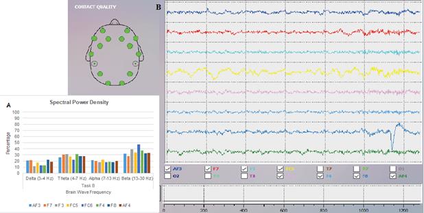
Figure 5 A) Spectral Power Density and B) EEG Record with the Bench Test: during task B of selective and sustained attention of the Virtual training program "Brain HQ" Module "I focus my attention" in a polydrug adolescent consumer. Graph and EEG record associated with channels AF3, AF4, F3, F4, F7, F8, FC5, FC6 showed a predominance of theta-θ beta-β waves.
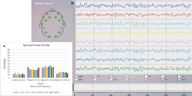
Figure 6 A) Spectral Power Density and B) EEG Record with the Bench Test: during task B of selective and sustained attention of the Virtual training program "Brain HQ" Module "I focus my attention" in a teenager of the Quasi-Control group. Graph and EEG record associated with channels AF3, AF4, F3, F4, F7, F8, FC5, FC6 showed a predominance of theta-θ alpha-α waves.
Discussion
The objective of this research was to record brain electrical activity (EEG) in attentional tasks (sustained and selective) in polydrug adolescent consumers using an Epoc Emotiv BCI (Brain Control Interface) instrument. The results obtained show the presence of theta-θ (4-7hz) beta-β (13-30hz) and theta-θ (4-7hz) delta-δ (3-4hz) waves in the group of polydrug adolescent users, in prefrontal and frontal areas associated with the channels AF3, AF4, F3, F4, F7, F8, FC5, FC6, and these in turn, related to the attentional process (Squire, Noudoost, Schafer & Moore, 2013). The presence of theta-θ (4-7hz) beta-β (13-30hz) waves in polydrug adolescent consumers is a finding that correlates with electroencephalographic recordings of subjects with ADHD who present a similar electrophysiological pattern. This indicates the possible existence of alterations in the attentional process in polydrug adolescent users (Adamaszek, Khaw, Buck, Andresen & Thomasius, 2010, Heming et al., 1994, Kiyatkin & Smimov, 2010, Lansbergen et al., 2011, Reid et al. ., 2006; Van et al., 2010). Polydrug use is a disorder characterized by behavioral disinhibition (Lacono et al., 2008) or decreased ability to regulate control over desire (Feil, Sheppard, Fitzgerald, Yucel, Lubman & Bradshaw, 2010), which in turn, is characteristic of subjects with ADHD.
The increase in theta-θ (4-7hz) delta-δ (3-4hz) waves identified in the present study in polydrug adolescent users, suggests a hypo activity in frontal and prefrontal zones associated with attentional slowdown, cortical atrophy, frontal dysfunction and poor performance in attentional tasks, results comparable to those found in the studies conducted by Adamaszek et al. (2010); Balconi and Finocchiaro (2015); Courtney and Polich (2010); Greenwald and Roehrs (2005); Quesada, Diaz, Herrera, Tamayo and Rubio (2007). In relation to the quasi-control group of adolescents, a predominance of theta-θ (4-7hz) alpha-α (7-13hz) waves was identified in prefrontal and frontal areas associated with the channels AF3, AF4, F3, F4, F7 , F8, FC5, FC6 in the majority of participants of the sample, especially in the groups of 12, 14, 15 and 16 years of age during the attentional tasks A and B. These waves are representative of the attentional process, a fact supported from different sources stating that the presence of theta-θ (4-7hz) alpha-α (7-13hz) waves is related to this process (Lee, 2013; Lomas et al., 2015). Although theta-θ waves (4-7hz) are observed when there is a decrease in the level of activation (Nicolas & Gómez, 2012), they are also present and associated with states that require a lot of concentration during the performance of tasks involving the attentional process and working memory (Carretie, 2009, Salgado, 2003).
According to the above, these results suggest that the consumption of psychoactive substances generate changes in adolescents' electrical brain activity. This activity is reflected in the attentional deficits observed in polydrug consumer adolescents and, in turn, constitutes a possible risk factor for the start and maintenance of substance use. Regarding response times, these were higher in adolescent consumers compared to adolescents in the quasi-control group during the performance of both attentional tasks A and B (Table 1). This finding could be associated with the presence of an alteration and / or slowing down in the processing of visual information in polydrug participants, which affects the performance in tasks of sustained and selective attention (Acosta et al., 2009, Fernández et al., 2011 Jacobus & Tapert, 2013; Mota & Corral, 2011).
This research contributes to the field of psychophysiology of brain electrical activity with data that allow and help characterize possible electrophysiological profiles associated with adolescent population consuming psychoactive substances. These profiles could be taken into account when evaluating, designing and planning new intervention strategies such as feedback for this type of population. This work constitutes a first effort to link the new brain electrophysiological recording technologies to the study of psychobiological aspects of the consumption of psychoactive substances.
One of the limitations of this study was the size of the sample, so it would be interesting for future research to use larger samples in order to contrast and / or replicate the results obtained in the present investigation.
REFERENCES
Acosta, J., Cervantes, M. L., & Puentes, P. (2009). Perfil del Mini-Mental en policonsumidores de 25-50 años del área metropolitana de la ciudad de Barranquilla-Colombia. Psicogente, 12(22), 316-325. doi: 10.17081/psico.12.22.1062. [ Links ]
Acosta, J., Cervantes, M. L., Pineda, W. F., Torre, G., López, L., & Cárdenas, B (2011). Policonsumo desde una perspectiva neuropsicológica. Psicogente, 14(25), 178-189. Recuperado de http://revistas.unisimon.edu.co/index.php/psicogente/article/view/1865/1781. [ Links ]
Adamaszek, M., Khaw, A. V., Buck, U., Andresen, B., & Thomasius, R. (2010). Evidence of Neurotoxicity of Ecstasy: Sustained Effects on Electroencephalographic Activity in Polydrug Users. PLoS ONE, 5(11), 1-6. doi:10.1371/journal.pone.0014097. [ Links ]
Allsop, D. J., & Copeland, J. (2016). Age at first cannabis use moderates EEG markers of recovery from cannabis. Journal of substance use, 21(4), 400-406. doi: 10.3109/14659891.2015.1040090. [ Links ]
American Psychriatric Association (APA). (2005). Manual Diagnostico y Estadístico de los Trastornos Mentales DSM IV-TR (Edición española). España: Masson. [ Links ]
Balconi, M., & Finocchiaro, R. (2015). Decisional impairments in cocaine addiction, reward bias, and cortical oscillation “unbalance”. Neuropsychiatric Disease and Treatment, 11, 777-786. doi: 10.2147/NDT.S79696. [ Links ]
Bauer, L. O. (2001). Predicting Relapse to Alcohol and Drug Abuse via Quantitative Electroencephalography. Neuropsychopharmacology, 25(3), 332-340. doi: 10.1016/S0893133X(01)00236-6. [ Links ]
Belin, D., Belin-Rauscent, A., Murray, J. E., & Everitt, B. J. (2013). Addiction: failure of control over maladaptive incentive habits. Current opinion in neurobiology, 23(4), 564572. doi: 10.1016/j.conb.2013.01.025. [ Links ]
Bobrov, P., Frolov, A., Charles, C., Fedulova, I., Bakhnyan, M., & Zhavoronkov, A. (2011). Brain-Computer Interface Based on Generation of Visual Images. Open Access Freely Available Online, 6(6), 1-12. doi: 10.1371/journal.pone.0020674. [ Links ]
Caballero, L. (2005). Adicción a cocaína: Neurobiología, clínica, diagnóstico y tratamiento. Madrid: Ministerio de Sanidad y Consumo. [ Links ]
Camelo, S., Rojas, D., Mejía, A., & Castro, R. (2015). Registro de la actividad eléctrica cerebral de la atención implicada en la conducción bajo el efecto del alcohol usando un instrumento BCI (Brain Control Interface). Revista Diversitas Perspectivas En Psicología, 11(2), 217-233. doi: 10.15332/s1794-9998.2015.0002.04. [ Links ]
Carretie, L. (2009). Psicofisiología. Madrid: Ed. Piramide. [ Links ]
Ceballos, N. A., Tivis, R., Prather, R., & Nixon, S. J. (2008). Transdermal Nicotine Administration and the Electroencephalographic Activity of Substance Abusers in Treatment. NIH Public Access, 2(4), 202-214. doi: 10.1097/ADM.0b013e31818b4e27. [ Links ]
Choi, D., Ryu, Y., Lee, Y., & Lee, M. (2011). Performance evaluation of a motor-imagerybased EEG-Brain computer interface using a combined cue with heterogeneous training data in BCI-Naive subjects. BioMedical Engineering OnLine, 10(91), 2-12. doi: 10.1186/1475-925X-10-91. [ Links ]
Colado, M. I. (2008). Éxtasis (MDMA): estudios neurobiológicos en el laboratorio. Trastornos adictivos, 10(3), 183-189. doi: 10.1016/S1575-0973(08)76364-5. [ Links ]
Colegio Colombiano de Psicólogos (2013). Deontología y bioética del ejercicio de la psicología en Colombia. Bogotá: El Manual Moderno S. A. [ Links ]
Colzato, L. S., Van den Wildenberg, W. P., & Hommel, B. (2009). Reduced Attentional Scope in Cocaine Polydrug Users. PLoS ONE, 4(6), 1-5. doi: 10.1371/journal.pone.0006043. [ Links ]
Connolly, C. G., Foxe, J. J., Nierenberg, J., Shpaner, M., & Garavan, H. (2012). The neurobiology of cognitive control in successful cocaine abstinence. Drug Alcohol Depend, 121(1-2), 45-53. doi: 10.1016/j.drugalcdep.2011.08.007. [ Links ]
Copersino, M. L., Heming, R. I., Better, W., Cadet, J. L., & Gorelick, D. A. (2009). EEG and cerebral blood flow velocity abnormalities in chronic cocaine users. Clinical EEG and Neuroscience, 40(1), 39-41. doi: 10.1177/155005940904000111. [ Links ]
Corominas, M., Roncero, C., Bruguera, E., & Casas, M. (2007). Sistema dopaminérgico y adicciones. Revista de neurología, 44(1), 23-31. Recuperado de https://dialnet.unirioja.es/servlet/articulo?codigo=2224138. [ Links ]
Coullaut-Valera, R., Arbaiza-Diaz del Rio, I., Arrúe-Ruilobal, R., Coullaut-Valera, J., & BajoBretón, R. (2011). Deterioro cognitivo asociado al consumo de diferentes sustancias psicoactivas. Actas Españolas de Psiquiatría, 39(3), 168-173. Recuperado de https://www.actaspsiquiatria.es/repositorio/13/71/ESP/13-71-ESP-168-173-776266.pdf. [ Links ]
Courtney, K. E., & Polich, J. (2010). Binge drinking effects on EEG in young adult humans. International Journal of Environmental Research and Public Health, 7(5), 2325-2336. doi:10.3390/ijerph7052325. [ Links ]
Crespo-Fernández, J. A., & Rodríguez, C. A. (2007). Bases neuroanatomicas, neurobiológicas y del aprendizaje de la conducta de adicción a la cocaína. Revista Latinoamericana de Psicología, 39(1), 83-107. doi: 10.14349/rlp.v39i1.572. [ Links ]
Cuervo, M. T., & Quijano, M. C. (2008). Alteraciones de la atención y su rehabilitación en trauma craneoencefálico. Pensamiento psicológico, 4(11), 167-182. [ Links ]
Cui, Y., Versace, F., Engelmann, J. M., Minnix, J. A., Robinson … Cinciripini, P. M. (2013). Alpha Oscillations in response to affective and Cigarette-related stimuli in smoker. Nicotine y Tobacco research, 15(5), 917-924. doi: 10.1093/ntr/nts209. [ Links ]
Daza, M. (2009). Efectos conductuales y neuroquímicos del consumo de éxtasis y cocaína en ratones adolescentes (Tesis doctoral). Departamento de Psicobiología, Universidad de Valencia, Valencia. Recuperado de http://www.tdx.cat/handle/10803/10203?show=full. [ Links ]
D’Souza, M. S., & Markou, A. (2011). Neuronal Mechanisms Underlying Development of Nicotine Dependence: Implications for Novel Smoking-Cessation Treatments. Addiction Science and Clinical Practice, 6(1), 4-17. Recuperado de https://www.ncbi.nlm.nih.gov/pmc/articles/PMC3188825/pdf/ascp-06-1-4.pdf. [ Links ]
Duvinage, M., Castermans, T., & Dutoit, T. (2012). A P300BASED quantitative comparison between the emotiv epoc headset and a medical EEG device. LNMB Lab, 1-6. doi: 10.2316/P.2012.764-071. [ Links ]
Ehlers, C. L., & Phillips, E. (2007). Association of EEG alpha variants and alpha power with alcohol dependence in Mexican American Young Adults. Alcohol, 41(1), 13-20. doi: 10.1016/j.alcohol.2007.02.001. [ Links ]
Emotiv inc (2011). Emotiv EPOC. Recuperado en: https://emotiv.gitbook.io/epoc-user-manual/epoc+_headset_details/universal_usb_receiver_dongle. [ Links ]
Esel, E. (2006). Neurobiology of Alcohol Withdrawal: Inhibitory and Excitatory Neurotransmitters. Turkish Journal of Psychiatry, 17(2), 1-9. Recuperado de https://pdfs.semanticscholar.org/aaa0/5a53fdc40f33a3d4ed182ca5050664e119f4.pdf. [ Links ]
Fagundo, A. B., Martin-Santos, R., Abanades, S., Farré, M., & Verdejo-García, A. (2008). Neuroimagen y adicción II: correlatos neuroantómicos y funcionales de la administración aguda, el craving y el consumo crónico de opiáceos, alcohol y cannabis. Revista Española de Drogodependencia, 32(2), 125-149. Recuperado de http://roderic.uv.es/bitstream/handle/10550/22378/v33n2_1.pdf?sequence=1&isAllowed=y. [ Links ]
Feil, J., Sheppard, D., Fitzgerald, P. B., Yucel, M., Lubman, D. I., & Bradshaw, J. L. (2010). Addiction, compulsive drug seeking, and the role of frontostriatal mechanisms in regulating inhibitory control. Neuroscience y Beobehavioral Reviews, 35(2), 248-275. doi: 10.1016/j.neubiorev.2010.03.001. [ Links ]
Fernández-Espejo, E. (2006). Neurobiología de la adicción en psicoestimulante. Revista de Neurología, 43(3), 147-154. Recuperado de https://idus.us.es/xmlui/bitstream/handle/11441/32261/neurobiologiadelaadiccion.pdf?sequence=1&isAllowed=y. [ Links ]
Fernández-Serrano, M. J., Pérez-García, M., & Verdejo-García, A. (2011).What are the specific Vs Generalized effects of drugs of abuse in neuropsychological performance. Neuroscience and Biobehavioral Reviews, 35(3), 377-406. doi: 10.1016/j.neubiorev.2010.04.008. [ Links ]
Flórez, F., Azorín, J. M., Úbeda, A., & Fernández, E. (2011). Development of a low-cost svm-based spontaneous braincomputer interfac. Biomedical Neuroengineering Group (NBIO), 415-421. doi: 10.5220/0003725704650469. [ Links ]
Fondo de la Naciones Unidas para la Infancia (Unicef). (2011). Estado Mundial de la Infancia: la adolescencia una época de oportunidades. Nueva York: Unicef. [ Links ]
Fondo de la Naciones Unidas para la Infancia (Unicef). (2013). Situación Actual con respecto al uso indebido de drogas. Informe de la Secretaria. Viena: Unicef. [ Links ]
Franken, I. H. (2003). Drug craving and addiction: integrating psychological and neuropsychopharmacological approaches. Progress in Neuro-Psychopharmacology and Biological Psychiatry, 27(4), 563-579. doi: 10.1016/S0278-5846(03)00081-2. [ Links ]
Garcia, J. M., Garcia, M., & Rivera, S. (2015). Potencial resiliente en familias con adolescentes que consumen y no consumen alcohol. Acta Colombiana de Psicología, 18(2), 163-172. doi: 10.14718/ACP.2015.18.2.14. [ Links ]
García, L. M., & Barriguete, B. (2012). Avance en la comprensión del fenómeno de las adicciones. México: Secretaría de Salud. [ Links ]
Gaviria, M. I. (2010). Interacciones neuroquímicas y comportamentales entre 3.4metilenodiosimetanfetamina (MDMA) y etanol: implicación en la neurotoxicidad de MDMA y en el consumo de etanol en roedores (Tesis doctoral). Facultad de Medicina, Universidad Complutense de Madrid, Madrid. [ Links ]
Gilpin, N. G., & Koob, G. F. (2008). Neurobiology of Alcohol Dependence: Focus on Motivational Mechanisms. Alcohol Research y Health, 31(3), 185195. Recuperado de https://www.ncbi.nlm.nih.gov/pmc/articles/PMC2770186/pdf/arh31-3-185.pdf. [ Links ]
Gobierno Nacional de la República de Colombia (2013). Estudio Nacional de Consumo de Sustancias Psicoactivas en Colombia. Bogotá: ALVI Impresores S.A.S. [ Links ]
Golarai, G., Ghahremani, D. G., Whitfield-Gabrieli, S., Reiss, A., Eberhardt, J. L., De Gabrieli, J., & Grill-Spector, K. (2007). Differential development of high-level visual cortex correlates with category-specific recognition memory. Nature Neuroscience, 10(4), 512-522. doi: 10.1038/nn1865. [ Links ]
González, L., Ortiz, T., Gutiérrez, C., & González, C. (2011). Estudio conjunto de magnetoencefaografía electroencefalogrfía en epilepsia (Tesis doctoral). Departamento de Psiquiatría. Universidad Complutense de Madrid, Madrid. Recuperado de http://eprints.ucm.es/12536/1/T32720.pdf. [ Links ]
Greenwald, M. K., & Roehrs, T. A. (2005). Mu-opioid Self-Administration vs Passive Administration in Heroin Abusers Produces Differential EEG Activation. Neuropsychopharmacology, 30(1), 212-221. doi:10.1038/sj.npp.1300596. [ Links ]
Guardia, J. (2001). Neuroimagen y drogodependencias. Trastornos adictivos, 3(2), 95-110. Recuperado de http://www.elsevier.es/es-revista-trastornos-adictivos-182-pdf-13015408. [ Links ]
Heming, R. I., Glover, B. J., Koeppl, B., Phillips, R. L., & London, E. D. (1994). Cocaine-Induced Increases in EEG Alpha and Beta Activity: Evidence for Reduced Cortical Processing. Neuropsychopharmacology, 11(1), 1-9. doi: 10.1038/npp.1994.30. [ Links ]
Jacobus, J., & Tapert, S. F. (2013). Neurotoxic effects of alcohol in adolescence. Annual Review of Clinical Psychology, 9, 703-721. doi: 10.1146/annurev-clinpsy-050212-185610. [ Links ]
Kauhanen, L., Jylanki, P., Lehtonen, J., Rantanen, P., Alaranta, H., & Sams, M. (2007). EEG-Based Brain-Computer Interface for Tetraplegics. Computational Intelligence and Neuroscience, 2007 (23864), 1-11. doi: 10.1155/2007/23864. [ Links ]
Kiyatkin, E. A., & Smimov, M. S. (2010). Rapid EEG desynchronization and EEG activation induced by intravenous cocaine in freely moving rats: a peripheral, nondopamine neural triggering. American journal of physiology, 298(2), R285-R300. doi:10.1152/ajpregu.00628.2009. [ Links ]
Kosten, T. R., & George, T. P. (2002). The Neurobiology of Opioid Dependence: Implications for Treatment. Science y Practice Perspectives, 1(1), 13-20. doi: 10.1151/spp021113. [ Links ]
Kreek, M. J., Levran, O., Reed, B., Schlussman, S. D., Zhou, Y., & Butelman, E. R. (2012). Opiate addiction and cocaine addiction: underlying molecular neurobiology and genetics. The Journal of Clinical Investigation, 122(10), 3387-3393. doi:10.1172/JCI60390. [ Links ]
Lacono, W., Malone, S. M., & McGue, M. (2008). Behavioral Disinhibition and the Development of EarlyOnset Addiction: Common and Specific influences. Annual Review of Clinical Psychology, 4, 325-348. doi: 10.1146/annurev.clinpsy.4.022007.141157. [ Links ]
Lansbergen, M. M., Dumont, G. J., Van Gerven, J. M., Buitelaar, J. K., & Verke, R. J. (2011). Acuteeffects of MDMA (3.4-methylenedioxymethamphetamine) on EEG oscillations: alone and in combination with ethanol or THC (delta9-tetrahydrocannabinol). Psychopharmacology, 213(4), 745-756. doi: 10.1007/s00213-010-2031-4. [ Links ]
Lansbergen, M. M., Arns, M., Van Dongen-Boomsma, M., Spronk, D., & Buitelaar, J. (2011). The increase in theta/ beta ratio on resting-state EEG in boys with attentiondeficit/hyperactivity disorder is mediated vy slow alpha peak frequency. Progress in Neuro-Psychopharmacology and Biological Psychiatry, 35(1), 47-52. doi: 10.1016/j.pnpbp.2010.08.004. [ Links ]
Lee, L. (2013). Mechanisms and Functions of theta Rhythms. Annual Review of Neuroscience, 36, 395-312. doi: 10.1146/annurev-neuro-062012-170330. [ Links ]
León, M. l., González, L. H., León, A., Armas, J. O., Urquiza, A., & Rodríguez, G. (2014). Bases Neurobiológicas de la adicción al alcohol. Revista Finlay, 4(1), 40-53. Recuperado de http://www.revfinlay.sld.cu/index.php/finlay/article/view/253/1257. [ Links ]
León, O. G., & Montero, I. (2003). Métodos de investigación en Psicología y Educación (3.ª ed.). Madrid: Mc Graw-Hill. [ Links ]
Littel, M., Franken, I. H., & Van Strien, J. W. (2009). Changes in the Electroencephalographic Spectrum in Response to Smoking Cues in Smokers and Ex-Smokers. Neuropsychopharmacology, 59(1), 43-50. doi: 10.1159/000205517. [ Links ]
Liu, X., Vaupel, D. B., Grant, S., & London, E. D. (1998). Effect of Cocaine-Related Environmental Stimuli on the Spontaneous Electroencephalogram in Polydrug Abusers. Neuropsychopharmacology, 19(1), 10-17. doi: 10.1016/ S0893-133X(97)00192-9. [ Links ]
Lomas, T., Ivtzan, I., & Cynthia, H. Y. (2015). A systematic review of the neurophysiology of mindfulness on EEG oscillations. Neuroscience y Biobehavioral Reviews, 57, 401410. doi: 10.1016/j.neubiorev.2015.09.018. [ Links ]
López-Caneda, E., Mota, N., Crego, A., Velásquez, T., Corral, M., Rodríguez, S., & Cadaveira, F. (2014). Anomalías neurocognitivas asociadas al consumo intensivo de alcohol (binge drinking) en jóvenes y adolescentes: una revisión. Adicciones, 26(4), 334-359. doi: 10.20882/adicciones.39. [ Links ]
Madoz-Gúríde, A., Ochoa, E., & Martínez, B. (2009). Consumo de cocaína y daño neuropsicológico: implicaciones clínicas. Revista de Medicina Clínica, 132(14), 555-559. Recuperado de https://www.researchgate.net/publication/246618526_Consumo_de_cocaina_y_dano_neuropsicologico_Implicaciones_clinicas. [ Links ]
Madoz-Gúrpide, A., & Ochoa-Mangado, E. (2012). Alteraciones de funciones cognitivas y ejecutivas en pacientes dependientes de cocaína: estudio de casos y controles. Revista de Neurología, 54(4), 199-208. [ Links ]
Mena, I., Dorr, A., Viani, S., Neubauer, S., Gorostegui, M. E., Dorr, M. P., & Ulloa, D. (2013). Efectos del consumo de marihuana en escolares sobre funciones cerebrales demostrados mediante pruebas neuropsicológicas e imágenes de neuro-SPECT. Salud mental, 36(5), 367-374. Recuperado de http://www.scielo.org.mx/pdf/sm/v36n5/v36n5a3.pdf. [ Links ]
Merzenich, M. (2003). Ejercicio “enfoco mi Atención” del Programa virtual de entrenamiento Brain HQ. Recuperado de https://es.brainhq.com/?fblogin=returning_fb#train/attention/0/0/0. [ Links ]
Moratalla, R. (2008). Neurobiología de la cocaína. Trastornos Adictivos, 10(3), 143-150. Recuperado de http://www.elsevier.es/es-revista-trastornos-adictivos-182-pdf-13128589. [ Links ]
Mota, N. G., & Corral, M. M. (2011). Estudio longitudinal del perfil neuropsicológico del consumo intensivo de alcohol entre jóvenes universitarios (Tesis doctoral). Departamento de Psicología Clínica y Psicobiología, Universidad de Santiago de Compostela, Santiago de Compostela. Recuperado de https://minerva.usc.es/xmlui/bitstream/handle/10347/3388/9788498876345_content.pdf?sequence=1. [ Links ]
National Intitute on Drug Abuse (2010). Cocaína: abuso y adicción. España. Recuperado de http://www.ndcrc.org/content/serie-de-reportes-de-investigaci%C3%B3ncoca%C3%ADna-abuso-y-adicci%C3%B3n. [ Links ]
Nestler, E. J. (2005). The Neurobiology of Cocaine Addiction. Science y Practice Perspectives, 3(1) 4-12. [ Links ]
Nicolas, L. F., & Gómez, J. (2012). Brain Computer Interfaces, a Review. Sensors, 12(2), 1211-1279. doi: 10.3390/s120201211. [ Links ]
Oficina de Naciones Unidas contra la droga y el delito (UNODC). (2016). Informe Mundial sobre las Drogas. Recuperado de https://www.unodc.org/doc/wdr2016/WDR_2016_ExSum_spanish.pdf. [ Links ]
Organización Mundial de la Salud. (2011). La prueba de detección de consumo de alcohol, tabaco y sustancias (ASSIST): Manual para uso en la atención primaria. Estados Unidos: OMS. [ Links ]
Pagani, L. S. (2014). Environmental tobacco smoke exposure and brain development: The case of attention déficit/ Hyperactivity disorder. Neuroscience y Biobehavioral Reviews, 44, 195-205. doi: 10.1016/j.neubiorev.2013.03.008. [ Links ]
Partilla, J. S., Dempsey, A. G., Nagpal, A, S., Blough, B. E., Baumann, M. H., & Rothman, R. B. (2006). Interaction of Amphetamines and Related Compounds at the Vesicular Monoamine Transporter. The journal of pharmacology and experimental therapeutics, 319(1), 237-246. doi: 10.1124/jpet.106.103622. [ Links ]
Pérez-Rubio, G., Urdapilleta, E., Camarena, A., ReséndizHernández, J. M., Méndez, M., … Falfán-Valencia, R. (2011). Visión general de la neurobiología y genética en la adicción a la nicotina. Neumología y cirugia del Tórax, 70(3), 179-187. Recuperado de http://www.medigraphic.com/pdfs/neumo/nt-2011/nt113g.pdf. [ Links ]
Phillips, R. L., Heming, R., & London, E. D. (1994). Morphine Effects on the Spontaneous Electroencephalogram in Polydrug Abusers: Correlations with Subjective Self-Reports. Neuropsychopharmacology, 10(3), 171-181. doi: 10.1038/ npp.1994.19. [ Links ]
Quesada, M. E., Diaz-Pérez, G. F., Herrera, M., TamayoPorras, M., & Rubio-López, R. (2007). Características del electroencefalograma cuantitativo y trastorno cognitivos en pacientes alcohólicos. Revista de Neurología, 44(2), 81-88. Recuperado de https://dialnet.unirioja.es/servlet/articulo?codigo=2232935. [ Links ]
Redolar, D (2008). Cerebro y adicción. Barcelona: Editorial UOC. [ Links ]
Reid, M. S., Flammino, F., Howard, B., Nilsen, D., & Prichep, L. S. (2006). Topographic Imaging of Quantitative EEG in Response to Smoked Cocaine Self-Administration in Humans. Neuropsychopharmacology, 31, 872-884. doi: 10.1038/sj.npp.1300888. [ Links ]
Ríos, M., Muñoz, J., & Paúl, N. (2007). Alteraciones de la atención tras daño cerebral traumático: evaluación y rehabilitación. Revista de Neurología, 44(5), 291-297. Recuperado de http://www.sld.cu/galerias/pdf/sitios/rehabilitacion-adulto/alteraciones_de_la_atencion_tras_dano.pdf. [ Links ]
Rodríguez, U., Carrillo, E., & Soto, E. (2005). Cannabinoides: neurobiología y usos médicos. Elementos: Ciencia y Cultura, 12(60), 3-9. Recuperado de http://www.redalyc.org/pdf/294/29406001.pdf. [ Links ]
Rosselli, M., & Ardila, A. (1996). Cognitive effects of cocaine and polydrug abuse. Journal of Clinical and Experimental Neuropsychology, 18(1), 122-135. doi: 10.1080/01688639608408268. [ Links ]
Sánchez, A., Vázquez, C., & Valiente, C. (2011). Atención selectiva como mecanismo de regulación emocional y factor de vulnerabilidad a la depresión (Tesis doctoral). Facultad de Psicología, Universidad Complutense de Madrid, Madrid. Recuperado de http://eprints.ucm.es/14460/1/T33365.pdf. [ Links ]
Santana, D., Ramírez, M., & Ostrosky-Solís, F. (2004). Novedades en tecnología de la rehabilitación: una revisión acerca de la interfaz cerebro-computadora. Revista de Neurología, 39, 447-450. Recuperado de https://www.researchgate.net/profile/Maura_Ramirez/publication/221657835_Novedades_en_tecnologia_de_la_rehabilitacion_Una_revision_acerca_de_interfaz_cerebro-computadora/links/0d1c84f54efc8ad4aa000000/Novedades-en-tecnologia-de-la-rehabilitacion-Una-revision-acerca-de-interfazcerebro-computadora.pdf. [ Links ]
Salgado, A. (2003). Evaluación psicofisiológica: Introducción a la Psicofisiología. España: Ed. Universidad Pontificia de Salamanca. [ Links ]
Silva, J. (2011). Métodos en neurociencias cognitivas. México: Manual Moderno. [ Links ]
Squire, R. F., Noudoost, B., Schafer, R. J., & Moore, T. (2013). Prefrotal Contributions to visual selective attention. Annual Review of Neuroscience, 36, 451-466. doi: 10.1146/annurev-neuro-062111-150439. [ Links ]
Talamillo, T. (2011). Manual básico para enfermeros en electroencefalografía. Enfermería Docente, 94, 29-33. Recuperado de http://www.juntadeandalucia.es/servicioandaluzdesalud/huvvsites/default/files/revistas/ED-094-07.pdf. [ Links ]
Urigüen, L., & Callado, L. F. (2010). Cocaína y cerebro. Trastornos Adictivos, 12(4), 129-134. doi: 10.1016/S1575-0973(10)70025-8. [ Links ]
Van Hell, H. H., Vink, M., Ossewaarde, L., Jager, G., Kahn, R. S., & Ramsey, N. F. (2011). Efectos crónicos del consumo de cannabis sobre el sistema de recompensa humano: un estudio de RMf. Psiquiatría Biológica, 18(2), 45-54. doi:10.1016/j.psiq.2011.08.003. [ Links ]
Weise, M., Eisenhofer, G., & Merke, D. P. (2002). Pubertal and Gender-Related Changes in the Sympathoadrenal System in Healthy Children. The Journal of Clinical Endocrinology y Metabolism, 87(11), 5038-5043. doi: 10.1210/jc.2002-0205905038. [ Links ]
Zhao, Q., & Zhang, L. (2007). Temporal and Spatial Features of Single-Trial EEG for Brain-Computer Interface. Computational Intelligence and Neuroscience. 1-14. doi: 10.1155/2007/37695. [ Links ]
How to quote this article: Díaz Baquero, A.A. & Camelo Roa, S.M. (2019). Actividad eléctrica cerebral de la atención en adolescentes policonsumidores usando un equipo BCI (brain control interface). Acta Colombiana de Psicología, 22(1), 175-188. doi: http://www.dx.doi.org/10.14718/ ACP.2019.22.1.9.
Received: November 07, 2017; Revised: March 12, 2018; Accepted: April 30, 2018
 Este es un artículo publicado en acceso abierto bajo una licencia Creative Commons
