


Services on Demand
Journal
Article
 English (pdf)
 Article in xml format
 Article references
 Send this article by e-mail
Indicators
-
 Cited by SciELO
 -
 Access statistics
Related links
-
 Cited by Google
 -
 Similars in
SciELO
Similars in
SciELO -
 Similars in Google
Share

 Permalink
Infectio
Print version ISSN 0123-9392
Infect. vol.18 no.4 Bogotá Sep./Dec. 2014
https://doi.org/10.1016/j.infect.2014.04.0020123-9392
CASE REPORT
http://dx.doi.org/10.1016/j.infect.2014.04.0020123-9392/
Liver abscess secondary to umbilical catheterization in a newborn
Gustavo Adolfo Carvajal-Barrios a, Ivohne Fernanda Corrales-Cobos a, b, María Carmenza Cuenca-Arias a, Gloria Amparo Troncoso-Moreno a, b, *
a Unidad de Cuidado Neonatal, Fundacion Cardoinfantil-IC, Bogotá, Colombia
b Universidad del Rosario, Bogotá, Colombia
Received 4 August 2013; accepted 8 April 2014 Available online 19 June 2014
Abstract
We present a case of liver abscess in a 34-week preterm infant after umbilical venous catheterization (UVC). The infant had clinical symptoms of intestinal sepsis, with an encapsulated hypodense hepatic focal lesion with air inside, as observed by abdominal tomography. Staphylococcus aureus was isolated in blood cultures. Liver abscess is a rare complication associated with umbilical catheterization and can be prevented by implementing an appropriate prevention program.
KEYWORDS
Liver abscess; Umbilical venous catheter; Parenteral nutrition; Premature
© 2013 ACIN. Published by Elsevier España, S.L.U. All rights reserved.
Absceso hepático asociado con cateterización umbilical en un neonato
Resumen
Presentamos un caso de un absceso hepático en una prematura de 34 semanas. La niña presentó síntomas clínicos de sepsis intestinal con una imagen de lesión hipodensa conteniendo aire en hígado en la tomografía de abdomen. Se aisló Staphylococcus aureus en cultivos de sangre. El absceso secundario a colocación de catéter venoso umbilical es infrecuente y puede ser prevenido con un programa preventivo adecuado.
PALABRAS CLAVE
Absceso hepático; Catéter venoso umbilical; Nutrición parenteral; Prematuro
© 2013 ACIN. Publicado por Elsevier España, S.L.U. Todos los derechos reservados.
0123-9392/© 2013 ACIN. Published by Elsevier España, S.L.U. All rights reserved.
Introduction
Liver abscess secondary to umbilical catheterization is an infrequent, preventable, and highly lethal complication, which increases hospital stays, morbidity and mortality. Itis generally caused by bacteria and fungi, but in some cases there is not microbiological isolation. 1,2 Its usual location isin the right hepatic lobe. 1-7 Fewer than 60 cases have been reported between 1972 and 2013, most of them in the United States (USA), India, France and Venezuela. 1,2 Associated risk factors are prematurity, immunodeficiency, malposition of the umbilical catheter and infusion of hypertonic fluids. 1-7
Description of the case
In February, 2011, an 8-day-old, 34-week preterm infant originating from Aruba was admitted to the neonatal intensive care unit (NICU). She was the product of the second pregnancy of a healthy 23 years old mother with prenatal care, delivered by emergency C-section due to preeclampsia. She received 2 doses of pulmonary maturation; the dose and type of steroid were not reported. Birth weight was 1654 g, length 42 cm, head circumference 31 cm, Apgar 9-9-10/10. The referring site began early per os (by mouth) and placed a umbilical venous catheter (UVC) for total parenteral nutrition (TPN). Hypoglycemia was corrected and indirect hyperbilirubinemia was treated with phototherapy for two days. Brain ultrasound showed grade I intraventricular hemorrhage and the echocardiogram was reported as normal. Continuous positive airway pressure (CPAP) was begun due to persistent apneas, and ampicillin was started and later replaced with flucloxacillin due to isolation of Staphylococcus aureus in blood. She developed abdominal distension without necrotizing enterocolitis (NEC) images, and per os was suspended. She developed anemia which required transfusions, and thrombocytopenia without repercussions or transfusions. Antibiotic treatment was steppedup to meropenem for 24 h, after which, she was referred to a tertiary level due to suspected NEC.
On admission to our NICU she was pale, with respiratory distress, without tachycardia, with systolic heart murmur, audible bowel sounds, and soft and distended abdomen with a visible venous network and a painful hepatomegaly 4 cm below the right costal margin, abdominal circumference of 32 cm, weight 2090 g and length 43 cm. She continued nothing by mouth, with intravenous fluids, oxygenand open orogastric tube. She was admitted with anemia and thrombocytopenia without clinical repercussions andwith elevated levels of erythroid sedimentation rate (ESR),C-reactive protein (CRP) and aspartate transferase (AST) enzymes. The renal and thyroid function was within normallimits for age, but she presented severe hypoglycemia and hypercalcemia, which were corrected. After 24 h of hospitalization, she developed bands on the CBC with an increase in the severity of thrombocytopenia without repercussion (seeTable 1). Congenital infection with syphilis, Toxoplasma, rubella, cytomegalovirus, herpes I and II or human immunodeficiency virus type 1 and 2 was ruled out. Two blood cultures were drawn and piperacillin, tazobactam and vancomycin were started.
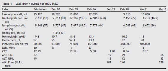
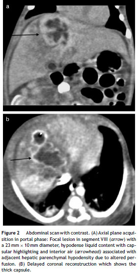
The initial abdominal ultrasound showed a focal hepaticlesion in segment IVA and VIII (see Fig. 1) and the abdominal scan with contrast showed an image compatible with an encapsulated hepatic abscess (see Fig. 2a and b). Methicillin sensitive S. aureus was isolated in blood andoropharynx (Bac/ALERT FA Plus machine; USA). Oxacillin scheme was included and subsequent negative blood cultures were obtained 72 h later. She showed good clinical and paraclinical progress with resolution of the anemia and thrombocytopenia, normalization of AST, ESR, and CRP (seeTable 1), and she continued with IV antibiotics without percutaneous drainage of the abscess.

The brain ultrasound, electrocardiogram and follow-upechocardiogram were normal. In addition, primary cellular immunodeficiency (PI), hypogammaglobulinemia and chronic granulomatous disease were ruled out in the patient and the mother. After finishing the 21-day IV antibiotic cycle, she completed six weeks with cephalexin. The clinical, para-clinical and imaging progress was adequate, with a decrease in the size of the hepatic lesion to 16 mm in segment VIII. The lesion disappeared in the next follow-up ultrasound.
Discussion
Hepatic abscess secondary to umbilical venous catheterization is an infrequent and lethal complication producedby an ascending infection which enters the portal system and usually affects the right lobe of the liver, due to its segmental vascular distribution. Fifty percent are solitary lesions. 1,2 They can be homogenous, solitary, well-defined circular masses or heterogenous, multiloculated,poorly defined, septated masses, with detritus or evenair inside. 1-7 The reported etiologic agents are 37% gram 258 positive cocci (distributed in S. aureus 21% and Staphylococcus epidermidis 16%) with a latent clinical evolution,such occurred in our case; gram negative bacilli in 36% with greater systemic involvement and higher mortality; fungi in 12.5% associated with a fatal outcome, and approximately 10% of cases without microbial isolation, with a subacute evolution. 1-7 Isolation in blood was performed by colorimetry on Bact/ALERT equipment, which has high analytical sensibility, growth efficiency and microbiologic recovery. Clinically, they develop abdominal distension, painful hepatomegaly and nonspecific signs of sepsis such as irritability, altered heart rate, and dysthermias related to altered acutephase reactants, leukocyte counts and platelet counts, as we observed in our patient.
The majority of cases reported until now were from USA, India, France and Venezuela, which reflects a greater reporting culture rather than a higher incidence. 1-7 The literature lists as risk factors: prematurity, low birth weight, immunelability secondary to prematurity, NEC, primary or acquired immunodeficiency. Chronic granulomatous disease should beruled out by using the flow cytometric dihydro-rhodamine 123 test, 8 as well as invasive procedures, such as TPN administration, infusion of hypertonic solutions and antibiotics. 1-7
This case report coincides with the literature in two risk factors: 34-week gestation prematurity and placement of a UVC with TPN and antibiotic infusion, with prematurity being non modifiable in the NICU. Precautions such asthe isolation of the catheter emplacement and a correct location at the vena cava, above the ductus venosus and suprahepatic veins and below the level of the right atrium, 9 with radiologic control to avoid hepatic intraparenchymal migration and infections related to the device, should betaken. These recommendations are in accord with the guidelines published in 2011 by the IDSA (Infectious Diseases Society of America) 10 and includes adequate hand washing, 11 and washing of surfaces and medical equipment in the NICU with 4% clorhexidine prior to the procedure, given the increased incidence in the NICU of colonization and infection by methicillin resistant S. aureus (MRSA) of intra orextra-institutional origin, which could be a cause of neonatal bacterial endocarditis. 12,13
Regarding the treatment received by our patient, IV antibiotic treatment for three weeks was considered due toher high risk secondary to prematurity, weight, and latentclinical, paraclinical and imaging involvement on admission.She completed a total of 6 weeks abx treatment per os with strict follow-up since she was not a candidate for percutaneous drainage due to an adequate response to medical management, abscess size less than 6 cm, location in the right lobe of the liver, no perforation and lack of associated immunodeficiency. 14
In conclusion, literature review and our experience withpresent case, indicate that hepatic abscess related to umbilical venous catheterization is an infrequent but lethal situation where clinician should be aware. A precise indication for catheter placement along with precautions such as a correct location of the catheter and strict radiologic follow-up, will decrease potential complications.
Ethical responsibilities
Protection of human and animal subjects. The authors declare that no experiments were performed on humans or animals for this study.
Confidentiality of data. The authors declare that they have followed the protocols of their work center on the publication of patient data
Right to privacy and informed consent. The authors have obtained the written informed consent of the patients or subjects mentioned in the article. The corresponding autho ris in possession of this document.
Conflict of interest
The authors declare not to have any conflict of interest.
Acknowledgements
The authors would like to thank the Department of Radiology from the "Fundación Cardio Infantil" who kindly gave access to the images of this case.
* Corresponding author. E-mail addresses: gtroncoso@cardionfantil.org, Glotromo33@yahoo.com (G.A. Troncoso-Moreno).
References
1. Palmero M, Matute A, León S, León K. Absceso hepático piógeno a propósito de un caso. Rev Obstet Ginecol Venez. 2005;65:1-4. [ Links ]
2. Palmero MI, Araujo O, Rodriguez S, Marrugo M. Abscesos hepáticos en el período neonatal: reporte de siete casos y revisión de literatura. Rev Obstet Ginecol Venez. 2008;68:109-13. [ Links ]
3. Jain P, Mishra A, Agarawal V. Ruptured liver abscess in a neonate. African J Paediatric Surg. 2012;9:80-2. [ Links ]
4. Simeunovic E, Arnold M, Sidler D, Moore SW. Liver abscess inneonates. Pediatr Surg Int. 2009;25:153-6. [ Links ]
5. M´hamdi K, Kabiri M, Karboubi L, Ghanimi Z, Barkat A. Neonatal liver abscess after umbilical venous catheter. Arch Pediatr.2013;20:196-8. [ Links ]
6. Mahajan V, Rahman A, Tarawneh A, Sant´anna GM. Liver fluid collection in neonates and its association with the use of a specific umbilical vein catheter: report of five cases. Paediatr ChildHealth. 2011;16:13-5. [ Links ]
7. Shah SI, Hudak J, Meng HD. Meta-analysis of factors associated with survival of neonatal hepatic abscess. Pediatr Infect Dis J.2010;5:215-20. [ Links ]
8. Jirapongsananuruk O, Malech HL, Kuhns DB, Niemela JE, Brown MR, Anderson-Cohen M, et al. Diagnostic paradigm for evaluation of male patients with chronic granulomatous disease, based on the dihydrorhodamine 123 assay. J Allergy Clin Immunol. 2003;111:374-9. [ Links ]
9. Sigman LJ, Custer J. Procedures. In: Tschudy M, Arcara K, editors. The Harriet lane handbook. 19th ed. 2012. p. 66. [ Links ]
10. O´Grady NP, Alexander M, Burns LA, Dellinger EP, Garland J, Heard SO, et al. Guidelines for the prevention of intravascular catheter-related infections. Am J Infect Control. 2011;39 Suppl.1:34. [ Links ]
11. Boyce JM, Pittet D, Healthcare Infection Control Practices Advisory Committee, undefined, HICPAC/SHEA/APIC/IDSA Hand Hygiene Task Force, undefined. Guideline for Hand Hygienein Health-Care Settings. Recommendations of the Health-care Infection Control Practices Advisory Committee and the HIPAC/SHEA/APIC/IDSA Hand Hygiene Task Force. Am J Infect Control. 2002;30:46. [ Links ]
12. Townell NJ, Munckhof WJ, Nimmo G, Bannan A, Holley A, Daniel A, et al. Community-associated methicillin-resistant Staphylococcus aureus endocarditis down under´: case series and literature review. Scand J Infect Dis. 2012;44:536-40. [ Links ]
13. Liu C, Bayer A, Cosgrove SE, Daum RS, Fridkin SK, Gorwitz RJ,et al. Clinical practice guidelines by the infectious diseases society of America for the treatment of methicillin-resistant Staphylococcus aureus infections in adults and children. Clin Infect Dis. 2011;52:55. [ Links ]
14. Bari S, Sheikh KA, Malik AA, Wani RA, Naqash SH. Percutaneous aspiration versus open drainage of liver abscess in children. Pediatr Surg Int. 2007;23:69-74. [ Links ]
