SciELO - Scientific Electronic Library Online

 
vol.23 issue2Procalcitonin test for diagnosis of sepsis or septic shock: systematic review of literature and metanalysis author indexsubject indexarticles search
Home Pagealphabetic serial listing  

Services on Demand

Journal

Article

Indicators

Related links

  • On index processCited by Google
  • Have no similar articlesSimilars in SciELO
  • On index processSimilars in Google

Share


Infectio

Print version ISSN 0123-9392

Infect. vol.23 no.2 Bogotá Apr./June 2019

https://doi.org/10.22354/in.v23i2.768 

ARTÍCULO ORIGINAL

Confirmation of the presence of Angiostrongylus cantonensis in lung tissue of the African giant snail (Lissachatina fulica) in Colombia

Confirmación de la presencia de Angiostrongylus cantonensis en tejido pulmonar de caracol gigante Africano (Lissachatina fulica) en Colombia

Alan Giraldo1  * 

Camilo Garzón1 

Andrés Castillo1  2 

Diego Fernando Córdoba-Rojas1 

1 Universidad del Valle, Facultad de Ciencias Naturales y Exactas, Departamento de Biología, Grupo de Investigación en Ecología Animal. Cali, Colombia.

2 Universidad del Valle, Facultad de Ciencias Naturales y Exactas, Departamento de Biología, Grupo de Investigación en Genética Molecular Humana. Cali, Colombia.


Abstract

Objetive:

To confirm the presence of Angiostrongylus cantonensis in Lissachatina fulica individuals in Colombia.

Methods:

19 individuals of L. fulica were collected in the city of Buenaventura, Valle del Cauca, Colombia. Lung tissue was extracted and two analysis methods were used: visual identification by histological observation, and identification through conventional and real-time PCR. The A. cantonensis detection rate was established taking into account each of the analysis techniques used.

Results:

Presence of A. cantonensis was confirmed in the lung tissue of L. fulica specimens collected in the city of Buenaventura, Valle del Cauca, Colombia. The detection rate based on visual identification through histological analysis was 89%, whereas it was 95% using conventional PCR, and 100% using real-time PCR.

Conclusion:

This study confirmed for the first time the presence of A. cantonensis associated with L. fulica specimens in Colombia. Therefore, eosinophilic meningitis could be considered an emerging disease in Colombia.

Key words: Parasitic disease; meningoencephalitis; mollusks; parasites; disease vectors; zoonoses

Resumen

Objetivo:

confirmar la presencia de Angiostrongylus cantonensis en individuos de Lissachatina fulica en Colombia.

Materiales y métodos:

se recolectaron 19 individuos de L. fulica en la ciudad de Buenaventura, Valle del Cauca, Colombia. Se extrajo el tejido pumonar y se utilizaron dos métodos de análisis: identificación visual por observación histológica e identificación a través de PCR convencional y en tiempo real. La tasa de detección de A. cantonensis fue establecida para cada una de las técnicas de análisis utilizadas.

Resultados:

se confirmó la presencia de A. cantonensis en el tejido pulmonar de los especímenes de L. fulica recolectados en la ciudad de Buenaventura, Valle del Cauca, Colombia. La tasa de detección alcanzanda a través de la técnica de identificación visual mediante análisis histológico fue del 89%, mientras que usando la técnica de PCR convencional fue del 95% y de PCR en tiempo real fue del 100%.

Conclusión:

este estudio confirma por primera vez la presencia de A. cantonensis asociado a especímenes de L. fulica en Colombia. Por lo tanto, se sugiere que la meningitis eosinofílica sea propuesta como una enfermedad emergente en Colombia.

Key words: enfermedad parasítica; meningoencefalítis; moluscos; parasitos; vectores de enfermedades; zoonosis

Introduction

Angiostrongylus cantonensis (Nematoda: Strongylida: Metastrongyloidea) is a helminth characterized by a resistant external cuticle, unsegmented body, and a completely developed gastrointestinal tract. It belongs to the Order Strongylida, Superfamily Metastrongyloidea, which includes Ancylostoma nematodes, a long, thin, fliform worms that reside in the host´s lungs (Vitta et al. 2011). The first report of the presence of Angiostrongylus in humans was made by Nomura and Lim (1945), who detected A. cantonensis in the cephalorachidian liquid of a Taiwanese patient with eosinophilic meningitis.

Like all Metastrongyloidea worm species, A. cantonensis adults inhabit the pulmonary arteries of rats and mice (Anderson 2000), where females lay eggs from which first sta ge (L1) larvae emerge. These L1 larvae migrate towards the pharynx and are swallowed and eliminated through feces. Once in the outside environment they invade a habitual intermediate host, among which are terrestrial mollusks such as the giant African snail (Lissachatina fulica). Larvae go through two molts inside this host over an approximate period of two weeks. When they reach the third stage (L3) they can infect their definitive host. Once inside the host, the L3 migrate to the brain where their development continues until reaching the young adult stage (L5). This process can occur within a 4-week period. The young adults return to the venous system to reach the pulmonary arteries, where after two weeks they reach sexual maturity and can begin laying eggs (Dorta et al. 2007).

Humans are considered accidental A. cantonensis hosts. This parasite can enter their system as a result of the ingestion of snails, of vegetables infected by snail secretions, or of other paratenic hosts such as terrestrial crabs or shrimp. A. cantonensis larvae can reach the brain, and on rare occasions the lungs or the eyes (Dorta et al. 2007, Thiengo et al. 2010). Once inside the central nervous system (CNS), the A. cantonensis larval movement produces meningoencephalitis with a proteiform clinical picture, characterized by its association with an important increase in blood eosinophils and cephalorachidian liquid (CRL); if it moves into the eyes it causes permanent blindness in the patient (Alicata 1991, Jarvi et al. 2012, Londoño et al. 2013).

In less than 10 years the giant African snail has been reported in over 70% of Colombia´s departments. It has been associated in particular with urban centers, in which it finds refuge and food in gardens, road medians, green areas, empty lots, abandoned building sites, or places where organic material has accumulated (De la Ossa et al. 2012, 2014, Bolivar et al. 2013, Giraldo et al. 2014, Avendaño & Linares 2015, Patiño & Giraldo 2017). Recently, Córdoba et al. (2017) reported the presence of three genera of nematode parasites (Angiostrongylus, Aelurostrongylus, and Strongyluris) associated with giant African snail specimens collected in nine cities of the Valle del Cauca, Colombia: Jamundí, Cali, Palmira, Buga, Tuluá, Bugalagrande, Cartago, Dagua, and Buenaventura. owever, these authors do not confirm the specific presence of A. cantonensis in the samples reviewed.

Methods

Samples

In order to confirm the presence of Angiostrongylus cantonensis in individuals of the giant African snail, 19 individuals of L. fulica were collected opportunistically in the continental area of the city of Buenaventura following the recommendations of manipulation stablished by the Ministry of Environment and sustainable development (MADS, 2011). Individuals were transported to the Animal Ecology Laboratory of the Universidad del Valle, where they were sacrificed and preserved following recommendations from MADS (2011).

Histological techniques

Lung tissue was extracted and a visual search was conducted using a stereoscopic microscope to quantify the number of free-living and cystic A. cantonensis larvae. Larvae were identified based on characteristics described for the species by Ash (1970) and Lv et al. (2009).

DNA extraction and PCR

To confirm molecularly the taxonomic identity, DNA extraction was done from lung tissue pieces as well as the nonencysted individuals using the “Wizard Genomic Purification” kit following of manufactural instruction. A. Cantonensis parasites were detected by both conventional and real-time PCR techniques using specific pair-primers designed for the ITS1 ribosomal DNA region of A. cantonensis

The pair-primers were obtained with the online software Primer3 (http://bioinfo.ut.ee/primer3-0.4.0/) using as reference nucleotide sequence: GU587760, available in the GenBank collection. The primers sequences were: AcanITS1Revers 5’-TTC ATG GAT GGC GAA CTG ATA G-3 ‘and AcanITS1Forward 5’-GCG CCC ATT GAA ACA TTA TAC TT-3’. One of the sample positives using the specific primers for A. cantonensis was sequenced by the Sanger method to verify the parasite taxonomic identity using blast analysis-NCBI. Afterward, this sample was used as positive control in the experiments. Also, the free PCR mixture was used as a negative control (Garzon et al. 2017).

Results

Using a histological technique for direct observation, a total of 17 cysts and three free-living A. cantonensis larvae were collected and identifed in 17 of the 19 analyzed samples (Figure 1), which is equivalent to an 89% detection rate. All samples had an identical nucleotide sequence and showed the highest percentage identity with the KP776415.1 A. cantonensis clone 4 (Sydney study) (100% identities, 0% gaps). The negative control failed to amplify in any of the test repetitions (Figure 2).

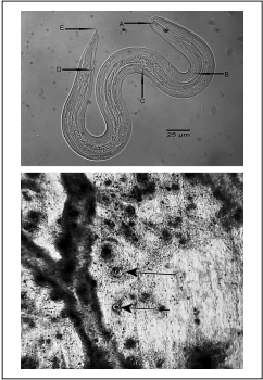
Figure 1 Upper panel: Free-living L3 Angiostrongylus cantonensis larva in Lissachatina fulica lung tissue. A. Rod-like structure with rounded ends in the spear. B. Excretory pore, C. Esophageal bulb, D. Anus, E. Pointed tip. Lower panel: Angiostrongylus cantonensis cysts in Lissachatina fulica lung tissue (arrows) 

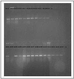
Figure 2 Endpoint PCR in 1% agarose gel. A fluorescent band could be observed for all samples except sample AC02. Negative controls failed to amplify. 

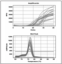
Figure 3 Upper panel: Real-time PCR results for DNA samples from Lissachatina fulica lung tissue containing Angyostrongylus cantonensis cysts. Lower panel: dissociation curves (Tm) of products amplifed by PCR. The Tm of amplifed products was between 76º and 77ºC, whereas a Tm peak was absent for the negative control 

Through conventional PCR we found that 18 of 19 analyzed samples were positive for A. cantonensis presence, equivalent to a 95% detection rate. Moreover, all tissue and free-living nematode samples were positive for A. cantonensis using real-time PCR (Figure 3), equivalent to a 100% detection rate. The analysis of the melting temperature (Tm) showed the same maximum emission value for all samples, which corresponded to the Tm of the primers used (Figure 3).

Discussion

The confirmation of A. cantonensis presence in the lung tissue of giant African snail specimens from the city of Buenaventura, Valle del Cauca, in addition to the great densities of this invasive mollusk reported in urban areas of the Valle del Cauca, Colombia, (Bolivar et al. 2013, Giraldo et al. 2014) and its presence in more than 70% of the country’s departments (De la ossa-Lacayo et al. 2012), could increase the probability of exposure of human beings to the parasite load of this snail, which includes eggs and larvae of A. cantonensis. Therefore, eosinophilic meningitis could be considered an emerging disease in Colombia, as has been reported for Ecuador, Brazil, and Venezuela (Espirito-Santo et al. 2013, Solórzano- Álava et al. 2014).

In Colombia, meningitis is attributed mainly to viral infections or bacterial infections (Tique et al. 2006), excluding the nematode A. cantonensis from the possible agents of infection. However, results from the present study confirm the need to alert medical entities, which should be prepared to carry out timely interventions in case of the development of an epidemic focal point in Colombia (see Londoño et al. 2013).

Real-time PCR detection of A. cantonensis is not yet widely used in its identification, but in the present study is has been proven reliable for the detection of A. cantonensis DNA in lung tissues from the giant African snails with a new specific pairprimers. Despite that the prevalence of A. cantonensis no show statistical difference among techniques, our findings suggest that the Real-time PCR method is more eficient to assess A. cantonensis infection than conventional PCR or histology, especially when there is a small number of parasites.

The differences among methods may be explained by the sensitivity of each method. The real-time PCR is a technique used to monitor the progress of a PCR reaction in real time. At the same time, a relatively small amount of DNA can be detected. Real-Time PCR is based on the detection of the fluorescence produced by a reporter molecule which increases, as the reaction proceeds. This occurs due to the accumulation of the PCR product with each cycle of amplification (Tamay de Dios et al. 2013).

In our experiment, the fluorescent reporter molecules that bind to the double-stranded DNA was SYBR® Green. So, minimal amounts of the nucleic acid of A. cantonensis were detected accurately in the lung tissue of giant African snails. Moreover, there was no need post PCR processing like the electrophoresis in a gel, which saves both resources and time, in contrast, conventional PCR and histology need most more time, and in the case of histology also, it is necessary an expert identify the parasite by observation under a stereoscope.

The implementation of the real-time multiplex PCR technique (Varela et al. 2017) or the technique used during the present study (see Garzon et al. 2017) could become diagnostic tools that would allow the identification of the prevalence of A. cantonensis in giant African snails for a given location, so that control efforts could be directed towards this invasive species to reduce the risk of development of an epidemic focal point of neuro-angiostrongyliasis.

In conclusion, the three techniques employed identified A. cantonensis in the tissue of A. fulica collected in the city of Buenaventura, Colombia. This study confirmed for the first time the presence of A. cantonensis associated with L. fulica specimens in Colombia using histological and molecular methods.

References

1 . Alicata JE. The discovery of Angiostrongylus cantonensis as a cause of human eosinophilic meningitis. Parasitol. Today. 1991; 7(6): 151-53. https://doi.org/10.1016/0169-4758(91)90285-V. [ Links ]

2. Ash L. Diagnostic morphology of the third-stage larvae of Angiostrongylus cantonensis, Angiostrongylus vasorum, Aelurostrongylus abstrusus, and Anaflaroides rostratus (Nematoda: Metastrongyloidea). J. Parasitol. 1970; 56(2): 249-53. https://doi.org/10.2307/3277651. [ Links ]

3. Avendaño JM, Linares E. Morfométrica del caracol gigante africano Achatina fulica (Gastropoda: Achatinidae) en Colombia. Cuadernos de Investigación UNED. 2015; 7(2): 287-93. [ Links ]

4. Bolívar-García W, Giraldo A, Cantera J. (Eds). Generación de información de línea base sobre la especie invasora Achatina fulica en cuatro municipios del Departamento del Valle del Cauca. Informe técnico final Convenio 028/2013. Corporación Autónoma Regional del Valle del Cauca - Grupo de Investigación en Ecología Animal, Departamento de Biología, Facultad de Ciencias Naturales y Exactas, Universidad del Valle. Cali. 2013; 73 p. [ Links ]

5. Cordoba-R D, Patiño-Montoya A, Giraldo A. Prevalence of nematodes associated with African Snail in Valle del Cauca, Colombia. Rev. MVZ Córdoba. 2017; 22(3): 6276-6286. [ Links ]

6. De la ossa-Lacayo A, De la Ossa J. Caracol africano gigante Achatina fulica Bowdich 1822 (Mollusca: Gastropoda-achatinidae) en zona urbana de Sincelejo y Sampués, Sucre, Colombia. Rev. Colombiana Cienc. Anim - RECIA. 2014; 6(2): 299-304. https://doi.org/10.24188/recia.v6.n2.2014.432Links ]

7. De la ossa-Lacayo A, De la Ossa J, Lasso C. Registro del caracol africano gigante Achatina fulica (Bowdich 1822) (Mollusca: GastropodaAchatinidae) en Sincelejo, Costa Caribe de Colombia. Biota colombiana 2012; 13(2):247-52. [ Links ]

8. Dorta AJ, Núñez FA, Pérez O, González ML, Magraner ME, Coifu RB, García EN, Interián T, Martínez JF, Sánchez E. Meningoencefalitis Eeosinofílica por Angiostrongylus Cantonensis. A un cuarto de siglo de una enfermedad emergente en las Américas. Rev. Ecuat. Neurol. 2007; 16(2): 103-13. [ Links ]

9. Espírito-Santo MCCD, Pinto PLS, Mota DJGD, Gryschek RCB. The first case of Angiostrongylus cantonensis eosinophilic meningitis diagnosed in the city of São Paulo, Brazil. Rev. Inst. Med. Trop. São Paulo. 2013; 55(2): 129-32. http://dx.doi.org/10.1590/S0036-46652013000200012Links ]

10. Garzón C, Castillo A, Giraldo A. Implementación de PCR tiempo real para la detección de Angiostrongylus cantonensis (Metastrongylidae) en caracol africano (Achatina fulica) en el Valle del Cauca. Trabajo de Grado. Programa Académico de Biología, Universidad del Valle. Colombia. 2017; 27 p. [ Links ]

11. Giraldo A, Bolívar-García W, Gómez M. (Eds.). Estudio y monitoreo de la especie desestabilizadora caracol gigante africano (Achatina fulica) en siete municipios priorizados del Valle del Cauca. Informe técnico final Convenio 054/2014, Capítulo 1, 8 - 48 pp. Corporación Autónoma Regional del Valle del Cauca - Grupo de Investigación en Ecología Animal, Departamento de Biología, Facultad de Ciencias Naturales y Exactas, Universidad del Valle. Cali. 2014; 127 p. [ Links ]

12. Jarvi SI, Fariasa MEM, Howea K, Jacquiera S, Hollingsworth R, Pitt W. Quantitative PCR estimates Angiostrongylus cantonensis (rat lungworm) infection levels in semi-slugs (Parmarion martensi). Mol. Biochem. Parasitol. 2012; 185(2): 174-76. https://doi.org/10.1016/j.molbiopara.2012.08.002Links ]

13. Londoño JD, Zamora A, Osorio J. Angiostrongylus cantonensis y el caracol gigante africano como causantes de meningitis eosinoflica. Rev. Fac. Nac. Salud Pública. 2013; 5(2):61-69. [ Links ]

14. Lv S, Zhang Y, Liu H, Zhang C, Steimann P, Zhou X, Utzinger J. Angiostrongylus cantonensis: morphological and behavioral investigation within the freshwater snail Pomacea canaliculata. Parasitol. Res. 2009; 104:1351-59. https://doi.org/10.1007/s00436-009-1334-zLinks ]

15. MADS. 2011. Resolución 0654 del abril 7 de 2011. Adopta medidas que deben seguir las autoridades ambientales para la prevención, control y manejo de la especie Caracol Gigante Africano (Achatina fulica). Ministerio de Ambiente y Desarrollo Sostenible, MADS, Colombia. [ Links ]

16. Nomura S, Lin PH. First case report of human infection with Haemostrogylus ratti Yokogawa. Taiwan No Ikai. 1945; 3: 589-92 [ Links ]

17. Patiño A, Giraldo A. Intrapopulational genic variation of the giant African Snail (Achatina fulica) in the Valle del Cauca. Rev. MVZ Córdoba. 2017; 21(2): 5924-36. [ Links ]

18. Solórzano-Álava LF, Martini-Robles L, Hernández-Álvarez H, Sarracent-Pérez J, Muzzio-Aroca J, Rojas- Rivero L. Angiostrongylus cantonensis: un parásito emergente en Ecuador. Rev. Cubana Med. Trop. 2014; 66(1): 20-33. [ Links ]

19. Tamay de Dios L, Ibarra C, Velasquillo C. Fundamentos de la reacción en cadena de la polimerasa (PCR) y de la PCR en tiempo real. Investigación en discapacidad. 2013; 2(2): 70-78. [ Links ]

20. Thiengo SC, Maldonado A, Motac EM, Torres EJL, Caldeirae R, Carvalho OS, Oliveira APM, Simões RO, Fernandez MA, Lanfredi RM. The giant African snail Achatina fulica as natural intermediate host of Angiostrongylus cantonensis in Pernambuco northest Brazil. Acta Trop. 2010; 115: 194-99. https://doi.org/10.1016/j.actatropica.2010.01.005Links ]

21. Tique V, Alvis N, Parodi R, Bustos A, Mattar S. Meningitis agudas en Córdoba, Colombia 2002 - 2004. Rev. Salud Pública. 2006; 8 (Suppl. 1):33-46. http://dx.doi.org/10.1590/S0124-00642006000400004Links ]

22. Varela RE, Arias JS, Velásquez LE. Standardization of a multiplex realtime PCR test for the identification of Angiostrongylus cantonensis, A.costaricensis and A. vasorum. Biomédica, 2018; 38(1):111-119. http://dx.doi.org/10.7705/biomedica.v38i0.3407. [ Links ]

23. Vitta A, Polseela R, Nateeworanart S, Tattiyapong M. Survey of Angiostrongylus cantonensis in rats and giant African land snails in Phitsanulok province, Thailand. Asian Pacific J. Trop. Med. 2011; 4(8):597-99. https://doi.org/10.1016/S1995-7645(11)60154-5Links ]

Cómo citar este artículo: A. Giraldo, et al. Confirmation of the presence of Angiostrongylus cantonensis in lung tissue of the African giant snail (Achatina fulica) in Colombia. Infectio 2019; 23(2): 129-132

Ethical considerations

Protection of human and animal subjects: This research was authorized by the Agencia Nacional de Licencias Ambientales through fle RCI0009-00-2016, and the DNA diagnostic test was delimited by guidelines from the 1993 resolution 8430 from the Health Ministry. This research do not use human material or data.

Confidentiality of data Not applicable

Funding This study was partly financed by the Animal Ecology Research Group and by the Molecular Pathology Laboratory of the Universidad del Valle.

Received: April 14, 2018; Revised: June 10, 2018; Accepted: August 11, 2018

* Autor para correspondencia. Correo electrónico: alan.giraldo@correounivalle.edu.co Departamento de Biología, Facultad de Ciencias Naturales y Exactas, Universidad del Valle. Calle 13 # 100-00, Cali, Valle. Colombia. Teléfono: 57 2 3212170

Conflict of interest statement

We declare that we have no conflict of interest

Creative Commons License This is an open-access article distributed under the terms of the Creative Commons Attribution License