SciELO - Scientific Electronic Library Online

 
vol.24 issue2HIV infection prevalence and associated risk factors in people who inject drugs in MedellinMicrobiological diagnosis of septic arthritis in children using blood culture bottles as an alternative method author indexsubject indexarticles search
Home Pagealphabetic serial listing  

Services on Demand

Journal

Article

Indicators

Related links

  • On index processCited by Google
  • Have no similar articlesSimilars in SciELO
  • On index processSimilars in Google

Share


Infectio

Print version ISSN 0123-9392

Infect. vol.24 no.2 Bogotá Apr./June 2020

https://doi.org/10.22354/in.v24i2.839 

ARTÍCULO ORIGINAL

Tourniquets used in peripheral venipuncture as a potential vehicle for transmission of microorganisms: scoping review

Torniquetes utilizados en la venopunción periférica como potencial vehículo de transmisión de microorganismos: revisión de alcance

Anabela Salgueiro-Oliveira1  2 

Vânia Oliveira1  2  * 

Paulo Costa1  2 

Fernando Gama3 

João Graveto1  2 

Pedro Parreira1  2 

Nádia Osório4  5 

1 Nursing School of Coimbra, Coimbra, Portugal

2 Health Sciences Research Unit: Nursing, Nursing School of Coimbra, Coimbra, Portugal

3 Coimbra Hospital and Universitary Centre, E.P.E., Coimbra, Portugal

4 College of Health Technology of Coimbra, Polytechnic Institute of Coimbra, Coimbra, Portugal

5 University of Coimbra - Molecular Physical-Chemistry, Coimbra Portugal


Abstract

Introduction:

The tourniquet used in venipuncture appears as a potential vehicle for the transmission of microorganisms that interferes with safety and the quality of clinical services.

Objective:

Mapping the scientific evidence on the microbiological contamination of the tourniquets used in peripheral venipuncture.

Methodology:

Scoping review following the Joanna Briggs Institute methodology.

Results:

20 studies have been included, in which of 1477 tourniquets were analyzed. The rates of microbiological contamination varied between 10-100% and 19 studies reported the presence of S. aureus, 11 of them detected methicillin-resistant strains with prevalence between 3.3-58.3%.

Conclusion:

The contamination rate in the majority of studies was ≥70%, including 4 studies which had sampled ≥100 tourniquets. The evidence of our study is that the tourniquets are reservoirs of potential pathogens and can be transmitted to patient on staff hands. We recommend studies that confirm the reusable tourniquets can be responsible to healthcare associated infections.

Keywords: Tourniquet; Venipuncture; Healthcare-associated infections

Resumen

Introducción:

El torniquete utilizado en la venopunción aparece como potencial vehículo para transmisión de microorganismos que entorpece la seguridad y calidad de los servicios clínicos.

Objetivo:

Mapear pruebas científicas sobre contaminación microbiológica de los torniquetes utilizados en la venopunción periférica.

Metodología:

Revisión de acuerdo con la metodología del Instituto Joanna Briggs.

Resultados:

Se han incluido 20 estudios, en los que se analizó un total de 1477 torniquetes. Las tasas de contaminación microbiológica variaron entre 10-100% y 19 estudios informaron la presencia de S. aureus, 11 detectaron cepas resistentes a meticilina con prevalencia entre 3.3-58.3%.

Conclusión:

La tasa de contaminación en mayoría de los estudios fue ≥70%, 4 estudios que habían muestreado ≥100 torniquetes. Nuestro estudio evidencia que los torniquetes son reservorios de patógenos y pueden transmitirse al paciente en manos del personal. Recomendamos estudios que confirmen que los torniquetes pueden ser responsables de las infecciones asociadas a la atención médica.

Palabras clave: Torniquete; Venopunción; Infecciones asociadas a la asistencia sanitaria

Introduction

Healthcare-associated infections and antimicrobial resistance have increased in recent years to become a major worldwide healthcare issue, leading to high morbidity and mortality rates, as well as increased costs related to the treatment of infected patients1.

Healthcare-associated pathogens often contaminate both porous and nonporous inanimate surfaces of medical equipment, which act as potential reservoirs for these infectious agents. For this reason, there is a risk of indirect transmisión via these contaminated medical devices, which are reused on several patients and between procedures, or health professionals’ hands2,3.

Improving quality in healthcare involves enhancing the quality and safety of the medical devices used by health professionals in different complex procedures4. Contamination of medical devices has been identified in outbreaks and crosstransmission of pathogens among hospitalized patients in different clinical settings5-8. Contamination occurs either by transfer of microorganisms contaminating health workers’ hands or direct patient shedding of microorganisms into the equipment used during care delivery9.

Multidrug-resistant (MDR) bacteria have been reported as contaminating commonly used medical devices10. It has been reported that both Gram-positive and Gram-negative bacteria are able to survive up to months on dry inanimate surfaces, with longer persistence under humid and lowertemperature conditions10. Factors that may affect the transfer of microorganisms from one surface to another and crosscontamination rates are type of microorganisms, source and destination surfaces, humidity level, and size of inoculum11,12.

Therefore, medical device contamination is a major public health concern as the reusable medical devices are being extensively used for diagnostic and therapeutic purposes6. Several studies show that highly portable medical devices are associated with high contamination rates, often linked with bacterial cultures that are MDR to conventional antibiotic therapy7-9.

This challenge acquires a new dimension in the case of tourniquets used to perform invasive procedures involving peripheral venous puncture since these devices are applied proximal to the desired puncture site13-15. Consequently, tourniquets used in peripheral venous puncture can be a potential source of microbial contamination15. The noncompliance with the guidelines for managing medical devices can pose a risk for the dissemination of microorganisms16. For this purpose, it is recommended that tourniquets be manufactured using a material with a low risk for microbial contamination15,17. Most recent guidelines recommend the use of single-patient tourniquets17.

After an extensive review of the literature, no studies were found that synthesize information relative to the contamination of tourniquets used during procedures involving peripheral venous puncture.

Scoping review method

A scoping review was conducted based on the methodology proposed by the Joanna Briggs Institute for Scoping Reviews18,19. This review intends to answer the following question: What is the most common microbiological contamination found in tourniquets used by health professionals during peripheral venipuncture (contamination rate, microorganisms found and resistance profile)? The search strategy was limited to MEDLINE (via PubMed) and CINAHL complete (via EBSCO) databases. Studies written in English, Spanish, French, and Portuguese were considered for inclusion in this review, until the year 2017. The keywords as search query used in were ”Tourniquet” AND “Microbial contamination” OR “Bacterial colonization” OR “Microorganisms” OR “Infection” OR “Pathogens” OR “Fomites”. The study hits from the search strategy were reviewed for inclusion and exclusion criteria. Studies were considered eligible for inclusion if it was possible to evaluate the microbiological contamination of the tourniquets. Data extraction was guided by checklist assessing clarity of aims and research questions. The most relevant information was extracted from each eligible study (authors, year, country, sampled tourniquets, contaminated tourniquets, isolated bacteria and antibiotic resistance profile).

Results

A total of 1,587 studies were identified, of which 530 were duplicates. The remaining 1,057 studies were analyzed by title/abstract, and 36 were included for full-text analysis. Of these, only 20 were included in this review. Ten studies were excluded due to lack of microbiological data and six due to lack of access to the full-text version and author’s response (Figure 1). Of the studies included, eight were conducted in the United Kingdom20-27, two in Brazil28,29, and two in the United States of America30,31. Additionally, this review included studies conducted in Germany32, Australia33, South Korea34, Pakistan35, Portugal36, Nigeria37, New Zealand38 and Turkey39. The included studies were published between 1986 and 2017. A total of 1,477 reusable tourniquets were analyzed, ranging from 10 to 241 tourniquets analyzed per study and only 6 studies that had samples sizes ≥100. The microbial contamination rate varied of 9% to 100%. Relevant data such as the number of sampled tourniquets, the number of contaminated tourniquets, the main isolated bacteria per number of tourniquets and significant antibiotic resistance profiles are shown below (Table 1). The Staphylococcus genus was the most prevalent bacterial genus in the tourniquets contamination, the coagulase-negative staphylococci had high representativeness in all studies20-39. The Staphylococcus aureus was found in nineteen studies, demonstrated to be present, as contaminating agent, in more than 250 tourniquets, its prevalence, in these studies varied between 1-80%20-31,33-39. Eleven studies showed the presence of methicillin-resistant Staphylococcus aureus, varying the rate of contamination between 3.3-58.3%20-22,25,26,29,30,33,35,38,39.

Figure 1 PRISMA Flow diagram (adapted) of the study selection process. 

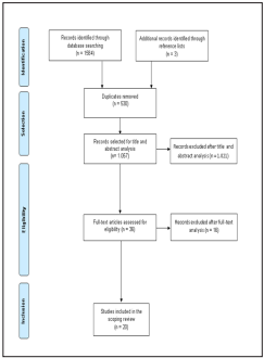
Table 1 Characterization of the include studies relatively to the microbial contamination in tourniquets, isolated species and significant antibiotic susceptibility profile 

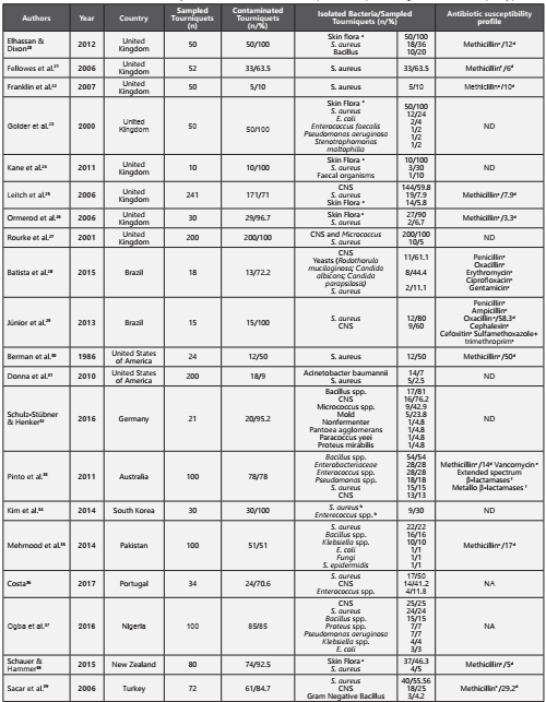
CNS - Coagulase-negative staphylococci; NA - Not available; ND - No detected methicillin-resistant strains aSkin Flora (several species in human skin); bSpecies quantified together per number of sampled tourniquets; cSusceptibility profile tested in Staphylococcus spp.; dPrevalence of Methicillin-resistant Staphylococcus aureus; e Susceptibility profile tested in Enterococcus spp.; f Susceptibility profile tested in Enterobacteriaceae.

In five studies, Enterococcus spp. was another microbial agent associated with tourniquets contamination, more tan 40 tourniquets23,24,33,34,36. Although only one study reported vancomycin-resistant Enterococci and it was found in 19 of contaminated tourniquets33.

In four studies, the genus Bacillus were found, responsible for 112 contaminated tourniquets20,32,33,35,37

Gram-negative bacteria were found in five studies, with 49 contaminated tourniquets23,32,33,35,37,39. Bacteria of the family Enterobacteriaceae namely Escherichia coli23,35,37, Klebsiella species35,37 and Proteus species32,37. Only one study reported contamination by Enterobacter cloacae resistant to extendedspectrum β-lactamases and metallo β-lactamases33.

Fungi were another important microbial group found in this review. In three studies28,32,35. Only one of the studies identified the type of fungi, confirming the presence of Rodothorula mucilaginosa, Candida albicans and Candida parapsilosis28.

Discussion

The main objective of this scoping review was to analyze and map studies examining the microbiological contamination of tourniquets used in peripheral venipuncture in order to understand the biological contamination in tourniquets and identify the most common microorganisms and their antibiotic susceptibility profile. Suspecting that the reusable tourniquets maybe important vehicles in the transmission of potential pathogens and since their site of use is close to the puncture site, many of the microorganisms present in there can be responsible for some bloodstream infections. Therefore we focused our microbiological analysis in the most relevant bacteria in this field.

Staphylococcus coagulase negative and S. aureus showed to be the most important pathogens contaminating tourniquets and other studies also emphasized its higher ability in survive for long periods of time in tourniquets, specially methicillin resistant species.We also observed a significant number of tourniquets contaminated with this type of the strains in several studies. Actually we know that the infections caused by this type of methicillin-resistant microorganisms are a serious public health issue due to the few therapeutic options available and their difficult eradication from hospital settings3,40-44. This resistance is mediated by gene mecA, which is carried by the mobile genetic element staphylococcal cassette chromosome mec (SCCmec). Methicillin-resistant Staphylococcus aureus produces an alternative penicillin-binding protein (PBP2A), which has low affinity for β-lactam antibiotics, resulting in oxacillin resistance. This resistance mechanism has been widely studied and is associated with several species of Staphylococcus both coagulase-negative and coagulase-positive40-44.

Other relevant species found to contaminate tourniquets was Enterococcus species. Besides of they are also able to survive in worst environmental conditions, this genus has increased considerably their antibiotic resistance making it difficult the treatment of enterococcal infections. The vancomycin-resistance is one of the most recently acquired resistances reported in this genus3,40,45,46. Glycopeptides, such as vancomycin, inhibit cell wall synthesis through their high affinity to the terminal d-alanyl-d-alanine group of peptidoglycan precursors. Until now, eight phenotypes of vancomycin-resistance have been identified, with the most common ones being vanA, vanB, and vanC. The manifestation of these phenotypes leads to changes in the peptidoglycan precursor, where the original D-alanyl-D-alanine terminus is replaced by Dalanyl-D-lactate or D-alanyl-D-serine, to which vancomycin has low binding affinity42,47-49. Only Pinto et al. in 2011 described the presence of vancomycin-resistant Enterococcus in tourniquets, however the other studies that detect this specie didn’t test the susceptibility profile of the strains23,24,33,34,36.

The Gram-negative bacteria namely Enterobacteriacceae was another group found in some studies as responsible to the contamination of tourniquets23,24,32,33,35,37,39. The production of β-lactamases is the most common mechanism of resistance studied in these family3,40,50-54. These enzymes are responsable for inactivating β-lactam antibiotics through the hydrolysis of their β-lactam ring. At present, the most worrying groups are the extended-spectrum β-lactamases and carbapenemases. The most common genes encoding extended-spectrum β-lactamases are blaTEM-1, blaSHV-1, and blaCTX-M. The most common in carbapenemases are encoded by the blaKPC, blaIMP, blaVIM, blaNDM, and blaOXA genes. These enzymes have an extended spectrum of activity and, when activated, complicate the treatment of the associated infections, reducing the therapeutic options available40,50-54. These types of antibiotic resistance mechanisms in isolates found contaminating tourniquets only were described in Pinto et al., in 2011 specifically in Enterobacter cloaceae. However, E. coli e Klebsiella normally carried out these genes and they are found in three studies23,35,37.

Fungal healthcare-associated infections have become a major challenge in clinical settings worldwide, with high morbidity and mortality rates55-57. The genus Candida is the leading cause of fungal infections in hospital environments, with values close to 80%55,56. Although Candida albicans is the most frequently isolated species in patients with invasive fungal infections, over the last decade, the epidemiology of this fungal genus has been changing, with an increase in the prevalence of the non-albicans species. This fact may be associated with the overuse of antibiotics and antifungals and increase of antibiotic resistance caused by bacterial adaptation, which complicate the treatment of this type of infections55-57. Three included studies in these review reported contamination of tourniquets, with fungi and yeast, specifically in Batista et al. 2015 that identified C. albicans and C. parapsilosis28,32,35.

Thus, these results show that reusable tourniquets used during clinical procedures are reservoirs of pathogens and they are moved from arm to arm and should be changed or disinfected regularly in order to ensure the safety and quality of healthcare. They showed to be potential fomites, proving with intervention studies, their possible contribution to healthcare-associated infections, specifically the bloodstream infections, disposable tourniquets may be more recommended.

Conclusion

This review intended to map studies that focus on the microbiological contamination of tourniquets used during peripheral venous puncture.

In the included studies, the contamination rate ranged from 9% to 100%, with 15 studies reporting rates equal or higher than 70%. The microorganisms responsible for these high contamination rates belong to different species, with the most prevalent being the Staphylococcus genus, followed by the Bacillus, Enterobacteriaceae family and Enterococcus species.

Nineteen studies identified Staphylococcus aureus as a common contaminant of tourniquets with more than 250 contaminated. The presence of resistance to methicillin was often associated with this species, in eleven studies the prevalence varied between 3.3-58.3%.

Patient safety can be at risk due to the high contamination rates found in tourniquets and their use in invasive procedures, such as peripheral venipuncture. For this reason, we suggest the disinfection procedure regularly in these medical devices or to adopt the disposable tourniquets. However, it will important provide evidences that the strains found in tourniquets are the same present in the catheter tips involved in bloodstream infections of the patient.

Bibliography

1. Programa de Prevenção e Controlo de Infeções e de Resistência aos Antimicrobianos. Programa de prevenção e controlo de infeções e de resistência aos antimicrobianos 2017. 2017. [ Links ]

2. Dinis A., Vale B. O papel das fomites na transmissão de doenças infeciosas. Saúde Infant. 2011;33(1):23-7. [ Links ]

3. Esteves D. Avaliação da viabilidade de amostras bacterianas em superfícies abióticas com a influência de fluídos biológicos. Universidade do Oeste Paulista - Unoeste, 2014. [ Links ]

4. Veiga-malta I. Preventing healthcare-associated infections by monitoring the cleanliness of medical devices and other critical points in a sterilization service. AAMI 2016 Conference & Expo. 2016. p. 45-52. [ Links ]

5. Seki M., Machida N., Yamagishi Y. Nosocomial outbreak of multidrugresistant Pseudomonas aeruginosa caused by damaged transesophageal echocardiogram probe used in cardiovascular surgical operations. J Infect Chemother. 2013;19(4):677-81, doi: 10.1007/s10156-012-0542-0. [ Links ]

6. Hassan M., Gonzalez E., Hitchins V., Ilev I. Detecting bacteria contamination on medical device surfaces using an integrated fiber-optic mid-infrared spectroscopy sensing method. Sens Actuators B Chem. 2016;231(1):646-54, doi: 10.1016/j.snb.2016.03.044. [ Links ]

7. Livshiz-Riven I., Borer A., Nativ R., Eskira S., Larson E. Relationship between shared patient care items and healthcare-associated infections: A systematic review. Int J Nurs Stud. 2015;52(1):380-92, doi: 10.1016/j.ijnurstu.2014.06.001. [ Links ]

8. Harris PNA., Ashhurst-Smith C., Berenger SJ., Shoobert A., Ferguson JK. Adhesive tape in the health care setting: another high-risk fomite? Med J Aust. 2012;196(1):34, doi: 10.5694/mja11.11211. [ Links ]

9. Mcnichol L., Lund C., Rosen T., Gray M. Medical adhesives and patient safety: state of the science consensus statements for the assessment, prevention, and treatment of adhesive-related skin injuries. Orthop Nurs. 2013;32(5):267-81, doi: 10.1097/NOR.0b013e3182a39caf. [ Links ]

10. Russotto V., Cortegiani A., Raineri SM., Giarratano A. Bacterial contamination of inanimate surfaces and equipment in the intensive care unit. J Intensive Care. 2015;3(54):1-8, doi: 10.1186/s40560-015-0120-5. [ Links ]

11. Dancer SJ. Importance of the environment in meticillin-resistant Staphylococcus aureus acquisition: the case for hospital. Lancet Infect Dis. 2008;8(1):101-13, doi: 10.1016/S1473-3099(07)70241-4. [ Links ]

12. Pittet D., Allegranzi B., Sax H., Dharan S., Pessoa-silva CL., Donaldson L., et al. Evidence-based model for hand transmission during patient care and the role of improved practices. Lancet Infect Dis. 2006;6(1):641-52. [ Links ]

13. Aftab H Bin., Zia B., Zahid F., Raheem A., Beg MA. Knowledge , attitude, and practices of healthcare personnel regarding the transmission of pathogens via fomites at a tertiary care hospital in Karachi, Pakistan. Open Forum Infect Dis. 2015;3(1):1-8, doi: 10.1093/ofid/ofv208. [ Links ]

14. Veiga BS., Henriques E., Fátima Barata., Fátima Santos., Isabel Silva Santos., Maria Manuela Martins., et al. Manual de Normas de Enfermagem Procedimentos Técnicos. 2011:1-285. [ Links ]

15. World Health Organization. WHO guidelines on drawing blood: best practices in phlebotomy. 2010:1-125. [ Links ]

16. World Health Organization. Decontamination and reprocessing of medical devices for health-care facilities. 2016:1-120. [ Links ]

17. Royal College of Nursing. Standards for infusion therapy. 2016:1-113. [ Links ]

18. Peters MDJ., Godfrey CM., BPharm HK., McInerney P., Parker D., Soares CB. Guidance for conducting systematic scoping reviews. Int J Evid Based Heal. 2015;13(1):141-6, doi: 10.1097/XEB.0000000000000050. [ Links ]

19. Jordan Z., Lockwood C., Munn Z., Aromataris E. The updated JBI model for evidence-based healthcare. 2016:1-12. [ Links ]

20. Elhassan HA., Dixon T. MRSA contaminated venepuncture tourniquets in clinical practice. Postgr Med J. 2012;88(1):194-7, doi: 10.1136/postgradmedj-2011-130411. [ Links ]

21. Fellowes C., Kerstein R., Clark J., Azadian BS. MRSA on tourniquets and keyboards. J Hosp Infect. 2006;64(1):86-8, doi: 10.1016/j.jhin.2006.05.003. [ Links ]

22. Franklin GF., Bal AM., McKenzie H. Phlebotomy tourniquets and MRSA. J Hosp Infect. 2006;65(2):173-5, doi: 10.1016/j.jhin.2006.11.002. [ Links ]

23. Golder M., Chan CLH., O’Shea S., Corbett K., Chrystie IL., French G. Potential risk of cross-infection during peripheral-venous access by contamination of tourniquets. Lancet. 2000;355(9197):44, doi: 10.1016/S0140-6736(99)04051-9. [ Links ]

24. Kane L., Krischock L., Lucas C. Phlebotomy tourniquets vectors for bacterial pathogens. Arch Dis Child. 2011;96(1):47-8. [ Links ]

25. Leitch A., Mccormick I., Gunn I., Gillespie T. Reducing the potential for phlebotomy tourniquets to act as a reservoir for meticillin-resistant Staphylococcus aureus. J Hosp Infect. 2006;63(1):428-31, doi: 10.1016/j.jhin.2006.03.006. [ Links ]

26. Ormerod JOM., Williams J., Lewis J., Dawson SJ. Risk of MRSA transmisión from tourniquets. J Hosp Infect. 2006;64(3):300-1, doi: 10.1016/j.jhin.2006.07.012. [ Links ]

27. Rourke C., Bates C., Read RC. Poor hospital infection control practice in venepuncture and use of tourniquets. J Hosp Infect. 2001;49(1):59-61, doi: 10.1053/jhin.2001.1038. [ Links ]

28. Batista KC de O., Tipple AFV., Leão-Vasconcelos LSN de O., Ribeiro EL., do Prado MA. Contamination of tourniquets for peripheral intravenous puncture. Acta Paul Enferm. 2015;28(5):426-32. [ Links ]

29. Dionísio J., Júnior DP., Gonçalves JC., Luíz A., Tinoco A., Coelho RO. Identificação e perfil de sensibilidade de bactérias em garrotes de uso hospitalar. Rev Ciênc Farm Básica Apl. 2013;34(2):269-73. [ Links ]

30. Berman DS., Schaefler S., Simberkoff MS. Tourniquets and nosocomial Methicillin-Resistant Staphylococcus aureus infections. N Engl J Med. 1986;315(8):514-5. [ Links ]

31. Donna M., Kevin J., David L. Acinetobacter baumannii and MRSA contamination on reusable phlebotomy tourniquets . Clin Lab Sci. 2010;23(3):151-6. [ Links ]

32. Schulz-Stübner S., Henker J. Tourniquet contamination in helicopter emergency medicine services in Germany. Infect Control Hosp Epidemiol. 2016;37(10):1262-4, doi: 10.1017/ice.2016.183. [ Links ]

33. Pinto AN., Phan T., Siarakas S., Gottlieb T. Reusable venesection tourniquets: a potential source of hospital transmission of multiresistant organisms. Med J Aust. 2011;195(5):276-9, doi: 10.5694/mja11.10333. [ Links ]

34. Kim JY., Ahn H., Lee E., Chae HB. Anesthesiologist’s hand hygiene and disinfection of reusable rubber tourniquet with alcohol swabs before intravascular cannulation. Korean J Anesth. 2014;67(1):9-10. [ Links ]

35. Mehmood Z., Mubeen SM., Afzal MS., Hussain Z. Potential risk of crossinfection by tourniquets: a need for effective control practices in Pakistan. Int J Prev Med. 2014;5(9):1119-24. [ Links ]

36. Costa P. Gestão de material clínico de bolso por enfermeiros: fatores determinantes e avaliação microbiológica. 2017. [ Links ]

37. Ogba OM., Selekeowei T., Otu-bassey I. Infection transmission potential of reusable plebotomy tourniquet in selected facilities in Calabar, Nigeria. Eur J Pharm Med Res. 2016;3(10):96-100. [ Links ]

38. Schauer CKMW., Hammer DA. Quantifying patient bacterial exposure risk from reusable phlebotomy tourniquets in a New Zealand secondary level hospital. J Infect Prev. 2015;16(6):262-5, doi: 10.1177/1757177415600242. [ Links ]

39. Sacar S., Turgut H., Kaleli I., Cevahir N., Asan A. Poor hospital infection control practice in hand hygiene , glove utilization , and usage of tourniquets. Am J Infect Control. 2006;34(1):606-9, doi: 10.1016/j.ajic.2006.02.006. [ Links ]

40. Santajit S., Indrawattana N. Mechanisms of antimicrobial resistance in ESKAPE pathogens. Biomed Res Int. 2016;2016(1):1-8, doi:10.1155/2016/2475067. [ Links ]

41. Gelatti LC., Sukiennik T., Becker AP., Inoue FM., do Carmo MS., Castrucci FM., et al. Sepsis due to community-acquired methicillin-resistant Staphylococcus aureus in southern Brazil. Rev Soc Bras Med Trop. 2009;42(4):458-60, doi: 10.1590/S0037-86822009000400019. [ Links ]

42. Ratti RP., Sousa CP. Staphylococcus aureus meticilina resistente (MRSA) e infecções nosocomiais. Rev Ciencias Farm Basica e Apl. 2009;30(2):137-43, doi: 10.1590/S0104-42301998000400002. [ Links ]

43. Catarina A. Infeção hospitalar por Staphylococcus aureus resistente à meticilina associada aos cuidados de enfermagem. Universidade do Minho, 2014. [ Links ]

44. Moreira RAS. Infeções nosocomiais e bactérias implicadas neste tipo de infeção. Universidade de Coimbra, 2016. [ Links ]

45. Furtado G., Martins S., Coutinho AP., Soares G., Wey S., Medeiros E. Incidência de Enterococcus resistente à vancomicina em hospital universitário no Brasil. Rev Saúde Pública. 2005;39(1):41-6, doi: 10.1590/S0034-89102005000100006. [ Links ]

46. Drees M., Snydman DR., Schmid CH., Barefoot L., Hansjosten K., Vue PM., et al. Prior environmental contamination increases the risk of acquisition of vancomycin-resistant Enterococci. Clin Infect Dis. 2008;46(1):678-85, doi: 10.1086/527394. [ Links ]

47. O’Driscoll T., Crank CW. Vancomycin-resistant enterococcal infections : epidemiology, clinical manifestations, and optimal management. Infect Drug Resist. 2015;8(1):217-30. [ Links ]

48. Gama BA. Análise da resistência antimicrobiana e de genes de virulência de Enterococcus spp . 2008. [ Links ]

49. Riboldi GP. Perfil de resistência antimicrobiana e análise genotípica de Enterococcus spp. isolados de alimentos em Porto Alegre, RS. 2007. [ Links ]

50. Azevedo SMM. Farmacologia dos Antibióticos Beta-lactâmicos. Universidade Fernando Pessoa, 2014. [ Links ]

51. Seibert G., Hörner R., Meneghetti BH., Righi RA., Lucia N., Dal F., et al. Nosocomial infections by Klebsiella pneumoniae carbapenemase producing enterobacteria in a teaching hospital. Einstein (São Paulo). 2014;12(3):282-6, doi: 10.1590/S1679-45082014AO3131. [ Links ]

52. Arnold RS., Thom K a., Sharma S., Phillips M., Johnson JK., Morgan DJ. Emergence of Klebsiella pneumoniae carbapenemase (KPC)- producing bacteria. South Med J. 2011;104(1):40-5, doi: 10.1097/SMJ.0b013e3181fd7d5a.Emergence. [ Links ]

53. Fernandes L. Resistência bacteriana aos betalactâmicos por mecanismo enzimático: uma revisão de literatura com enfoque nas betalactamases de espectro estendido. Universidade Estadual da Paraíba, 2014. [ Links ]

54. Wollheim C. Epidemiologia molecular de Escherichia coli e Klebsiella spp produtoras de beta-lactamase de espectro ampliado. Universidade de Caxias do Sul, 2009. [ Links ]

55. Colombo AL., Guimarães T. Epidemiologia das infecções hematogénicas por Candida spp. Rev Soc Bras Med Trop. 2003;36(5):599-607. [ Links ]

56. Khouri S., Ruiz S., Domaneschi C., Hahn RC., Christiano B., Paula CR. Evaluation of infections by Candida at a university hospital of Vale do Paraíba region, São Paulo State, Brazil: species distribution, colonization, risk factors and antifungal susceptibility. Rev Pan-Amaz Saude. 2016;7(2):51-7, doi: 10.5123/S2176-62232016000200006. [ Links ]

57. Moreira LS., Cristina A., Cesar O., Figueira FR., Di C., Costa P., et al. Estudo da resistência aos A antifúngicos de leveduras isoladas de candidúrias de um hospital de médio porte. Rev Univap. 2017;23(43):44-52. [ Links ]

Cómo citar este artículo: A. Salgueiro-Oliveira, et al. Tourniquets used in peripheral venipuncture as a potential vehicle for transmission of microorganisms: scoping review. Infectio 2020; 24(2):92-97

Acknowledgments and Funding The authors would like to acknowledge the support provided by the Health Sciences Research Unit: Nursing (UICISA: E) of the Nursing School of Coimbra (ESEnfC) and Escola Superior de Tecnologia da Saúde de Coimbra. The author Paulo S. Costa (SFRH/BD/136487/2018) would like to thank the Portuguese National Funding Agency for Science, Research and Technology (FCT) for the financial support granted through the 2018 Ph.D. Scholarship program.

Ethical disclosure

Protection of human and animal subjects. This research do not use animal nor human material or data.

Confidentiality of data. Not aplicable

Received: January 11, 2019; Accepted: December 13, 2019

* Autor para correspondencia. Correo electrónico: vaniasoliveira19@gmail.com Avenida Bissaya Barreto, Apartado 7001, 3046-851, Coimbra, Portugal. Telephone: +351 239 802 850 / 239 487 200; Fax: +351 239 442648

Conflict of interest statement.

None to declare

Creative Commons License This is an open-access article distributed under the terms of the Creative Commons Attribution License