SciELO - Scientific Electronic Library Online

 
vol.25 issue3Rectal colonization by Enterobacterales producing multiple carbapenemases: a co-production case reportAn unexpected pathogen causing multiple intra-abdominal abscesses in an immunocompetent patient author indexsubject indexarticles search
Home Pagealphabetic serial listing  

Services on Demand

Journal

Article

Indicators

Related links

  • On index processCited by Google
  • Have no similar articlesSimilars in SciELO
  • On index processSimilars in Google

Share


Infectio

Print version ISSN 0123-9392

Infect. vol.25 no.3 Bogotá July/Sept. 2021  Epub May 15, 2021

https://doi.org/10.22354/in.v25i3.948 

CASO CLINICO

Mycetoma caused by Phellinus species: a rare case from Brazil

Micetoma causado por especies de phellinus: un caso raro de Brasil

Luís Arthur Brasil Gadelha-Farias12* 

Lucas Mustafa-Aguiar3  4 

Gabriela Studart-Galdino3  5 

Silviane Praciano-Bandeira1  6 

Giovany Gomes-Capistrano3  7 

Paulo Roberto Arruda-Tavares3  8 

Glaucia Morgana de Melo Guedes3  9 

Jaime David Acosta-España10 

1 Faculty of medicine, Universidade Federal do Ceará (UFC), Fortaleza, Ceará, Brazil.

2 https://orcid.org/0000-0002-8978-9903

3 Internal medicine department, Hospital Universitário Walter Cantídeo (HUWC), Fortaleza, Ceará, Brazil.

4 https://orcid.org/0000-0003-3842-74595

5 https://orcid.org/0000-0003-2760-3162

6 https://orcid.org/0000-0002-4051-5645

7 https://orcid.org/0000-0002-9078-6208

8 https://orcid.org/0000-0003-2989-8144

9 https://orcid.org/0000-0002-5469-8120

10 Hospital Gíneco Obstétrico Nueva Aurora, Quito, Ecuador. Universidad de las Américas, Quito-Ecuador. https://orcid.org/0000-0001-5299-2732


Abstract

Mycetoma is a chronic and slow-developing granulomatous disease characterized by the triad of large painless tumour-like subcutaneous swellings, the formation of sinuses, and discharge that usually contains grains. Phellinus spp. are saprophytic wood-decaying filamentous basidiomycetes. They are an under-recognised cause of invasive fungal infections and are rarely reported worldwide. We report a 59-year-old male patient with mycetoma caused by Phellinus spp. The diagnosis was confirmed with clinical examination, magnetic resonance imaging (MRI) study, soft tissue and bone biopsy culture, and polymerase chain reaction. To the best of our knowledge, this is the first reported case of mycetoma due to Phellinus spp. without chronic granulomatous disease (CGD).

Keywords: Fungal infections; Basidiomycota; Phellinus; Mycetoma

Resumen

El micetoma es una enfermedad granulomatosa crónica y de lento desarrollo caracterizada por la tríada de grandes inflamaciones subcutáneas similares a tumores indoloras, la formación de los senos nasales y secreción que generalmente contiene granos. Phellinus spp. son basidiomicetos filamentosos saprofitos que descomponen la madera. Son un poco reconocido causa de infecciones fúngicas invasivas y rara vez se informan en todo el mundo. Presentamos un paciente masculino de 59 años con micetoma causado por Phellinus spp. El diagnostico se confirmó con examen clínico, estudio de resonancia magnética (RM), cultivo de biopsia de tejido blando y óseo y reacción en cadena de la polimerasa. A lo mejor que sepamos, este es el primer caso reportado de micetoma debido a Phellinus spp. sin enfermedad granulomatosa crónica (EGC).

Palabras clave: Infecciones por hongos; Basidiomycota; Phellinus; Micetoma

Introduction

Mycetoma is a chronic and slow developing granulomatous disease characterized by large painless tumour-like subcutaneous swellings, formation of sinuses, and discharge that usually contains grains. Although it may occur in various countries, it is endemic in Sudan, Mexico, and India, and frequent in Brazil and Argentina1. It can be caused by either fungi or bacteria. The feet and legs are the most commonly affected regions, but any body part can be affected1,2. We report the case of a 59-year-old male patient with mycetoma caused by Phellinus spp., a basidiomycete genus. The diagnosis was confirmed with clinical examination, magnetic resonance imaging (MRI), soft tissue and bone biopsy, culture, and polymerase chain reaction. There have been case reports of chronic granulomatous disease (CGD) complicated with subcutaneous abscess due to Phellinus spp. (3,4 Fungal infections related to Phellinus spp. are an emerging disease in CGD patients, but this remains a rare condition in patients without CGD5. To the best of our knowledge, this is the first reported case of mycetoma due to Phellinus spp. without CGD.

Case report

Our patient presented with a 3-year history of foot swelling and progressive increase in size. He also complained about episodes of pain and the emergence of yellow discharges containing grains. He had previous history of pre-diabetes. Subsequently, the patient reported a wound caused by a piece of wood in childhood. Physical examination revealed a diffuse firm swelling mainly on the lateral aspect of the right foot, and three discharging sinuses (Figure 1).Admission tests revealed discrete anaemia (haemoglobin 12.6 g/dL). Erythrocyte sedimentation rate (70 mm) and C-reactive protein (5.4 mg/L) levels exceeded the normal range.

Figure 1. 

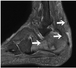
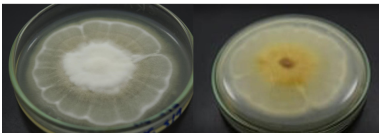
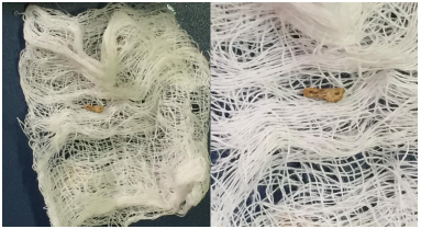
MRI of the right foot showed overgrowth of soft tissues, bone oedema, and small rounded hyperintensity surrounded by a low-signal-intensity rim with a hypointense dot in the centre, called a dot-in-circle sign (Figure 2). Diagnosis of mycetoma was made and a soft tissue and bone biopsy cultures were performed to identify the etiologic agent. The culture revealed a mycelial colony with bright white and yellow with abundant aerial mycelium on potato dextrose agar (Figure 3). Polymerase chain reaction was performed by sequencing the ITS region, using the panfungal primers ITS1 and ITS4. After obtaining the sequences, the NCBI Blast tool was used and indicated Phellinus spp. Therapy was started with voriconazole (6 mg/kg every 12 hours for 2 days followed by a maintenance regimen of 4 mg/kg every 12 hours for 7 days) and amphotericin B lipid-complex (5 mg/kg/day for 4 weeks followed by a maintenance regimen of 5 mg/kg/week for 6 months). After 30 days, he exhibited clinical improvement and the piece wood spontaneous came out (Figure 4). Six months later, the patient remains asymptomatic.

Figure 2. 

Figure 3. 

Figure 4. 

Discussion

To our knowledge, there is no prior data about Phellinus spp. infection with isolated mycetoma in the medical literature. Information regarding treatment is also scarce, but it is believed that lipid formulations of amphotericin B and azoles (such as voriconazole) may be helpful, especially in severe disease. According to current literature, amphotericin B-based regimens may be a better alternative, but voriconazole has also been successfully used to treat Phellinus spp. infections5. Additionally, identification of pathogen species is difficult because many isolates fail to sporulate and remain sterile in routine mycology media5.

In Brazil, mycetoma cases range between 101 and 500 per year1. MRI is useful for clinical diagnosis of fungal infection, since it allows evaluation of the invasion and extent of the lesion in most cases. The dot-in-circle sign is indicative of the presence of fungal grains, but not suggestive of the species. Small lesions present better prognosis. Long-term treatments are common, and sometimes ineffective1. Although surgical treatment may be necessary in many cases, it was not necessary in our patient.

We present a rare etiological agent for mycetoma. CGD should be investigated in any patient with identified Phellinus spp. infection, although X-linked mutations cannot be evaluated in many health centres. More sudies are needed to better understand the management and prognosis of infections caused by Phellinus spp.

References

1. Zijlstra EE, van de Sande WWJ, Welsh O, et al. Mycetoma: a unique neglected tropical disease. Lancet Infec Dis 2016; 16(1): 100-112. [ Links ]

2. Van de Sande W, Fahal A, Ahmed SA, et al. Closing the mycetoma knowledge gap. Med Mycol 2018; 56(suppl_1): S153-S164. [ Links ]

3. Shigemura T, Nakazawa Y, Amano Y, et al. Subcutaneous abscess due to the basidiomycete Phellinus mori in a patient with chronic granulomatous disease. Infection 2015; 43(3): 371-375. [ Links ]

4. Ramesh M, Resnick E, Hui Y, et al. Phellinus tropicalis abscesses in a Patient with Chronic Granulomatous Disease. J Clin Immunol 2013; 34(2): 130-133. [ Links ]

5. Haidar G, Zerbe CS, Cheng M, et al. Phellinus species: An emerging cause of refractory fungal infections in patients with X-linked chronic granulomatous disease. Mycoses 2016; 60(3): 155-160. [ Links ]

Cómo citar este artículo: L.A.B. Gadelha-Farias, et al. Mycetoma caused by phellinus species: a rare case from Brazil. Infectio 2021; 25(3): 197-199

Ethical disclosure

Sources of funding for our research. Authors

Protection of human and animal subjects. Consent Written informed consent was obtained from the patient for publication of this case report and accompanying images.

Confidentiality of data. No data that identifies patient are revealed

Received: September 14, 2020; Accepted: December 04, 2020

* Autor para correspondencia. Correo electrónico: luisarthurbrasilk@hotmail.com Hospital São José de Doenças Infecciosas (HSJ), Nestor Barbosa St., Number 315, Fortaleza, 60455-610, Brazil

Conflict of interest.

None

Creative Commons License This is an open-access article distributed under the terms of the Creative Commons Attribution License