


 English (pdf)
 Article in xml format
 Article references
 Send this article by e-mail
 Cited by SciELO
  Cited by Google
  Similars in
SciELO
Similars in
SciELO  Similars in Google
 Permalink
INTRODUCTION
The genus Dendropsophus includes 96 species of small, pond-dwelling tree frogs distributed from northern Argentina and Uruguay through tropical South America and Central America to the lowlands of southern Mexico (Frost 2015). There are nine species groups recognized in the genus Dendropsophus, with several species still unassigned to a group (Faivovich et al. 2005). The Dendropsophus microcephalus group, the most speciose in Dendropsophus, presently includes 36 species, most of them from the lowlands of South and Central America (Duellman 2001, Faivovich et al. 2005, Frost 2015). D. microcephalus (Cope, 1886) is a small, slender, yellowish frog with a widespread distribution in subhumid and humid lowlands of Central America and northern South America, with some scattered populations in the Amazon basin and southern Brazil (Duellman 2001, Savage 2002).
The tadpoles of the D. microcephalus group are considered to have extreme reduction and modification of the elements of the oral disc (Duellman 2001). Based on external observations alone, the tadpole of D. microcephalus is considered to lack papillae, labial tooth rows (Duellman & Fouquette 1968:527, Altig & McDiarmid 1999:37, Duellman 2001:214, Savage 2002:216, Lynch 2006:447), and dermal ridges (Pugliese et al. 2000:78, Pugliese et al. 2001:687). This tadpole is also considered to have a tube (referred to hereafter as the oral tube) around the mouth that can be protruded and retracted (Vera Candioti 2007:48, 144, Haas et al., 2014:17); when retracted, the oral tube forms an elevated U-shaped yoke around the mouth. It is considered that the oral tube constitutes a modified oral disc (i.e., they are homologous).
Some of the accepted claims concerning the oral morphology of the tadpole of D. microcephalus have been challenged. Altig & McDiarmid (1999:37) considered that an oral tube is absent in this species and Wassersug (1980:5), based on observations of the closely related and morphologically similar D. phlebodes (Stejneger, 1906) (Duellman & Fouquette 1968, Duellman 2001), considered that the oral disc is absent. In addition, an external examination of the tadpole's oral apparatus shows that parts of the jaw sheaths are covered by skin, suggesting that skin could also be hiding the papillae, labial tooth rows, and/or labial ridges. Clearly, a detailed examination of the oral apparatus on D. microcephalus is in order.
In this study, I obtained histological sections of the oral apparatus in a tadpole of D. microcephalus in order to 1. assess whether papillae, labial ridges, and labial tooth rows are present, 2. determine if there is an oral tube that protrudes and retracts, and that, when retracted, forms an elevated U-shaped yoke around the mouth, and 3. assess whether the oral tube is homologous to the oral disc.
MATERIALS AND METHODS
Serial sagittal sections (10 LI) of the mouth and surrounding areas were obtained from one tadpole of D. microcephalus (stage 29; UMMZ 139780) from Tabasco, Mexico. For comparisons, I sectioned one tadpole of D. mathiassoni (Cochran and Goin, 1970) (Stage 30; J. D. Lynch teaching collection, University of Nebraska, Lincoln) from Departamento del Meta, Colombia and one of D. rhodopeplus (Gunther, 1858) (stage 40; UMMZ 151677) from Napo, Ecuador. These two species were selected because they have external oral morphology similar to that of D. microcephalus (Duellman 1972, Lynch & Suarez-Mayorga 2011) and similar developmental stages were available from collections.
Staging of tadpoles follows that of Gosner (1960). Histology follows the method of Wessner (1960). Histological sections were stained with Hematoxilin and Eosin. Terminology of external oral morphology of the tadpole follows that of Altig & McDiarmid (1999). The term "oral apparatus" is defined as the sum of all the structures (e.g., U-shaped elevation, free-edged labia, labial tooth rows, and jaw sheaths) present on and around, and close to, the mouth, irrespective of shape or number. The term "oral disc" is defined as a round or elliptical area that is made evident, completely or partially, by crevices, papillae, and/or labia. Primary homologies (de Pinna 1991) were assessed following the criteria proposed by Remane (1956). Figure 1 corresponds to Duellman's Fig. 83 (2001), altered based on the histology results. Acronyms: ICN = Instituto de Ciencias Naturales, Universidad Nacional de Colombia; UMMZ = University of Michigan Museum of Zoology.
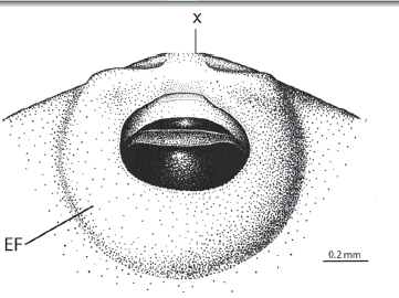
Figure 1 Illustration of the external oral morphology in a tadpole of D. microcephalus. Redrawn and modified based on Duellman (2001; Fig. 83). EF = external fold; X = approximate place of sagittal section.
RESULTS
A sagittal section through the mouth of a tadpole of Dendropsophus microcephalus-(UMMZ 139780) (Fig. 2a). The mouth is located at the anterior end of the head. Upper, lower, and lateral labia are present. The free edges of the labia are flexed towards the center of the mouth. The free edge of the upper labium is short, thin, and flexible while those of the lower and lateral labia are long, thick, turgid, and filled with connective tissue. The distal end of the lower labium (i.e., that closest to the mouth) presents a concave surface that is covered by a multilayered filamentous epithelium composed by four to five layers. The muscle mandibulolabialis is inserted on the lower labium. The upper labium presents one low transverse ridge about half way between its distal edge and the upper jaw sheath. Papillae and labial tooth rows are absent.
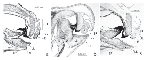
Figure 2 Sagittal sections through the mouth of tadpoles of a. D. microcephalus (stage 29), b. D. mathiassoni (stage 30), and c. D. rhodopeplus (stage 40). EF = external fold; LL = lower labium; UL = upper labium; R = tooth ridge; ras = m. rectus abdominis superficialis; ml = m. mandibulolabialis.
A fold of skin (hereafter as external fold) covers the labia, ridge, and peripheral parts of the jaw sheaths. The external fold emerges from the areas surrounding the oral apparatus, extends anteriorly, and leaves a small, round central opening over the mouth (Fig. 1). The part of the external fold covering the lower labium is long and smooth while that covering the upper labium is short and wrinkled. The muscle rectus abdominis superficialis is inserted on the most anterior end of the part of the external fold that covers the lower labium (Fig. 2a). The external fold is thick and filled with loose connective tissue.
DISCUSSION
This study shows that there is more to the morphology of the oral apparatus of the tadpole of D. microcephalus than has been recognized from external examination alone. First, there are upper, lower, and lateral labia with free edges. Second, the labia are flexed anteromedially towards the center of the mouth. Third, there is a ridge present on the upper labium. Fourth, the external fold completely covers the labia and labial ridge, and partially covers the jaw sheaths.
My findings support the claim that labial papillae and labial tooth rows are absent, but show that there is one labial ridge. This study supports the claim that there is a U-shaped oral tube around the mouth that is formed by the flexed labia of the oral disc (i.e., homologous to the disc). The oral tube is U-shaped because the lower and lateral labia are longer than the upper labium. This study does not support the claim that the oral tube extends and retracts because the lower and lateral labia are turgidly packed with connective tissue; rather, my observations suggest that the rigid oral tube would appear to protrude when the muscle rectus abdominis superficialis pulls back the external fold (see Altig and McDiarmid's [1999] for a similar hypothesis). This study does not support the claim that the U-shaped yoke around the mouth is formed by the retraction of the oral tube because the oral tube is rigid; rather, my observations suggest that the U-shaped yoke results from the fact that the flexed labia create differences in bulkiness beneath the external fold (i.e., bulkier posteriorly and laterally than anteriorly).
The tadpoles of D. mathiassoni and D. rhodopeplus are considered to lack labial papillae and tooth rows (Duellman 1972, Lynch & Suarez-Mayorga 2011). This study shows that the detailed morphology of the oral apparatuses of these species is nearly identical to that of D. microcephalus (Figs. 2b, c) since they have labia with their free edges flexed anteromedially, a ridge on the upper labium, and an external fold hiding the labia and ridge. This study shows that only in D. microcephalus is there a concave area on the lower labium.
Several other species of the D. microcephalus group (i.e., D. bipunctatus (Spix, 1824), D. haddadi (Bastos & Pombal, 1996), D. meridianus Lutz 1954, D. minusculus Rivero 1971, D. nanus Boulenger 1889, D. oliverai Bokermann 1963, D. phlebodes, D. pseudomeridianus Cruz et al. 2000, D. rubicundulus Reinhardt & Lutken 1862, D. sanborni Schmidt 1944, D. studerae Carvalho-e-Silva et al. 2003) are considered to have tadpoles lacking labial papillae and tooth rows, and to present a U-shaped yoke around the mouth (Bokermann 1963, Duellman & Fouquette 1968, Starrett 1973; Lavilla 1990, Cruz & Dias 1991, Cruz et al. 2000, Pugliese et al. 2000, Pugliese et al. 2001, Carvalho-e-Silva et al. 2003, Lorenco-de-Moraes et al. 2012, Abreu et al. 2013). However, it is unknown whether these species have the same detailed oral morphology present in D. mathiassoni, D. microcephalus, and D. rhodopeplus. It is also unknown whether the morphology of the oral apparatus is informative for the systematics and taxonomy of Dendropsophus and the D. microcephalus group.
Vera Candioti (2007) suggested that the oral tube of D. microcephalus is suctorial. Although I did not examined the function of the oral tube, the presence of a concave area covered by long filamentous cells on the lower labium near the mouth is consistent with the idea that the tube is involved in feeding. Presently, the functions, if any, of the external fold and oral tube are unknown.

