


Servicios Personalizados
Revista
Articulo
 Inglés (pdf)
 Articulo en XML
 Referencias del artículo
 Enviar articulo por email
Indicadores
-
 Citado por SciELO
 -
 Accesos
Links relacionados
-
 Citado por Google
 -
 Similares en
SciELO
Similares en
SciELO -
 Similares en Google
Compartir

 Permalink
Universitas Psychologica
versión impresa ISSN 1657-9267
Univ. Psychol. vol.12 no.2 Bogotá may./agos. 2013
Assessment of Intellectual and Visuo Spatial Abilities in Children and Adults with Williams Syndrome*
Evaluación de habilidades intelectuales y visuoespaciales de niños y adultos con Síndrome de Williams
Nunes M M **,
Honjo R S ***,
Dutra R L, Amaral V S, Amaral V A S, Oh H K ****,
Bertola D R *****,
Albano L Μ J ******,
Assumpção Júnior F Β *******,
Kim C A ********
Universidade de Sao Paulo, Brasil
Teixeira M C T V *********
Universidade Presbiteriana Mackenzie, Sao Paulo, Brasil
* Financing: This work was supported by FAPESP number 2008/55391-6. Conflict of interests: None. Enderezo para correspondencia: A/C Chong Ae Kim. Unidade de Genética do Instituto da Crianca. Av. Dr Enéas Carvalho de Aguiar, 647. Sao Paulo, SP CEP: 05403-000.
** Aluna de Doutorado do Instituto da Crianca da Facultade de Medicina da Universidade de Sao Paulo, Brasil.
*** Alunas de Doutorado da Unidade de Genética do Instituto da Crianca da Facultade de Medicina da Universidade de Sao Paulo, Brasil.
**** Universidade de Sao Paulo, Brasil. Professor da Faculdade de Medicina Alternativa de Jeonju - Coreia do Sul.
***** Médica Assistente da Unidade de Genética do Instituto da Crianca da Facultade de Medicina da Universidade de Sao Paulo, Brasil.
****** Médica Assistente aposentada da Unidade de Genética do Instituto da Crianca da Facultade de Medicina da Universidade de Sao Paulo, Brasil.
******* Professor Associado do Instituto de Psicologia da Universidade de Sao Paulo, Brasil.
******** Professora Livre Docente da Unidade de Genética do Instituto da Crianca da Facultade de Medicina da Universidade de Sao Paulo, Brasil.
********* Professora Adjunta I do Programa de Pós-Graduacao em Disturbios do Desenvolvimento do Centro de Ciencias Biológicas e da Saúde da Universidade Presbiteriana Mackenzie. E-mail: mctvteixeira@gmail.com
Recibido: junio 20 de 2012 | Revisado: julio 21 de 2012 | Aceptado: agosto 10 de 2012
Para citar este artículo:
Nunes, M. M., Honjo, R. S., Dutra, R. L., Amaral, V. A. S., Oh, H. K., Bertola, D. R., et al. (2013). Assessment of intellectual and visuo-spatial abilities in children and adults with Williams syndrome. Universitas Psychologica, 12(2), 581-589.
Abstract
The Williams-Beuren syndrome (SWB), also known as Williams syndrome, is a contiguous gene deletion of the region 7q.11.23. The main clinical characteristics are typical faces, supravalvular aortic stenosis, failure to thrive, short stature, transient neonatal hypercalcemia, delayed language, friendly personality, hyperacusis and intellectual disability. The diagnosis of SWB is confirmed by the detection of micro deletion by different techniques of molecular cytogenetics, FISH, MLPA or polymorphic markers. This study assessed the verbal intelligence quotient (IQ) and performance and visuo-spatial skills in children and adults with WBS. The composed group was of 31 WBS patients (19 M and 12 F), whose ages ranged from 9 to 26 years (M 14.45 y). All patients had the diagnosis confirmed molecularly. The tests used were the WISC-III, WAIS-III and Rey-Osterrieth Complex Figure Test. The results indicated a total IQ ranged from 51 to 86 (M 63): 22 with mild intellectual disability, 4 with moderate intellectual disability, 4 borderlines and 1 below the normal media. All patients had marked visual-spatial deficits. The results suggest nonverbal reasoning, visuo-spatial perception, spatial representation, working memory, motor planning and executive functions are very affected in this group.
Key words authors: Williams Syndrome, intelligence, assessment, cognition, visual-spatial abilities.
Key words plus: Genetic Disorder, Biomarkers, Intelligence, Quantitative Research.
Resumen
El síndrome de Williams-Beuren (SWB), también conocido como síndrome de Williams, es un síndrome de deleción de genes contiguos de la región 7q.11.23. Se caracteriza por dimorfismo facial típico asociado a anomalías cardiovasculares, personalidad amigable, hiperacusia y deficiencia intelectual. El diagnóstico del SWB es confirmado por la detección de microdeleción a partir de las diferentes técnicas de citogenética molecular: FISH, marcadores polimórficos o MLPA. Este estudio evaluó el cociente intelectual verbal y manipulativo, así como las habilidades visuoespaciales en niños y adultos con SWB. El grupo estuvo formado por 31 pacientes con SWB (19 de sexo masculino y 12 de sexo femenino), cuyas edades variaron entre 9 y 26 años (media 14.45 años). Todos los pacientes tenían el diagnóstico confirmado molecularmente. Los test utilizados fueron las escalas WISC-III, WAIS-III y el Test Figuras Complejas Rey-Osterrieth. Los resultados indicaron un cociente intelectual que osciló de 51 a 86 (media 63), distribuido así: 22 con deficiencia intelectual leve, 4 con deficiencia intelectual moderada, 4 limítrofes, 1 en la media inferior. Todos los pacientes presentaron déficit visuoespacial. Los resultados sugieren que el razonamiento no verbal, la percepción visuoespacial, la representación espacial, la memoria de trabajo, la planificación motora y las funciones ejecutivas están muy comprometidos en el grupo estudiado.
Palabras clave autores: Síndrome de Williams, inteligencia, evaluación, cognición, habilidades visuoespaciales.
Palabras clave descriptores: Alteración genética, biomarcador, inteligencia, investigación cuantitativa.
doi:10.11144/Javeriana.UPSY12-2.aiva
Introduction
Williams-Beuren syndrome (WBS), also known as Williams syndrome, is a neurodevelopmental disorder caused by a submicroscopic deletion of long arm of chromosome 7 (7q11.23). The commonly deleted region is 1.55 Mb (90%) and 1.84 Mb (8%) and 28 genes have been mapped within this region (Schubert, 2009). Its estimated prevalence ranges from 1:7500 to 1:20000 live births (Meyer-Lindenberg, Mervis & Berman, 2006).
The affected subjects have a wide range of medical diseases and a unique behavioral and cognitive profile. The main physical characteristics are typical faces, supravalvular aortic stenosis, failure to thrive, short stature, transient neonatal hypercalcemia and delayed language and motor development (Lenhoff, Wang, Greenberg & Bellugi, 1997; Marriage & Scientist, 1995). Behaviorally, WBS subjects have a hyper sociability, empathy in social relationships, a generalized anxiety disorder, phobias (Klein-Tasman & Mervis, 2003). A cognitive and behavioral phenotype of WBS is characterized by presenting a varying degree of intellectual disability, deficits in visuo-spatial skills and executive functions (working memory and planning), specific language skills and better performance in expressive language than in the receptive language (Farran & Jarrold, 2003; Martens, Wilson & Reutens, 2008; Moretti-Ferreira & Giacheti, 2007).
The mean cognitive level is within the range of mild to moderate retardation, with some peaks and valleys in mental domains, particularly severe visuospatial construction deficits accompanied by a relative strength in expressive language and relatively spared face and object recognition. In addition, subjects have a strong attraction to music and a strong auditory fascination alongside, extreme hyperacusis and phonophobia (Mervis et al., 2000).
Because WBS is etiologically homogeneous, it serves as an excellent model for the study of the biological developmental processes underlying sensory processing in humans. The findings may also have important implications for other developmental psychiatric disorders associated with pathological sensory processing and sensitivity, such as autism, schizophrenia, post-traumatic stress disorder and attention deficit hyperactivity disorder.
Insufficient studies have examined constructional abilities in children with atypical development (La Femina, Senese, Grossi & Venuti, 2009). Some aspects of cognition affected in individuals with WS are the attention and immediate visual memory skills. And one of the instruments designed to assess these abilities is the Rey-Osterrieth Complex Figure Test. In addition to these skills, the test also enables you to check visuospatial abilities, planning, organizing and problem solving (Fernando, Chard, Butcher & McKay, 2003; Lu, Boone, Cozolino & Mitchell, 2003).
Although the figures of the test have a significant number of details, such as the number of angles, layout and proportions, the quality of copies depends on good visual memory skills that allow the execution of the copy in an organized manner (Oliveira, Rigoni andreatta & Moraes, 2004). In Brazil the works exploring visual-spatial abilities in children and adults with WBS are scarce. The objective of this study was to evaluate the verbal and performance intelligence quotient and the visuospatial bilities of children and adults with WBS.
Method
Participants and procedures
This study was approved by the Ethics Committee for Analysis and Research Project — CAPPesq — Faculty of Medicine, University of Sao Paulo (n: 806/06) and the parents signed a consent form.
31 WBS patients participated in the research (19 M and 12 F) and their ages ranged from 9 to 26 years (with a media of 14,45 y), 15 of the evaluated patients were enrolled in special classes, regular class and 11 in 5 do not attend school. The main demographic and educational characteristics of the participants are shown in Table 1.
The diagnosis of WBS was confirmed by techniques of Fluorescence in Situ Hybridization (FISH), microsatellite markers analysis or Multiplex Ligation-dependent Probe Amplification (MLPA) in all patients. Of the 31 subjects evaluated, 23 had a deletion of 1.55 Mb and 3 had a deletion of 1.83 Mb; it was not possible to determine the size of the deletion for the remaining 5 because the tests were performed by FISH.
The intellectual function were evaluated using the Wechsler Intelligence Scale for Children—Third Edition (WISC III) (Wechsler, 2002) for ages 6 and 16years and for those aged greater than or equal to 17years, the Wechsler Intelligence Scale for Adult was used — Third Edition (WAIS-III) (Wechsler, 1997. The subtests included were 5 for verbal IQ (information, comprehension, similarities, arithmetic and vocabulary) and 5 subtests for performance IQ (picture completion, picture arrangement, block design, object assembly and coding).
The Wechsler scale was used to provide parameters on the scale and verbal business and the overall scale derived from these two scales: Intellectual Quotient (IQ), Verbal (VIQ) and Performance (PIQ) and Total (TIQ).
The assessment of visuo-spatial abilities, memory attention, planning and working memory, was assessed from the Rey-Osterrieth Complex Figure Test (ROCFT) (Oliveira, 1999) to assist in "differential diagnosis between mental retardation and constitutional deficit acquired as a result of traumatic brain injury" (Oliveira, 1999.p.9). He also examines the organization, planning and problem solving skills (Fernando et al., 2003) and immediate memory (Lu et al., 2003). It is widely used to explore nonverbal memory.
Individuals were evaluated individually and all tests were applied and corrected according to the evaluation system established by the manual of each instrument.
The results for each of the tests were described by their mean and standard deviation. The data collected were subject to descriptive statistics. To perform the calculations we used the SPSS 13.0 software, WISC-III and WAIS-III.
Results and discussion
Verbal Abilities
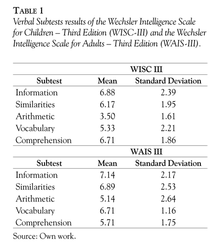
The patients' VIQ of the scale WISC-III ranged from 57 to 87 and these data are consistent with previous studies that quote a range of 41 to 80 (Tassabehji, 2003). The highest mean score was 6.88 for the information subtest and the lowest was 3.50 for the arithmetic subtest. When comparing these results it was concluded that both showed low values, as normal scores are greater than 10 points (Table 1).
In the scale WAIS-III the VIQ ranged from 57 to 87. The highest mean score was 7.14 for the information subtest and the lowest was 5.14 for the arithmetic subtest. Comparing these results it was concluded that both showed low values, as normal scores are greater than 10 points (Table 1).
We infer that WBS patients scored higher on the information subtest because it assessed content knowledge (including object names and historical facts) and knowledge acquired through individual experiences, neither of which depends exclusively on academic knowledge.
The scores were lower for the arithmetic subtest because it assesses reasoning skills and the ability to solve numerical problems and therefore it requires attention and concentration. This result is consistent with the literature showing that patients with this syndrome have attention deficits. A longitudinal study of learning disabilities found that learning to read, to write and to learn arithmetic requires a large amount of commitment. These learning processes are easier during the early school years and become stagnant through adulthood (Udwin, Davies & Howlin, 1996).
Non-verbal Abilities
The results of this test showed that the PIQ of the scale WISC-III ranged from 45 to 79; the mean was between 6.42 in the Picture completion subtest and 3 in the block design: All the subtest scores were lower than expected, as the normal result is 10. These results coincide with previous studies that document WBS patients' abilities in these areas (Montgolfier-Aubron et al., 2000) (Table 2).
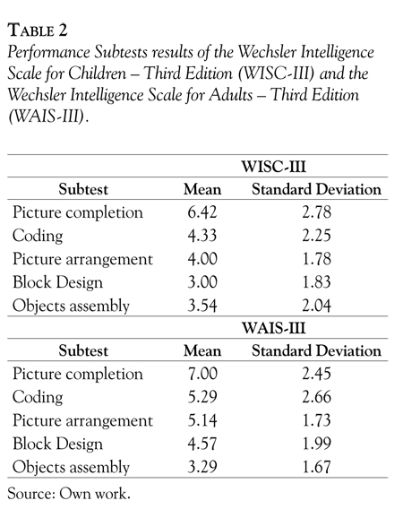
In the scale WAIS-III the PIQ ranged from 61 to 87; the mean obtained was between 7 in the Picture completion subtest and 3.29 in the objects assembly. All subtest scores were lower than expected, as the normal result is 10. These results coincide with previous studies documenting WBS patients' abilities in these areas (Montgolfier et al., 2000) (Table 2).
Consistent with the extant literature, patients scored lower overall on the PIQ subtests than in the VIQ subtests (Oliveira, 1999). More specifically, patients performed worst on tests of objects assembly and block design. These findings are consistent with previous literature that has also documented deficiencies in WBS patient's perceptual organization and their ability to perceive and to analyze ways of reasoning through visuo-spatial or visuomotor test (Tassabehji, 2003) (Table 3).
The studies conducted by Jordan, Reiss, Hoffman and Landau (2002) showed that patients with WBS had difficulty with visuo-constructive skills and show impairment on the part of their brain, suggesting that part of the brain is committed to the process of visual integration.
Total IQ, Verbal IQ and Performance IQ
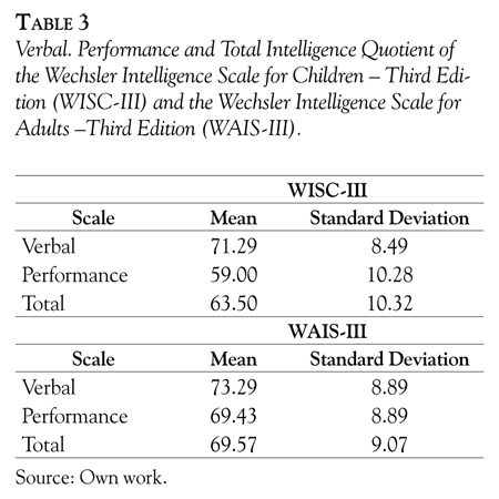
The patients' IQ scores ranged from 50 and 85 points in the test WISC-III and 61 and 86 in the test WAIS-III. These results were lower than expected, compared to the normal result for TIQ, which is of 90 points. These findings are consistent with the literature, which show that the IQ of WBS patients varies between 50 and 70, from moderate to mild mental retardation, respectively (Bellugi, Lichtenberger, Jones, Lai & St. George, 2000; Udwin et al., 1996).
In the test WISC-III the VIQ scores ranged between 57 and 87 points with a media of 71.29 and in the test WAIS-III the scores ranged between 86 and 64 points with a media of 69.43.
The results of the WISC-I11n the non-verbal IQ scores ranged from 45 and 83 points with a media of 59 and in the test WAIS-III ranged from 61 and 87 points with media of69.43. Although VIQ scores were higher than PIQ scores, both were lower than the normal score of 90 points (Table 3).
According to Jarrold, Baddeley and Hewes (1998) some tests have revealed significantly greater impairment in visuo-constructive tasks, as compared to verbal ability in WBS patients, implying dissociation of these tasks.
Our results were similar to those described in the literature, in which WBS patients exhibit greater impairment in PIQ than VIQ (Jarrold et al., 1998; Udwin & Yule, 1991).
Rey-Osterrieth Complex Figure Test
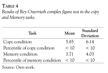
In the task which assessed their ability to copy a figure, the patients scored between 0 and 23 points, with a mean of 5.85 and sd of 6.14. Their memory test results ranged from 18,5 points, with a mean of 3.71 and sd of 4.03. Both results were much lower than expected; the normal score is 50 points (Table 4). The primitive forms showed a distorted integration of test figures, an impaired copy and memory of the overall structure of figures with simplification and loss of details. This performance is typical faulty visuo-spatial processing of WBS child.
A major feature of the cognitive profile in WBS is the marked impairment of visuo-spatial abilities, as demonstrated on drawing or copying tasks (Sempaio, et.al., 2008; Wang & Bellugi, 1993) and on motor tasks requiring visuo-spatial guidance, such as walking over uneven surfaces or down steps (Withers, 1996).
By contrast, face processing and object recognition are relatively spared (Bellugi, Bihrle, Neville, Jernigan & Doherty, 1992; Landau, Hoffman & Kurz, 2006). Form-, color- and face processing functions are associated with the ventral ("what") visual stream, with predominant input from the parvo-cellular pathway; spatial-integrative and motion-processing functions are associated with the dorsal ("where") extrastriatal stream linked to the magno-cellular pathway. Thus, the split in WBS between the extremely poor visuo-spatial abilities and the relatively preserved face and object processing skills, suggests a neural processing abnormality limited to the dorsal stream (Atkinson et al., 2001). This assumption was supported by the study of Meyer-Lindenberg et al. (2004) wherein high functioning subjects with WBS performed similarly well to controls in matching shapes, but significantly worse in assembling the shapes into squares. On functional magnetic resonance imaging (fMRI), the ventral stream was equally activated in both groups.
However, subjects with WBS showed hypo-activation in the dorsal stream areas adjacent to the intraparietal sulcus, which takes part in perceptual-motor coordination and visual attention control. Accordingly, on tasks of attention to objects vs. location, brain structure analyses revealed a reduced gray matter volume in the intraparietal sulcus. It is suggested, from path analysis, that the structural anomaly in the intraparietal sulcus may be responsible for the deficits in the dorsal stream of the visual system (Meyer-Lindenberg et al., 2004).
The subjects with WBS are also characterized by a deficiency in perception of global spatial arrangements, with relatively preserved perception of local spatial arrangements.
This pattern can explain their poor drawing abilities (Bihrle, Bellugi, Delis & Marks, 1989) as well as their poor performance on block design task, in which the subject is required to organize blocks into a global pattern (Landau et al., 2006). For instance, when children with WBS are asked to draw a house, they tend to place the windows and doors as separate entities from the house itself. By contrast, in a typical drawing of a child with Down syndrome, the house elements will be simplified, but there will be a better gestalt relationship among them (Bellugi, Lichtenberger, Mills, Galaburda & Korenberg, 1999).
In summary, the main cognitive phenotype of WBS consists of impairment in most visuo-spatial abilities with a local bias and deficiency in the perception of global spatial arrangements; face processing and object recognition are relatively spared. The phenotype is attributed in part to a deficiency in the dorsal visual stream alongside a relatively intact ventral visual stream.
The percentile of both was below 10. In evaluating the minute, we can see that in print they took longer to perform the test average was 3.7 with a standard deviation of 0.02. In memory, the average was 1.49 with a standard deviation of 0.02. We can infer that this has happened because of the attention turned off and can also be associated with psychiatric comorbidities. Subjects with WBS have visuo-spatial short-term memory deficits, compared to the typically developing controls and to subjects with Down syndrome (Bellugi et al., 1992; Jordan et al., 2002; Wang & Bellugi, 1994).
Physiologically, there is a reduction in the volume of the parahippocampal gyrus, an essential component of the neural system underlying the visuo-spatial memory (Garrett, Menon, MacKenzie & Reiss, 2004). The short-term memory deficit might be partly explained by the impaired visuo-spatial perception typical of these children, but it apparently extends beyond it (Jarrold, Baddeley & Hewes, 1999). Whether the deficit is related to coding, storage or retrieval processes is not yet known.
Vicari, Bellucci and Carlesimo (2003) revealed a failure to integrate results from the composite space among patients. Toga et al. (2006) attempted to clarify the understanding of the cognitive profile of Down's syndrome using neuroimaging and observed a decrease in the subcortical white matter, with increased cortical folds and a reduced volume of gray matter in the parietal sulcus-occipital. Tassa-behji (2003) evaluated the cognitive deficits in two groups of patients, Down syndrome and WBS and found that individuals with Down syndrome could draw the pictures without emphasizing the details, whereas patients with WBS could only focus on the details and were unable to give real shape to the figure.
The visual ability of facial recognition and the verbal ability to describe emotional expressions have been described in patients with WBS (Bellugi et al., 2000). Structurally, the high-resolution MRI studies revealed that subjects with WBS have a disproportionately large volume and an increased density of gray matter in areas known to be important for face processing (Garrett et al., 2004). The larger the gray matter volume in the fusiform gyrus area, the better their face recognition on the Benetton test (Jones, Rossen, Hickok, Jernigan & Bellugi, 1995).
Mimura, Hoeft, Kato, et al. (2010) investigated the possible role of orbitofrontal cortex (OFC) in WBS during positive and negative valence face processing. The results suggested that the activation for positive faces in WBS was equivalent in the medial and lateral OFC, which was also different from the activation pattern of the control group. The study concluded that the results of OFC activation pattern offer additional plausible biological correlates for the positive attribution bias and characteristic hypersociability seen in individuals with WBS). It is possible that the preserved functioning of the fusiform and frontal regions, including the anterior cingulate cortex, which has strong connections to the limbic system, may mediate the increased social interest and attention to faces characteristic of subjects with WBS. This assumption is in line with the robust ERP N200 amplitudes found in scalp regions adjacent to the anterior cingulate cortex during face-processing tasks (Tassabehji, 2003). The impairments in the visual cortical regions may account for the disrupted global coherence and visuo-spatial aspects of face and gaze processing in WBS, as manifested by the diminished accuracy and the longer response time on behavioral measures.
In summary, although face processing is relatively spared in WBS and may indicate an intact ventral stream, it is apparently executed via atypical neural processing mechanisms that rely on component strategies, instead of holistic ones. The face processing in subjects with WBS benefited from the high attention resources, which may be associated with the increased social appetite, typical of the syndrome.
Conclusions
The TIQ ranged from 51 to 86 (media 63): 22 with mild intellectual disability, 4 with moderate mental retardation, 4 borderline and 1 below the normal mean. All patients showed marked visuo-spatial deficits. The visuo-spatial representational skills, such as mental image rotation or assembling abilities, are complex mental processes concerning the ability to operate upon information while making reference (La Femina et al., 2009). It is necessary to understand in more detail the process underlying of these deficits visuo-spatial. Future studies can be conducted to disentangle the specific cognitive processes implied in the task execution to internal representations loaded from long-term memory. We emphasize on the importance of future neuro-psychological studies in Brazilian individuals with WBS because the current literature consists of few studies in this area.
References
Atkinson, J., Anker, S., Braddick, O., Nokes, L., Mason, A. & Braddick, F. (2001). Visual and visuo-spatial development in young children with Williams syndrome. Developmental Medicine and Child Neurology, 43(5), 330-337. [ Links ]
Bellugi, U., Bihrle, A., Neville, H., Jernigan, T. L. & Doherty, S. (1992). Language, cognition and brain organization in a neurodevelopmental disorder. In M. Gunnar & C. Nelson (Eds.), Developmental behavioral neuroscience (pp. 201-232). Hillsdale, NJ: Erlbaum. [ Links ]
Bellugi, U., Lichtenberger, L., Jones, W., Lai, Z. & St. George, M. (2000). The neurocognitive profile of Williams syndrome: A complex pattern of strengths and weaknesses. Journal of Cognitive Neuroscience, 12(1), 7-29. [ Links ]
Bellugi, U., Lichtenberger, L., Mills, D., Galaburda, A. & Korenberg, J. R. (1999). Bridging cognition, the brain and molecular genetics: Evidence from Williams syndrome. Trends in Neuroscience, 22(5),197-207. [ Links ]
Bihrle, A. M., Bellugi, U., Delis, D. & Marks, S. (1989). Seeing either the forest or the trees: Dissociation in visuospatial processing. Brain and Cognition, 11(1), 37-49. [ Links ]
Farran, E. K. & Jarrold, C. (2003). Visuospatial cognition in Williams Syndrome: Reviewing and accounting for the strengths and weaknesses in performance. Developmental Neuropsychology, 23(1-2), 173-200. [ Links ]
Fernando, K., Chard, L., Butcher, M. & McKay, C. (2003). Standardization of the Rey Complex Figure Test in New Zealand children and adolescents. New Zealand Journal of Psychology, 32(1), 33-38. [ Links ]
Jarrold, C., Baddeley, A. D. & Hewes, A. K. (1998). Verbal and nonverbal abilities in the Williams syndrome phenotype: Evidence for diverging developmental trajectories. Journal of Child Psychology and Psychiatry and Allied Disciplines, 39(4), 511-523. [ Links ]
Jarrold, C., Baddeley, A. D. & Hewes, A. K. (1999). Genetically dissociated components of working memory: Evidence from Down's and Williams syndrome. Neuropsychologia, 37(6), 637-651. [ Links ]
Jones, W., Rossen, M. L., Hickok, G., Jernigan, T. & Bellugi, U. (1995). Links between behavior and brain: Brain morphological correlates of language, face and auditory processing in Williams syndrome. Society for Neuroscience Abstracts, 21, 1926. [ Links ]
Jordan, H., Reiss, J., Hoffman, J. E. & Landau, B. (2002). Intact perception of biological motion in the face of profound spatial deficits: Williams syndrome. Psychological Science, 13(2), 162-167. [ Links ]
Klein-Tasman, B. P. & Mervis, C. B. (2003). Distinctive personality characteristics of 8-, 9- and 10-year-olds with Williams syndrome. Developmental Neuropsychology, 23(1-2), 269-290. [ Links ]
La Femina, F., Senese, V. P., Grossi, D. & Venuti, P. (2009). A battery for the assessment of visuo-spatial abilities involved in drawing task. The Clinical Neuropsychologist, 23(4), 691-714. [ Links ]
Landau, B., Hoffman, J. E. & Kurz, N. (2006). Object recognition with severe spatial deficits in Williams syndrome: Sparing and breakdown. Cognition, 100(3), 483-510. [ Links ]
Lenhoff, H. M., Wang, P. P, Greenberg, F. & Bellugi, U. (1997). Williams syndrome and the brain. Scientific American, 277(6), 68-73. [ Links ]
Lu, P. H., Boone, K. B., Cozolino, L. & Mitchell, C. (2003). Effectivenes of Rey Osterrieth Complex Figure Test and the Meyers and Meyers recognition trial: The detection of suspect effort. The Clinical Neuropsychologist, 17(3), 426-440. [ Links ]
Marriage, J. & Scientist, A. (1995). Central auditory hyperacusis in Williams syndrome. In U. Bellugi & C. A. Morris (Eds.), Williams syndrome: From cognition to gene. Abstracts from the Williams Syndrome Association Professional Conference. Genetic Counseling, 6(Special Issue), 131-192. [ Links ]
Martens, M. A., Wilson, S.J. & Reutens, D.C. (2008). Research Review: Williams syndrome: a critical review of the cognitive, behavioral and neuroanatomical phenotype. Journal of Child Psychology and Psychiatry, 49(6), 576-608. [ Links ]
Mervis, C. B., Robinson, B. F., Bertrand, J., Morris, C. A., Klein-Tasman, B. P. & Amstrong, S. C. (2000). The Williams Syndrome Cognitive Profile. Brain and Cognition, 44(3), 604-628. [ Links ]
Meyer-Lindenberg, A., Mervis, C. B. & Berman, K. F. (2006). Neural mechanisms in Williams syndrome: A unique window to genetic influences on cognition and behaviour. Nature Reviews Neuroscience, 7(5), 380-393. [ Links ]
Meyer-Lindenberg, A., Kohn, P., Mervis, C. B., Kippenhan, J. S., Olsen, R. K., Morris, C. A., et al. (2004). Neural basis of genetically determined visuospatial construction deficit in Williams syndrome. Neuron, 43(5), 623-631. [ Links ]
Mimura, M., Hoeft, F., Kato, M., Kobayashi N, Sheau K, et al. (2010). A preliminary study of orbitofrontal activation and hypersociability in Williams Syndrome. Journal of Neurodevelopmental Disorders, 2(2), 93-98. [ Links ]
Montgolfier-Aubron, I., Burglen, L., Chavet, M. S., Tevissen H., Perrot, C., Baudon, J. J., et al. (2000). Early revealing of Williams-Beuren syndrome by digestive disorders. Archives of Pediatrics and Adolescent Medicine, 7(10), 1085-1087. [ Links ]
Oliveira, M. S. (1999). Figuras Complexas de Rey: teste de cópia e de reprodução de memória de figuras geométricas complexas. Manual André Rey (Revisáo técnica Teresinha Rey & Lucia CF Franco. Tradução Teresinha Rey & Lucia CF Franco). Sao Paulo: Casa do Psicólogo. [ Links ]
Oliveira, M S., Rigoni, M S. andretta, I. & Moraes, J. (2004). Validação do Teste Figuras Complexas de Rey na População Brasileira. Avaliação Psicológica; 3(1), 33-38. [ Links ]
Rossi, N. F., Moretti-Ferreira, D. & Giacheti, C. M. (2007). Perfil comunicativo de individuos com a Síndrome de Williams-Beuren. Revista da Socieda-de Brasileira de Fonoaudiologia, 12(1), 1-9. [ Links ]
Sampaio A, Sousa N, Fernández M, Henriques M, Gongalves OF. (2008). Memory abilities in Williams syndrome: dissociation or developmental delay hypothesis? Brain and Cognition. 66(3), 290-297. [ Links ]
Tassabehji, M. (2003). Williams-Beuren syndrome: A challenge for genotype-phenotype correlations. Human Molecular Genetics, 15(12), 229-237. [ Links ]
Toga, A. W., Gaser, C., Luders, E., Thompson, P. M., Lee, A. D., Dutton, R. A., et al. (2006). Increased local gyrification mapped in Williams syndrome. Neurolmage, 33(1), 46-54. [ Links ]
Udwin, O., Davies, M. & Howlin, P. (1996). A longitudinal study of cognitive abilities and educational attainment in Williams syndrome. Developmental Medicine and Child Neurology, 38(11), 1020-1029. [ Links ]
Udwin, O. & Yule, W. (1991). A cognitive and behavioural phenotype in Williams syndrome. Journal of Clinical and Experimental Neuropsychology, 13(2), 232-244. [ Links ]
Vicari, S., Bellucci, S. & Carlesimo, G. A. (2003). Visual and spatial working memory dissociation: Evidence from Williams syndrome. Developmental Medicine and Child Neurology, 45(4), 269-273. [ Links ]
Wang, P. P. & Bellugi, U. (1993). Williams syndrome, Down syndrome and cognitive neuroscience. American Journal of Diseases of Children, 147(11), 1246-1251. [ Links ]
Wang, P. P. & Bellugi, U. (1994). Evidence from two genetic syndromes for a dissociation between verbal and visual-spatial short-term memory. Journal of Clinical and Experimental Neuropsychology, 16(2), 317-322. [ Links ]
Wechsler, D. (2002). WISC-lll: Escala de Inteligencia Wechsler para Criangas (Adaptação brasileira da 3s ed.). Sao Paulo: Casa do Psicólogo. [ Links ]
Wechsler, D. (1997). WAlS-lll: Escala de lnteligencia Wechsler para Adultos (Adaptação brasileira da 3s ed.). Sao Paulo: Casa do Psicólogo. [ Links ]
Withers, S. (1996). A new clinical sign in Williams syndrome. Archives of Disease in Childhood, 75(1), 89-90. [ Links ]
